SPRING WEBINAR WITH DR. BRUCE DONOFF
•Transferir como PPT, PDF•
11 gostaram•2,803 visualizações

EDIC is pleased to announce a webinar with Dr. R. Bruce Donoff, the Dean at Harvard Dental School. Dr. Donoff’s presentation will cover the risk factors for inferior alveolar and lingual nerve injury after third molar extraction, as well as the proper documentation and follow up of nerve injuries. Dr. Donoff will also discuss the potential for recovery from paresthesia after surgical intervention. The webinar will be held on May 10, 2011 at 7:00 PM.

Recomendados
Recomendados
Mais conteúdo relacionado
Mais procurados
Mais procurados (20)
Destaque
Destaque (20)
Semelhante a SPRING WEBINAR WITH DR. BRUCE DONOFF
Semelhante a SPRING WEBINAR WITH DR. BRUCE DONOFF (20)
Último
Último (20)
SPRING WEBINAR WITH DR. BRUCE DONOFF
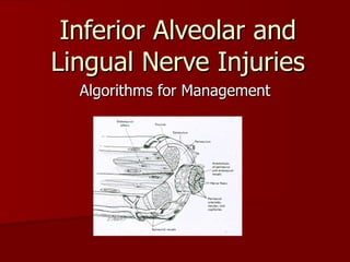
- 1. Inferior Alveolar and Lingual Nerve Injuries Algorithms for Management
- 10. The Five Radiographic Signs 1 Fig. 1: Darkening of the Root Fig. 2: Deflection of of the Roots Fig. 3: Interruption of White Line Fig. 4: Diversion of the IA Canal Fig. 5: Narrowing of the Root The Five Radiographic Signs 1 Fig. 1: Darkening of the Root Fig. 2: Deflection of the Roots Fig. 3: Interruption of White Line Fig. 4: Diversion of the IA Canal Fig. 5: Narrowing of the Root
- 11. Radiographic Risk Factors X-ray finding Sens (%) Spec (%) PPV (%) NPV (%) PPV (%) NPV (%) Diversion 50 82 34 89 2.7 99 Darkening 65 73 31 93 2.3 99 Interruption 80 54 25 93 1.7 99 Any finding 100 33 22 100 1.4 100
- 32. Using Ultrasound to Visualize the Lingual Nerve Presented by James Olsen HSDM 2006 January 20, 2006 http://i.cnn.net/cnn/2003/HEALTH/08/27/ultra.stethoscope/story.portable.ultrasound.jpg
- 33. Mandibular Nerve Branches A. Auriculotemporal B. Lingual C. Inferior Alveolar D. N. to the Mylohyoid E. Mental F. Buccal http://www.meddean.luc.edu/lumen/MedEd/GrossAnatomy/h_n/cn/cn1/images/cnb3.jpg
- 34. Lingual Nerve Anatomy The lingual nerve crosses the submandibular duct twice in the paralingual space. http://www.sciential.net/images/Clemente2f.jpg
- 35. Lingual Nerve Anatomy Pogrel MA, Renaut A, Schmidt B, Ammar A. The relationship of the lingual nerve to the mandibular third molar region: an anatomic study. J Oral Maxillofacial Surgery, 1995, pp. 1178-81.
- 36. Lingual Nerve Injury Loss of taste from anterior 2/3 of tongue ipsilateral to the lesion (special sensory component of CN VII) Loss of general sensation from the tongue (general sensory component of CN V3). http://info.med.yale.edu/caim/cnerves/cn7/cn7_graphics/fig7_25.gif
- 37. Treatment of Lingual Nerve Injuries Robinson PP, Alison RL, Julian MY, Smith KG. Current management of damage to the inferior alveolar and lingual nerves as a result of removal of third molars. British Journal of Oral and Maxillofacial Surgery , 2004; 42, 285-292
- 38. Case Report *Images courtesy of Dr. Donoff
- 39. Case Report
- 40. Imaging of Lingual Nerve Injuries CT
- 41. Imaging of Lingual Nerve Injuries Miloro M, Halkias LE, Slone HW, Chakeres DW. Assessment of the Linual Nerve in the Third Molar Region Using Magnetic Resonance Imaging. J Oral Maxillofacial Surgery , 1997; 55:134-137. MRI MRI
- 42. Using Ultrasound to Visualize the Lingual Nerve http://www.bbc.co.uk/health/images/300/ultrasound.jpg
- 43. Ultrasonography Altinok T, Baysal O, Karakas HM, Sigirci A, Alkan A, Kayhan A, Yologlu S. Ultrasonographic assessment of mild and moderate idiopathic carpal tunnel symdrome. Clinical Radiology, 2004 Oct; 59(10): 916-25
- 44. Ultrasonography De Kool BS, Van Neck JW, Blok JH, Walbeehm ET, Hekking IV, Gerhard H. Ultrasound imaging of the rabbit peroneal nerve. Journal of the Peripheral Nervous System , 10 (4), 369-374. PN, peroneal nerve; TN, tibial nerve; BF, biceps femoris muscle; SM, semimembranosus muscle; F, femur
- 46. Study HST transducer (25mm, 10-5 MHz). Sonosite 180 Plus
- 47. Study Yorkshire cadaver pig head OMFS Endoscopic Research Center
- 48. Study Surgery, Anesthesia, & Experimental Techniques in Swine by M. Michael Swindle, DVM
- 49. Study Yorkshire cadaver pig head
- 50. Study Yorkshire cadaver pig head
- 51. Study
- 52. Study
- 53. Study
- 54. Table 1: Summary Diagnostic Outcomes for Three Evaluators Average 17 / 27 (63%) Pig Key Evaluator #1 (Radiologist) Evaluator #2 (Senior Surgeon) Evaluator #3 (Surgical Fellow) 1 Right Partial Transection Intact Intact Partial Transection Left Nerve Intact Full Transection Intact Full Transection 2 Right Partial Transection Partial Transection Intact Full Transection Left Full Transection Full Transection Full Transection Intact 3 Right Nerve Intact Intact Intact Intact Left Partial Transection Partial Transection Partial Transection Partial Transection 4 Right Full Transection Full Transection Partial Transection Full Transection Left Nerve Intact Intact Intact Partial Transection 5 Right Full Transection Partial Transection Full Transection Full Transection Number of Correct Diagnoses (%) 6 (66.7%) 6 (66.7%) 5 (55.6%)
- 55. Results
- 56. Results
- 57. Table 2: Average distance of lingual nerve from alveolus as measured with ultrasound Pig Ave. distance from alveolus (mm) 1 Right 0.0 Left 1.867 2 Right 2.756 Left 0.533 3 Right 0.0 Left 1.956 4 Right 0.978 Left 0.356 5 Right 0.711 Average 1.02
- 58. Results Yorkshire cadaver pig head
- 59. Case Report *Images courtesy of Dr. Donoff
- 65. Susarla et al. JOMFS 65:1070, 2007
- 66. Susarla et al.JOMFS 65:1070, 2007
- 67. Susarla, et al. JOMFS 65:60, 2007
- 68. Susarla, et al. JOMFS 65:60, 2007
- 69. Hillerup and Stoltze. IJOMFS 36:1139, 2007
- 70. Hillerup and Stoltze IJOMFS 36: 1139, 2007
- 71. Hillerup and Stoltze IJOMFS 36:884, 2007
- 72. Hillerup and Stoltze IJOMFS 36:884, 2007