Mais conteúdo relacionado
Semelhante a Nature10209 (20)
Mais de Dickdick Maulana (20)
Nature10209
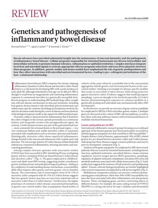
- 1. REVIEW doi:10.1038/nature10209
Genetics and pathogenesis of
inflammatory bowel disease
Bernard Khor1,2,3*, Agnès Gardet1,2* & Ramnik J. Xavier1,2,4
Recent advances have provided substantial insight into the maintenance of mucosal immunity and the pathogenesis
of inflammatory bowel disease. Cellular programs responsible for intestinal homeostasis use diverse intracellular and
intercellular networks to promote immune tolerance, inflammation or epithelial restitution. Complex interfaces integrate
local host and microbial signals to activate appropriate effector programs selectively and even drive plasticity between
these programs. In addition, genetic studies and mouse models have emphasized the role of genetic predispositions and
how they affect interactions with microbial and environmental factors, leading to pro-colitogenic perturbations of the
host–commensal relationship.
I
nflammatory bowel disease (IBD) comprises the chronic relapsing cohorts of the same ethnicity is probably due to the concomitant
inflammatory disorders Crohn’s disease and ulcerative colitis. Family aggregation of both genetic and environmental factors in the case-
history is a risk factor for developing IBD, with a peak incidence in control studies4. Smoking is an example of a disease-specific modifier
early adult life, although individuals of any age can be affected. IBD is that seems to exacerbate Crohn’s disease while being protective
thought to result from an inappropriate and continuing inflammatory against ulcerative colitis. Evidence suggests that smoking impairs
response to commensal microbes in a genetically susceptible host. autophagy, a process thought to be involved especially in Crohn’s
Recent progress in understanding IBD pathobiology offers insight disease, demonstrating how exposure to a disease modifier in a
into relevant disease mechanisms in mucosal immunity, including genetically predisposed individual may mechanistically affect IBD
how genetic factors interact with microbial and environmental cues development5.
within tissue-specific contexts, the biological checkpoints involved, the In this Review, we provide an overview of genes and susceptibility
selective decisions made during the course of disease and how plasticity loci implicated in IBD by GWAS and other genetic studies. Candidate
of the biological response results in the capacity for different phenotypes. genes are discussed in the context of IBD-relevant pathways, as well as
Ulcerative colitis is characterized by inflammation that is limited to how these molecular pathways interact with environmental factors to
the colon: it begins in the rectum, spreads proximally in a continuous modulate intestinal homeostasis.
fashion and frequently involves the periappendiceal region. By
contrast, Crohn’s disease involves any part of the gastrointestinal tract Genes and pathways in IBD
— most commonly the terminal ileum or the perianal region — in a International collaborative research groups focusing on an unbiased
non-continuous fashion and, unlike ulcerative colitis, is commonly appraisal of the human genome have been particularly successful in
associated with complications such as strictures, abscesses and fistulas. identifying genes and genetic loci that contribute to IBD susceptibility1,2.
Histologically, ulcerative colitis shows superficial inflammatory Despite distinct clinical features, approximately 30% of IBD-related
changes limited to the mucosa and submucosa with cryptitis and crypt genetic loci are shared between ulcerative colitis and Crohn’s disease,
abscesses. The microscopic features of Crohn’s disease include thickened indicating that these diseases engage common pathways and may be
submucosa, transmural inflammation, fissuring ulceration and non- part of a mechanistic continuum (Fig. 1).
caseating granulomas. Analyses of the genes and genetic loci implicated in IBD show several
Among complex diseases, genome-wide association studies pathways that are crucial for intestinal homeostasis, including barrier
(GWAS) have been successful in IBD, identifying 99 non-overlapping function, epithelial restitution, microbial defence, innate immune
genetic risk loci, including 28 that are shared between Crohn’s disease regulation, reactive oxygen species (ROS) generation, autophagy,
and ulcerative colitis1,2 (Fig. 1). The genes implicated in childhood- regulation of adaptive immunity, endoplasmic reticulum (ER) stress and
onset and adult-onset IBD overlap, suggesting similar contributory metabolic pathways associated with cellular homeostasis (Fig. 2). Early
genetic predispositions and pathophysiological pathways. Adding to studies have suggested the existence of both protective and predisposing
the complexity of understanding disease mechanisms, a susceptibility alleles6. Disease-relevant biological pathways are further highlighted
allele often requires other genetic and non-genetic cues to manifest when several components are implicated as risk factors together (Fig. 3).
disease. The concordance rate in monozygotic twins of 10–15% in Multidisease comparative analysis can uncover common disease-
ulcerative colitis compared with 30–35% in Crohn’s disease suggests causing genes and pathways. More than 50% of IBD susceptibility loci
that non-genetic factors may have an even more important role in have also been associated with other inflammatory and autoimmune
ulcerative colitis than in Crohn’s disease3. Furthermore, the higher diseases. These overlapping genes can have contrasting effects in
penetrance of common Crohn’s-disease-associated polymorphisms different diseases. For example, the same coding variant of PTPN22
in genetic case-control studies than in population-based studies of (R620W) is a strong risk factor for type 1 diabetes and rheumatoid
1
Gastrointestinal Unit and Center for the Study of Inflammatory Bowel Disease, Massachusetts General Hospital, Harvard Medical School, Boston, Massachusetts 02114, USA. 2Center for
Computational and Integrative Biology, Massachusetts General Hospital, Harvard Medical School, Boston, Massachusetts 02114, USA. 3Pathology Service, Massachusetts General Hospital, Boston,
Massachusetts 02114, USA. 4The Broad Institute of MIT and Harvard, 7 Cambridge Center, Cambridge, Massachusetts 02142, USA.
*These authors contributed equally to this work.
1 6 J U N E 2 0 1 1 | VO L 4 7 4 | NAT U R E | 3 0 7
© 2011 Macmillan Publishers Limited. All rights reserved
- 2. INSIGHT REVIEW
a Crohn’s disease Ulcerative colitis Figure 1 | Genetic architecture of IBD-linked
susceptibility loci. a, GWAS have identified
6 gene deserts 71 risk loci in Crohn’s disease and 47 risk loci in
eQTL effects 9 gene deserts ulcerative colitis (P value of association < 5×10⁻⁸).
cis/trans ncRNA Predicted ORF >5 genes Of these, 28 risk loci exhibit shared associations
1 11 4 ncRNA (defined as P < 5×10⁻⁸ for either Crohn’s disease
cis >5 genes 3 6 or ulcerative colitis, and P < 1×10⁻⁴ for the other
8 3
3 form of IBD). Approximately half of the loci
11 cis
trans 17 20
implicated in Crohn’s disease and ulcerative
41 21 colitis are associated with cis- and/or trans-
<5 genes <5 genes
54
expression quantitative trait loci (eQTL) effects
trans 32
10
(left panels). Genes whose expression are affected
by these variants could also be involved in IBD
eQTL Loci composition eQTL Loci composition pathogenesis. The loci composition (right panels)
shows the number of genes that either lie within
or segregate in linkage disequilibrium with IBD-
b implicated loci (coefficient of correlation r2 > 0.8).
These loci are structurally heterogeneous, and are
intracellular logistics tolerance
interferon response carbohydrate metabolism
associated with widely ranging numbers of genes.
barrier function chemotaxis solute carrier
Loci not associated with any genes, known as gene
deserts, frequently contain non-coding transcripts
iron metabolism dendritic cell plasticity polarized secretion microtubules/centrosome
innate defence
or predicted open reading frames (ORFs), and can
exocytosis
miRNA/lincRNA DNA methylation be associated with trans-eQTL effects.
inflammation mediators negative regulators of immunity b, Recurring terms illustrating biological
fibrogenesislymphocyte activation
adenosine
solute transport processes implicated by at least three genes
inflammasome represented in IBD loci; font sizes are proportional
restitution IL-23–IL-23R phagocytosis antimicrobial peptides
IgA/Breg cells
actin cytoskeleton
Paneth cells
DNA/RNA binding to the number of genes associated with each
respective process. Breg cells, B regulatory cells;
cell migration adaptive immunity regulators ER, endoplasmic reticulum; GPCR, G-protein-
TH17/Treg cells GPCR signalling ROS ER stress coupled receptor; IL, interleukin; lincRNA,
large intervening non-coding RNA; miRNA,
+Ly6Chigh/Gr1−Ly6Clow autophagy
hypoxia
Gr1 pathogen sensing microRNA; ncRNA, non-coding RNA; NF-κB,
antigen presentation lipid metabolism goblet cells/mucin nuclear factor-κB; ROS, reactive oxygen species;
stress response signature NF-κB activation/inhibition TH17 cells, T helper 17 cells; Treg cells, T regulatory
mycobacteria restriction factors cells.
arthritis, but is protective against Crohn’s disease7. These data suggest early-onset IBD12. Other interleukin-10 receptor (IL-10R) signalling
that crucial clues to disease biology may reside in understanding the components have also been implicated by GWAS, including STAT3,
function of these shared genes. Several loci containing genes such as TYK2, JAK2 and IL10 itself, in concordance with the notion that both
MST1, IL2, CARD9 and REL are shared between ulcerative colitis and rare and common variants may highlight the same pathway. Although
the associated complication primary sclerosing cholangitis (PSC)8. these components can also function in other contexts — for example,
This overlap may help to identify subsets of patients with ulcerative the transcription factor STAT3 and the kinase proteins TYK2 and JAK2
colitis who are at risk of PSC. Risk loci for Crohn’s disease present an are involved in the signalling of the interleukins IL-6, IL-22 and IL-23
unexpected overlap with susceptibility regions for Mycobacterium leprae — these results illustrate the value of genetic studies in determining
infection, including genes such as NOD2, C13orf31 and LRRK2 (ref. 9). not just single genes, but also disease-relevant pathways. Recent
Although absent from the leprosy GWAS, other Crohn’s-disease- resequencing studies in IBD recovered both known and new variants
associated genes are also implicated in host responses to mycobacterial of CARD9, NOD2 and IL23R, with independent effects on disease risk.
infection, including CARD9, LTA, ITLN1 and IRGM1. Thus, studies The IL23R variants were protective, supporting previous findings of
to delineate immune responses to antigens from, and infection by, a common protective IL23R allele and illustrating how studies of rare
mycobacteria, or other microbes that elicit similar host cell responses, variants can reinforce GWAS findings6. Furthermore, T helper 17
may also be pertinent to Crohn’s disease. (TH17) cells generated ex vivo from subjects with a variant IL23R allele
Genetic variants associated with IBD can vary in frequency (R381Q) show decreased production of the pro-inflammatory cytokine
depending on the cohort ethnicity, raising the possibility that some IL-17A in response to IL-23 stimulation, emphasizing the importance
such variants may have emerged in the context of historical selective of IL-23-related pathways in human IBD13.
pressures. Although this notion remains to be demonstrated in IBD, Early functional studies attempting to determine causality have
lessons from other autoimmune and infectious contexts lend support. largely focused on coding variants, although non-coding single
For example, variants of apolipoprotein L1 and the inhibitory Fc nucleotide polymorphisms (SNPs) can be associated with qualitative
receptor FcγRIIb that confer protection against trypanosomiasis and and quantitative changes. Alternative splicing exemplifies a qualitative
malaria, respectively, are more common in populations endemically change affected by non-coding modifications. In the context of
exposed to these pathogens, but these variants also confer increased regulating immune responses, IL23R and NOD2 can encode truncated
susceptibility to focal segmental glomerulosclerosis and systemic lupus variants that inhibit their signalling pathways14,15. Furthermore,
erythematosus (SLE), respectively10,11. genetic changes may affect transcription-factor-binding sequences,
Current GWAS are typically powered to characterize variants of locus accessibility, translational efficiency and trans-regulators such
>1% frequency and do not include the contributions from rare variants as non-coding RNAs and microRNAs (miRNAs). In this regard, a
(<1% frequency). Exome sequencing can be useful for identifying rare Crohn’s-disease-associated synonymous variant in IRGM (c.313C>T)
variants, whereas whole-genome sequencing is of value in elucidating perturbs regulation by miR-196A and miR-196B, and is associated
modifier loci. If pedigrees are available, rare variant discovery can with altered IRGM expression in patients with Crohn’s disease
be further targeted by fine mapping, as shown by the identification who bear this SNP16. Cis- or trans-expression quantitative trait loci
of IL10RA polymorphisms associated with the development of (eQTL) are detected for approximately half of the IBD risk regions,
3 0 8 | NAT U R E | VO L 4 7 4 | 1 6 J U N E 2 0 1 1
© 2011 Macmillan Publishers Limited. All rights reserved
- 3. REVIEW INSIGHT
IBD-related processes
Microbiota, Epithelial barrier
diet GNA12*, HNF4A, CDH1,
ERRFI1, MUC19, ITLN1*
Restitution
REL, PTGER4, NKX2-3, STAT3,
ERRFI1, HNF4A, PLA2G2A/E
Solute transport
SLC9A4, SLC22A5, SLC22A4*,
Microbial
sensors
P P G AQP12A/B, SLC9A3, SLC26A3
Paneth cells
ITLN1*, NOD2*, ATG16L1*, XBP1*
Innate mucosal defence
Recruitment
NOD2*, ITLN1*, CARD9*,
of mediators
REL, SLC11A1, FCGR2A*/B
ILC
Immune cell recruitment
Signal
CCL11/CCL2/CCL7/CCL8,
amplification
CCR6, IL8RA/IL8RB, MST1*
Antigen presentation
ERAP2*, LNPEP, DENND1B
B cell TH17 Treg cell
Transducers IL-23/TH17
and effectors IL23R*, JAK2, TYK2*, STAT3,
ICOSLG, IL21, TNFSF15*
IgA
T-cell regulation
Plasma cell NDFIP1, TNFSF8, TAGAP, IL2, IL2RA
TNFRSF9, PIM3, IL7R*, IL12B, IL23R*,
PRDM1, ICOSLG, TNFSF8, IFNG, IL21
B-cell regulation
IL5, IKZF1, BACH2, IL7R*, IRF5
Immune tolerance
Cellular responses IL10, IL27*, SBNO2, CREM,
IL1R1/IL1R2, NOD2*
Autophagy ER stress Intracellular logistics Cell migration
ATG16L1*, IRGM, NOD2*, CPEB4, ORMDL3, VAMP3, KIF21B, TTLL8, ARPC2, LSP1, AAMP
LRRK2, CUL2, PARK7, DAP SERINC3, XBP1* FGFR1OP, CEP72, TPPP
UC
CD
Apoptosis/necroptosis Carbohydrate metabolism Oxidative stress UC/CD
FASLG, THADA*, GCKR*, SLC2A4RG PRDX5, BACH2, ADO, GPX4, GPX1*, SLC22A4, LRRK2, cis-eQTL
DAP, PUS10, MST1* NOD2*, CARD9*, HSPA6, DLD, PARK7, UTS2*, PEX13
*Coding mutation
Figure 2 | A model for IBD pathways based on GWAS. Intestinal affect the balance of these signals. Genes in linkage disequilibrium (r2 > 0.8)
homeostasis involves the coordinated actions of epithelial, innate and adaptive with IBD-associated single nucleotide polymorphisms (SNPs) were manually
immune cells. Barrier permeability permits microbial incursion, which is curated and classified according to their function(s) in the context of intestinal
detected by the innate immune system, which then orchestrates appropriate homeostasis and immunity. Text colour indicates whether the genes are linked
tolerogenic, inflammatory and restitutive responses in part by releasing to risk loci associated with Crohn’s disease (CD; black), ulcerative colitis (UC;
extracellular mediators that recruit other cellular components, including blue) or both (red). Asterisk denotes corresponding coding mutations; cis-
adaptive immune cells. Genetic variants, the microbiota and immune factors eQTL effects are underlined. G, goblet cell; P, Paneth cell.
indicating that allele-specific gene-expression changes contribute to Epithelial encounters and pathogenicity
disease risk (Fig. 1). Furthermore, IBD-implicated loci contain more The intestinal mucosa exists in a functional equilibrium with the
than 10 miRNA-encoding sequences and 39 large intervening non- complex luminal milieu, which is dominated by a spectrum of microbial
coding RNAs (lincRNAs), 5 of which interacted with the histone species and their products. Maintaining this functional balance is
methyltransferase polycomb repressive complex 2 (PRC2), supporting central to preserving normal mucosal physiology, with perturbations
the notion that regulation of gene expression by miRNAs and lincRNAs contributing to the pathophysiology of many gastrointestinal disorders,
may be mechanistically relevant in IBD17. including IBD. In addition to nutrient absorption, intestinal epithelial
So far, GWAS account for 23% and 16% of the heritability in Crohn’s cells (IECs) perform both barrier and signal-transduction functions,
disease and ulcerative colitis, respectively1,2. Although these may be with the capacity to sense luminal contents through surface receptors
underestimates owing to the net effect of common variants that are and, in return, secrete regulatory products that can orchestrate an
individually too small to calculate accurately; the missing heritability appropriate response in the underlying lamina propria.
may further comprise genetic, epigenetic and non-genetic (including Molecular details of the epithelial barrier and the structure of tight
environmental) components. Genetic factors such as rare variants, junctions, which are crucial to its integrity, have been characterized.
private mutations, structural variants and interactions between genes Abnormal intestinal permeability has been observed in IBD patients
are not well captured by GWAS. Nevertheless, a key success of GWAS and in some of their first-degree relatives. Genes within several
in IBD has been the ability to provide insight into disease pathobiology IBD-associated loci indicate a role for barrier integrity in disease
by highlighting key molecular pathways. predisposition, implicating candidate genes such as CDH1, GNA12 and
1 6 J U N E 2 0 1 1 | VO L 4 7 4 | NAT U R E | 3 0 9
© 2011 Macmillan Publishers Limited. All rights reserved
- 4. INSIGHT REVIEW
Epithelial junctions Innate sensors GPCRs
LPA
PLA2G2E Mincle MDP ssRNA ω−3 fatty acid Kynurenic acid H+ SCFA
LRRK2
PTGER4 Gα12 HNF-4α GPR120 GPR35 GPR65 GPR109A GPR43
CARD9
ROS NOD2
?
ERRFI1 EGFR Apical junction PI(3)K β2-Arrestin cAMP
complex NF-κB IL-10 IRF3
Glucose uptake NF-kB Inflammation
resolution
IL-10 signalling TH17 program T-cell negative regulators B cells
Cytokines/
IL-6R IL-27Rα/IL-27 TNFSF15 TCR EGR2 IKZF1 CREM CD40 BCR CD22
ligands
STAT3 STAT1 IL-23R
JAK2 CBL-B IL-10 NOTCH1 IL-2 PTPN22 SIAE
MAF TYK2
STAT3
IL-10 PRDM1
IL-2
RORγt BACH2
ZFP36L1
AHR REL Sialyl-IgG
IL-10Rα ZFP36L2
BATF
(ORMDL3)
IRF4
Anti-inflammatory
IgA
responses
IL-17
IL-22
CCR6
Figure 3 | Genetic variants in IBD signalling modules. Schematic of BCR, B-cell receptor; cAMP, cyclic AMP; EGFR, epidermal growth factor
selected signalling pathways involved in the maintenance of intestinal receptor; ERRFI1, ERBB receptor feedback inhibitor 1; Gα12, G protein
homeostasis, including epithelial junctional complex assembly, innate immune subunit α12; GPCR, G-protein-coupled receptor; HNF-4α, hepatocyte nuclear
recognition of pathogen-associated motifs, GPCRs and immune defence, factor-4α; LPA, lysophosphatidic acid; MDP, muramyl dipeptide; NF-κB,
anti-inflammatory interleukin-10 (IL-10) signalling, TH17-cell differentiation, nuclear factor-κB; PI(3)K, phosphatidylinositol-3-OH kinase; PLA2G2E,
inhibitory pathways in lymphocyte signalling, and B-cell activation and phospholipase A2, group IIE; PTGER4, prostaglandin E receptor 4; SCFA,
IgA antibody responses. Proteins encoded by genes identified as being in short-chain fatty acid; SIAE, sialic acid acetylesterase; ssRNA, single-stranded
linkage disequilibrium with IBD-risk SNPs (r2 > 0.8) are highlighted in red. RNA; TCR, T-cell receptor.
PTPN2. Genetic studies have shown that truncated forms of the adherens to the role of these cells in crypt homeostasis and maintenance of the
junction protein E-cadherin (encoded by CDH1) are associated with intestinal stem-cell niche, they also secrete antimicrobial effectors
Crohn’s disease, and intestinal biopsies from patients with Crohn’s disease that prevent microbial invasion and control the composition of the
carrying these mutant alleles show inappropriate protein localization and gut microflora. These effectors include lysozyme, RegIIIγ, secreted
cytosolic accumulation18. Activation of the G protein Gα12 (encoded by phospholipase A2 (which degrades bacterial membrane phospholipids)
GNA12) leads to phosphorylation of the tight junction proteins ZO-1 and defensins HD5 and HD6 (pore-forming hydrophobic peptides that
and ZO-2, resulting in destabilization of cell junctions in epithelial cell can integrate into bacterial membranes, resulting in lysis). Production
lines19. In vitro studies show that the protein tyrosine phosphatase family of AMPs is regulated by Toll-like receptor (TLR) and NOD2 signals
member PTPN2 protects against interferon-γ (IFN-γ)-induced epithelial triggered by commensal flora. Paneth cell defects and susceptibility
permeability; concordantly, Ptpn2-deficient mice show increased to intestinal inflammation have been uncovered in mice deficient in
susceptibility to experimental colitis20,21. several Crohn’s-disease-associated genes, including Nod2, Atg16l1
Genetic studies have associated IBD with several transcription factors and Xbp1 (refs 27–29). These results highlight pathways important to
involved in epithelial regeneration, such as HNF4A and NKX2-3, which Paneth cell biology, such as the regulation of AMP production (Nod2),
control crypt cell proliferation and IEC differentiation, respectively22–24. granule exocytosis (Atg16l1) and the ER stress response (Xbp1). Similar
Spontaneous colitis did not occur in all animal models with IEC-specific phenotypes have been observed in human disease, such that patients
deletion of Hnf4a, suggesting that further environmental triggers are with Crohn’s disease carrying the ATG16L1 (T300A) mutation show
required for disease22,23. STAT3, the gene encoding which lies within an Paneth cell granule abnormalities. These findings suggest that defects
IBD-implicated locus, is activated in epithelial cells from patients with in Paneth cell biology may define a subset of patients with Crohn’s
IBD, and IEC-specific Stat3 deletion affects epithelial repair25. disease.
The intestinal barrier is enhanced by the presence of a pre-epithelial Cells with high synthetic capacity and secretory activity, such as
layer formed primarily of mucus glycoproteins, trefoil peptides, IgA Paneth cells and goblet cells, have high baseline levels of ER stress,
and antimicrobial peptides (AMPs). Goblet cells generate the mucus leading to activation of the unfolded protein response (UPR), which
layer, a protective polysaccharide bilayer rich in cationic proteins, controls cellular programs that allow proper protein processing.
the inner layer of which is essentially devoid of microbes. Patients The UPR is mainly cytoprotective, although it can signal apoptosis
with IBD frequently have a compromised mucus layer and increased after sustained ER stress. Increased intestine epithelial ER stress and
mucolytic bacteria; mucus layer defects are also observed in Muc2−/− susceptibility to colitis have been observed in mice with overactivation
and IEC-specific C1galt1−/− mice, which develop spontaneous colitis26. of, or perturbations in, the UPR pathway, including Muc2 missense
Interestingly, some patients with ulcerative colitis show defective mutation, Agr2−/−, Ern2−/− (also known as Ire1b−/−), IEC-specific Xbp1−/−
intestinal O-glycosylation resembling that seen in C1galt1−/− mice26. and Mbtps1-hypomorphic mice29,30. Similarly, studies in primary
Paneth cells are located in the crypts of the small intestine. In addition IECs from patients with IBD show activated ER stress responses,
3 1 0 | NAT U R E | VO L 4 7 4 | 1 6 J U N E 2 0 1 1
© 2011 Macmillan Publishers Limited. All rights reserved
- 5. REVIEW INSIGHT
and hypomorphic variants of XBP1 have been associated with risk vascular-endothelial-growth-factor-secreting Ly6Clow monocytes are
of IBD29. Overall, these results indicate that genetic variants that mobilized during the resolution phase of inflammation39. Neutrophils
perturb mechanisms that protect against ER stress can affect intestinal may also contribute to the resolution of inflammation, for example, by
homeostasis in IBD. In addition to its effects on cell viability, ER stress synthesizing anti-inflammatory mediators such as lipoxin A4. Studies
also activates autophagy and IL-23 release, suggesting that sustained showing impaired secretion of lipoxin A4 in mucosal tissues from
ER stress may engage inflammatory circuits that are subsequently patients with ulcerative colitis support the relevance of such mechanisms
propagated by T cells31. in IBD.
In addition to limiting bacterial translocation across the mucosal IL-22 is emerging as an important cytokine in epithelial homeostasis,
barrier, IECs promote intestinal homeostasis by regulating innate showing protective activity in different models of colitis through its
and adaptive immune responses. Illustrating this point, IECs produce stimulatory effect on antimicrobial and reparative processes. Produced
intestinal alkaline phosphatase, which can mediate lipopolysaccharide by several cell types, such as ILCs, lymphoid tissue induced (LTi)
detoxification. Resolvin-E1, which is generated in part through cells, TH17 cells and γδ T cells, most intestinal IL-22 at steady state is
the action of epithelial cyclooxygenase-2, attenuates neutrophil produced by ILCs expressing the transcription factor RORγt40,41. Studies
transmigration and upregulates epithelial expression of intestinal in patients with Crohn’s disease have shown decreased frequencies of
alkaline phosphatase during the restitutive response, a process termed IL-22-secreting ILCs in the lamina propria42. Together, these findings
epithelial imprinting32. IECs can also modulate adaptive immune suggest a central role for ILCs (and other IL-22-producing cells) in
responses, driving the differentiation of anti-inflammatory T regulatory regulating intestinal homeostasis, which remains to be characterized
(Treg) cells by releasing the vitamin A metabolite retinoic acid and the in IBD.
cytokines thymic stromal lymphopoietin (TSLP) and transforming
growth factor-β (TGF-β)33. Breakdown in such epithelial defence NOD2 and IBD
mechanisms could lead to pathological intestinal inflammation. NOD2 was the first gene to be associated with IBD, and thereafter sev-
eral genes that interact epistatically with NOD2 signalling were also
Checkpoints in the innate immune response implicated. NOD2 recognizes the peptidoglycan product muramyl
The physical barrier of the intestinal epithelium is complemented by dipeptide (MDP), which modulates both innate and adaptive immune
a well-evolved mucosal innate immune system, which is populated responses43 (Fig. 4). For example, MDP stimulation induces autophagy,
by cells poised to defend against pathogenic incursions and curtail which controls bacterial replication and antigen presentation, and
inflammatory responses to maintain a state of hyporesponsiveness to acts on dendritic cells in conjunction with TLR ligands to promote
commensal bacteria. Dendritic cells, macrophages, innate lymphoid TH17-cell differentiation44,45. NOD2 may also contribute to immune
cells (ILCs) and neutrophils are crucial cellular components of the innate tolerance. These effects are impaired in cells from patients with the
immune system during infection or inflammation. Supporting the Crohn’s-disease-associated NOD2 mutation 3020insC. Furthermore,
notion that defective innate immune responses can lead to IBD, patients NOD2 can participate in distinct MDP-independent pathways such
with innate immunodeficiencies such as chronic granulomatous disease as regulation of the T-cell response and the type I IFN response to
and Hermansky–Pudlak syndrome, which is associated with defective single-stranded RNA (ssRNA) stimulation, indicating that gut micro-
responses to bacterial DNA motifs (CpG oligonucleotides) specifically bial ssRNAs may exist and have immunomodulatory properties46.
in plasmacytoid dendritic cells, tend to develop IBD34. Similarly, patients The relative contributions of these cytosolic MDP-sensing pathways
with Crohn’s disease have defective innate immune responses, including vary greatly between cell types (Fig. 4). Further studies are needed to
attenuated macrophage activity in vitro, and impaired neutrophil uncover the effect of disease-associated NOD2 alleles in different cell-
recruitment and exogenous Escherichia coli clearance in vivo35. specific programs, and unravel the precise role(s) of NOD2 in IBD.
Intestinal dendritic cells constitute a central interface for monitoring Other families of innate immune receptors linked to intestinal inflam-
the environment and relaying signals to initiate appropriate adaptive mation and immunity include NOD-like receptors (NLRs) and RIG-I-
immune responses33. Dendritic cell subsets are specialized and like receptors (RLRs). These receptors recognize microbial motifs or
respond to endogenous and exogenous stimuli such as microbial damage-associated molecular patterns and can activate the inflam-
motifs, fatty acids, oxidized lipids and vitamin D by selectively masome, thus appropriate regulation of these pathways is required
engaging pro-inflammatory, anti-inflammatory, epithelial restitutive for intestinal homeostasis. For example, mouse knockout studies of
or T-cell education programs, as well as inducing IgA production33,36. Nlrp3 or RIG-I (also known as Ddx58) show increased susceptibility
For example, Treg-cell differentiation can be promoted by tolerogenic to experimental colitis47. Conversely, sustained overactivation of NLRs
dendritic cells induced by TSLP, TGF-β and retinoic acid, all of which can also have detrimental effects, as illustrated by activating mutations
are made by IECs and stromal cells; these dendritic cells express the in NOD2 and NLRP3 giving rise to Blau syndrome and cryopyrino
integrin CD103 but not the chemokine receptor CX3CR1 (ref. 33). By pathies, respectively.
contrast, dendritic cells expressing E-cadherin are a pro-inflammatory
subset that promotes TH17-cell differentiation (see ref. 37 and page 298 CARD9 and IBD
for further details). Bacterial flagellins can override dendritic cell CARD9 is an IBD-implicated adaptor protein that integrates signals
tolerogenic programs by stimulating TLR5 and inducing the release from many innate immune receptors that recognize viral, bacterial and
of pro-inflammatory mediators from hyporesponsive lamina propria fungal motifs. Depending on the stimulus, CARD9 interacts with dis-
CD11chigh dendritic cells, pointing to a broader role for flagellated tinct signalling complexes and activates different pathways to modulate
bacteria in IBD38. This specific immunostimulatory role for TLR5 may cytokine environments appropriately48,49. In particular, recognition of
be particularly relevant in IBD, as seroreactivity to the bacterial flagellin fungal motifs in human dendritic cells leading to CARD9 and dectin-1
CBir1, observed in approximately 50% of patients with Crohn’s disease, signalling results in the broad activation of members of the nuclear
correlates with a complicated clinical course. factor-κB (NF-κB) transcription factor family, whereas CARD9 and
Intestinal homeostasis is maintained in part by the actions of resident dectin-2 signalling selectively activates the IBD-implicated NF-κB fac-
macrophages that have enhanced phagocytic and bactericidal activity tor REL, enhancing the production of TH17-polarizing cytokines such as
and decreased production of pro-inflammatory cytokines. Specialized IL-1β and the IL-23 p19 subunit50. Defective CARD9 function leads to
macrophage subsets are also involved; tumour-necrosis factor-α the immune disorder mucocutaneous candidiasis, at least in part owing
(TNF-α)-secreting and IL-1β-secreting Ly6Chigh monocytes are to failure to promote an adequate TH17 immune response. These data
recruited in the initial phase of microbial challenge or tissue illustrate how innate immune signalling molecules, including NOD2
injury, whereas reparative IL-10-secreting, TGF-β-secreting and and CARD9, can act as central hubs to integrate diverse signals and
1 6 J U N E 2 0 1 1 | VO L 4 7 4 | NAT U R E | 3 1 1
© 2011 Macmillan Publishers Limited. All rights reserved
- 6. INSIGHT REVIEW
a selectively activate specific effector pathways; in the polymicrobial
context of the gut, it seems reasonable that defects at such nodal points
AMPs would constitute key predispositions to IBD.
Paneth cells Redox equilibrium in IBD
Defensins
The reduction and oxidation (redox) state of the gut depends on an
Antibacterial equilibrium between oxidants, such as free radicals, ROS or reactive
activity nitrogen species, and antioxidant mechanisms, such as the glutathione
peroxidase (GPX) and glutathione S-transferase enzymes. This redox
state affects many signal-transduction pathways, such as NF-κB signal-
ling and AMP activity51. Supporting the importance of antioxidant path-
b Antigen-presenting cell
ways in intestinal homeostasis, mice deficient in both Gpx1 and Gpx2
Acute activation Chronic activation develop spontaneous colitis. IBD genetic studies have implicated loci
IFN-β containing GPX1 and GPX4, further highlighting the relevance of these
MDP mechanisms in disease (Fig. 2). Among the oxidants, ROS represent an
RIP2 independent
IRF4, IL-10, TGF-β, IL-1RA, important class of effector molecules generated by mitochondrial and
ssRNA/RSV IRAK-M dependent
non-mitochondrial sources. ROS are non-toxic at basal levels and are
MDP Self-tolerance
Cross-tolerance
even required to maintain the intestinal stem-cell niche. In the context
Antibacterial autophagy TLR2, TLR4 ligands, of innate immunity, ROS have important antimicrobial activity, and
Antigen presentation IL-1β contribute to intracellular signalling, promoting the production of pro-
IL-1β, TNF-α, IL-6, IL-10
inflammatory cytokines. Furthermore, ROS generated by epithelial cells
after infection can transmit signals to adjacent cells in a paracrine man-
RIP2 dependent
ner, allowing the local coordination of chemokine production52. Genes
MDP +TLR2 IL-23 TH17
MDP ligand
within several IBD-associated loci may either regulate ROS production
or protect against oxidative stress (Fig. 2). In particular, NOD2, CARD9
and IFN-γ-regulated leucine-rich repeat kinase 2 (LRRK2) all contrib-
c T cell
ute to ROS production43,53,54. In addition to pro-inflammatory pathways,
ROS are also involved in Treg-cell polarization and function55,56. Thus,
TH1 model RIP2 understanding the role of disease variants will require a broader under-
independent
standing of the cell- and tissue-specific effects of ROS.
IL-2 (REL dependent) No intestinal inflammation Autophagy and IBD
IFN-γ secretion after transfer into Rag1–/– mice
Genetic analyses have shown an unsuspected role for autophagy in innate
immunity and IBD, implicating two component genes, ATG16L1 and
IRGM, in IBD pathogenesis57–59. Autophagy is involved in intracellular
Humans, impaired by 3020insC
homeostasis, contributing to the degradation and recycling of cytosolic
Mice, impaired by Nod2−/−
Humans and mice, impaired by 3020insC and Nod2−/−, respectively contents and organelles, as well as to resistance against infection and
the removal of intracellular microbes (Fig. 5). ATG16L1 is essential for
all forms of autophagy, and the coding mutation T300A is associated
Figure 4 | Cell-intrinsic functions of NOD2. NOD2 is activated by the with increased risk of Crohn’s disease. Despite ubiquitous expression of
bacterial peptidoglycan muramyl dipeptide (MDP). Cell-specific NOD2 ATG16L1, defects associated with ATG16L1 polymorphisms have so far
functions are shown, distinguishing between those functions impaired
been described only within the gut, probably owing to the high microbial
in cells from humans with the Crohn’s-disease-associated mutation
3020insC (red), from Nod2-deficient mice (blue), or from both (black). a, load in this tissue. Subsequent evidence for MDP stimulation of NOD2-
In Paneth cells, Nod2 deficiency leads to attenuated antibacterial activity activated autophagy illustrates a link between genetic risk loci, and high-
in the intestinal crypts and decreased expression of α-defensin 4 (encoded lights the importance of defining disease-associated pathways and the
by Defcr4, also known as Defa4) and α-defensin-related sequence 10 potential of new roles for known genes44,45. Epithelial cells and dendritic
(DEFCR-RS10, also known as DEFA-RS10). b, MDP-stimulated release of cells containing Crohn’s-disease-associated ATG16L1 and NOD2 vari-
pro-inflammatory NF-κB-dependent cytokines (such as IL-1β, TNF-α and ants show defects in antibacterial autophagy44,45,60. In dendritic cells, these
IL-6), as well as secretion of IL-23 (which promotes TH17 differentiation) defects are associated with an impaired ability to present exogenous anti-
after co-stimulation with MDP and TLR2 ligands, is decreased in antigen- gens to CD4+ T cells44. These results illustrate a close relationship between
presenting cells from Nod2-deficient mice or 3020insC human donors.
NOD2, ATG16L1 and autophagy, affecting intracellular processing and
MDP stimulation also leads to NOD2-activated autophagy and antigen
presentation. In mice, the activation of antigen-presenting cells by ssRNA
communication with the adaptive immune system, suggesting that
or respiratory syncytial virus (RSV) stimulates secretion of type I interferon genetic polymorphisms may affect both pathways concomitantly.
(IFN-β) in a NOD2-dependent, receptor-interacting protein-2 (RIP2)- Abnormalities consistent with Crohn’s disease have been observed
independent fashion. In contrast to the pro-inflammatory effects, chronic in mice with defects in autophagy, including hypomorphic Atg16l1
NOD2 activation (right) by MDP induces both self-tolerance and cross- (Atg16l1HM) and IEC-specific Atg5-deficient mice27. Paneth cells either
tolerance to IL-1β, and TLR2 and TLR4 ligands. This is dependent on from Atg16l1HM mice or from patients with Crohn’s disease who have
IRF4 in mice and humans, and also on IL-10, TGF-β, IL-1RA and IL-1R- the ATG16L1 (T300A variant) allele show aberrant granule size, number
associated kinase M (IRAK-M) in humans. MDP-induced tolerance is lost and location, and reduced AMP secretion; notably, they also show gain
in Nod2-deficient mice and in patients with the 3020insC variant. NOD2- of function, as evidenced by upregulated peroxisome proliferator-
dependent release of IL-10 after MDP stimulation has been demonstrated
activated receptor signalling27. The landmark findings that gnotobiotic
to be specific to humans and is impaired in 3020insC cells. MDP-stimulated
release of several cytokines, including IL-10, IL-1β, TNF-α and IL-6, is (germ-free) Atg16l1HM mice lost these Paneth cell anomalies and their
dependent on RIP2. c, In mice, NOD2 mediates IFN-γ secretion and REL- sensitivity to dextrate sulphate sodium (DSS)-induced colitis, and that
dependent IL-2 production in T cells in response to Toxoplasma gondii these abnormalities were restored by norovirus infection provide a
infection. Also, Nod2 deficiency attenuates the ability of T cells to cause definitive demonstration of how host–microbial interactions contribute
experimental colitis after transfer into Rag1-deficient hosts. to the pathophysiology of IBD61.
3 1 2 | NAT U R E | VO L 4 7 4 | 1 6 J U N E 2 0 1 1
© 2011 Macmillan Publishers Limited. All rights reserved
- 7. REVIEW INSIGHT
Effectors and regulators of adaptive immunity polarization in mice and to autoimmunity in patients69. IBD-associated
Homeostasis in the gut involves a balance between anti-inflammatory loci also contain other members of the ITCH pathway, including
and pro-inflammatory signals, such that inflammatory disease results NDFIP1 and TNFAIP3. Defects in these proteins are associated with
from an inadequate Treg-cell response in the face of an overly exuberant inappropriate T-cell activation, skewed TH-cell polarization and
response largely involving TH1 and TH17 cells in Crohn’s disease and pathological intestinal inflammation, consistent with the hypothesis
TH2 cells in ulcerative colitis. Intestinal inflammation resulting from a that NDFIP1 and TNFAIP3 are disease-contributory genes70.
failure to maintain this balance is exemplified by patients with immune The soluble mediators secreted by Treg cells are also released by other,
dysregulation, polyendocrinopathy, enteropathy, X-linked (IPEX) FOXP3− regulatory T-cell subsets in the gut, such as T regulatory 1 (Tr1)
syndrome or with WAS (also known as WASP) deficiency, who have cells. IL-10 release can be induced by IL-27 in several subsets, including
deficient Treg-cell function. Furthermore, TH-cell polarization in IBD both pro-inflammatory TH1 and anti-inflammatory Tr1 subsets. IL-27
is unlikely to be a simple divergence between a few disparate T-cell is made by antigen-presenting cells, illustrating another homeostatic
fates, but rather to include diverse sub-programs that can be selectively interaction between innate and adaptive immune cells71. Interestingly,
activated by antigen-presenting cells, cytokine milieu, microbial factors GWAS implicate both IL10 and IL27 in IBD, suggesting that this may
and metabolic programs. This notion is supported by the findings that represent a central axis of immune regulation in the context of IBD.
T cells expressing both IFN-γ and IL-17 are detected during all stages of In addition to the contribution of T-cell subsets, experimental
Crohn’s disease, and it evolves our understanding of pro-inflammatory evidence suggests that B-cell defects may contribute to the development
and anti-inflammatory cell types and pathways. of colitis in several ways, including impaired IgA production and
Recent studies indicate that Treg cells and TH17 cells may arise from a antigen presentation, effects on early B-cell selection, and the perturbed
common precursor, consistent with the observation that TGF-β helps production of pro- and anti-inflammatory mediators. Supporting the
to direct differentiation of both subsets62. The generation of two subsets importance of IgA production in IBD, recent GWAS of selective IgA
with opposing activities from a common precursor is reminiscent of deficiency showed genes also implicated in IBD, namely ORMDL3, REL
differential responses of precursor cells along morphogen gradients and PTPN22 (ref. 72).
during development, suggesting that similar morphogen gradients Immunoglobulins can also have immune modulatory activity,
may work in the gut in vivo. In this regard, TGF-β alone drives Treg- which is highlighted by IBD genetic studies. Peripheral B-cell toler-
cell differentiation, with retinoic acid exerting a synergistic effect; ance can be maintained by signalling through the lectin CD22, which
the differentiation and function of mucosal Treg cells depends on the binds immunoglobulin bearing α2,6-linked sialic acid73. This epitope
transcription factors BLIMP1 and IRF4 (ref. 63). Given the abundance is generated in part through the action of sialic acid acetylesterase
of TGF-β in intestinal tissues, this may contribute to baseline (SIAE). Sequencing studies in small cohorts detected rare SIAE vari-
homeostasis, for example, by promoting Treg-cell differentiation in ants that disrupted enzyme function and secretion in patients with
naive lamina propria CD4+ T cells. However, in conjunction with other IBD and other autoimmune diseases73. In addition, sialylated IgG may
signals, including cytokines, metabolites and microbial signals, TH17- signal through DC-SIGN (dendritic-cell-specific ICAM3-grabbing
cell differentiation is promoted instead. Experiments demonstrating the non-integrin) on myeloid cells, leading to increased expression of
crucial role of IL-23, IL-6 and IL-17 in the development of experimental inhibitory FcγRIIβ (the gene encoding which lies in an ulcerative-
colitis support a role for TH17 cells in disease propagation64. Illustrating colitis-implicated locus) on macrophages. These data demonstrate
some of the intercellular interactions that affect the TH17–Treg-cell axis, pathways by which immunoglobulins can exert anti-inflammatory
γδ T cells can drive the TH17 program and contribute to experimental activities and highlight components that genetic studies suggest may
colitis, and are in turn suppressed by Treg cells65,66. Furthermore, Treg cells be perturbed in IBD.
can support the development of TH17 cells by maintaining decreased The functional relevance of anti-inflammatory B regulatory (Breg)
levels of IL-2 in the local milieu67. cells has been demonstrated in several mouse models of inflammatory
Transcriptional programs helmed by the Treg- and TH17-cell-lineage- diseases, including colitis; Breg cells from patients with SLE also show
defining transcription factors FOXP3 and RORγt work together impaired function74. Breg cells differentiate after stimulation in the
with a network of transcription factors, which can in turn respond context of either anti-CD40 antibody or TLR ligands, and secrete anti-
to lineage-inducing cytokines and microbial factors. Transcription inflammatory cytokines such as TGF-β and IL-10. Defects in Breg-cell
factors can mediate dichotomous functions depending on the cellular development or function might lead to failure to upregulate IL-10,
(and probably cytokine) context; for example, STAT3 drives TH17- leading to attenuated suppression of CD4+ T-cell production of IFN-γ
cell differentiation, but is also required for anti-inflammatory IL-10 and TNF-α, consistent with a broader role of Breg cells in autoimmunity
signalling through distinct pathways, inducing repressors such as and inflammatory disease.
strawberry notch homologue 2 (SBNO2). The aryl hydrocarbon Other cell types that help to regulate gut immunity include intra
receptor (AHR) is a nuclear receptor that is essential for IL-22 epithelial lymphocytes, which comprise many subsets such as CD8αα+
production and also enhances IL-17 production, albeit to a lesser extent γδ T cells (which show both cytoprotective and cytolytic activities) and
than RORγt-driven pathways, illustrating how distinct transcription CD8αα+ αβ T cells (which are thought to be regulatory cells that require
factors may drive separate functions in the same cell68. AHR responds TGF-β for development)75. Enrichment analysis of expression profiles
to polycyclic hydrocarbons, suggesting that xenobiotic stimuli may further suggests that a subset of IBD-implicated genes is expressed
modulate IL-22 and IL-17 production. Many of the genes required for in natural killer T (NKT) cells, which can detect infection though
Treg- and TH17-cell differentiation have been implicated in IBD (Fig. 3). microbial lipids presented by CD1d or through atypical endogenous
CCR6, which lies in a locus associated with Crohn’s disease, encodes a lipids, such as isoglobotriaosylceramide, which accumulate after
chemokine receptor that is important for lymphocyte homing to the gut microbial–TLR signalling76. Furthermore, NKT cells from patients with
as well as for the development of intestinal lymphoid follicles — areas ulcerative colitis produce IL-13 and show enhanced cytotoxicity, further
important for the production of T-cell-independent IgA, which affects indicating that a perturbed NKT-cell response to as yet unidentified
the microbiota composition of the host33. Thus, the gut may use TGF-β bacterial ligands may be pro-colitogenic.
pathways to poise T cells to carry out both pro- and anti-inflammatory Genetic studies may offer insight into how this balance is disturbed,
programs depending on the local presence of cytokines and microbial leading to pathological inflammation. Interestingly, perturbation by
products. blocking cytotoxic T-lymphocyte antigen 4, an important inhibitory
Illustrating the concept that hyper- or hypo-activation can affect the molecule expressed on activated T cells, commonly resulted in colitis
outcome of T-cell differentiation, defects in ITCH, a HECT-type E3 in patients, again suggesting the poised state of activation of T cells in
ubiquitin ligase involved in T-cell activation, lead to impaired Treg-cell the gut77.
1 6 J U N E 2 0 1 1 | VO L 4 7 4 | NAT U R E | 3 1 3
© 2011 Macmillan Publishers Limited. All rights reserved
- 8. INSIGHT REVIEW
Mucosal ecology and immune responses in disease
The gut microflora is a community that has co-evolved with the
host and confers beneficial effects, including helping to metabolize
nutrients, modulate immune responses and defend against pathogens.
However, dysregulation of normal co-evolved homeostatic relationships
between gut bacteria and host immune responses can lead to intestinal
inflammation. Indeed, accumulating evidence suggests that luminal
flora is a requisite, perhaps even a central factor in the development
of IBD.
Efforts to correlate changes in enteric microbial communities with
disease are complicated by the great interindividual variation, such that PRR
Ubiquitylation
even monozygotic twins may share only 40% of faecal phylotypes 78. Vacuole
recognition
Polarized secretion
However, clustering the abundance of genes in certain categories in internalization
of mediators
a species-independent fashion shows high interindividual similarity,
Proteasome
suggesting that the microbiome can be perceived as a conserved ER
functional entity79. Differences in the abundance of both bacterial
species and functional gene categories (such as bacterial motility, sugar Autophagy Inflammasome
and iron metabolism) can differentiate patients with IBD from healthy
ROS
individuals, demonstrating IBD-related changes in gut microbial
Pro-inflammatory
ecology80 (C. Huttenhower, personal communication). and anti-inflammatory
Even within an individual, intestinal microbial communities Mitochondrion responses Transcriptional
are dynamic and influenced by host factors, dietary effects and the Fusion with lysosome alterations
microbes themselves. Many of the specific examples have emerged
from studies in mice. Illustrating how microbial communities can be
Figure 5 | Intracellular defence programs in microbial recognition. Host
affected by host responses, infection-induced inflammation results in an
cells have evolved processes by which they restrict the availability of
oxidative metabolic shift in Salmonella enterica serovar Typhimurium intracellular permissive niches to microbes. Microbial recognition by PRRs,
(S. Typhimurium), which the bacterium uses in conjunction with such as NOD proteins and TLRs, activates key immediate host programs,
host-derived ROS to create a growth advantage over fermenting leading to polarized secretion of pro-inflammatory mediators (directed to
bacteria81. Microbe–host relationships are tightly interrelated, such either the luminal or basolateral surface). Bacteria can either be maintained
that host factors can induce functional changes in the microflora that, in subcellular compartments such as microbe-containing vacuoles, or
in return, affect host biology. TLRs recognize microbial motifs and have escape into the cytoplasm, where they can be ubiquitylated and targeted for
a crucial role in determining mucosal susceptibility to injury and repair degradation. Both subsets can be targeted by the autophagy pathway, which is
responses. Impaired TLR signalling due to Myd88 deficiency in non- also regulated by other host defence mechanisms such as oxidative stress and
obese diabetic (NOD) mice induces changes in microbiota community inflammasome activation.
structure that protect against diabetes82. Conversely, impaired innate
immune function in T-bet−/−Rag1−/− (also known as Tbx21−/−Rag1−/−) GPR65 (also known as TDAG8) and GPR35, which can be activated by
mice and Tlr5−/− mice leads to the generation of a pathogenic microbiota ω-3 fatty acids, extracellular protons and kynurenic acid, respectively,
that causes colitis and metabolic syndrome, respectively, even in with anti-inflammatory effects89. Of interest, kynurenic acid and other
genetically normal hosts83,84. Similarly, the gut microbiota induces a anti-inflammatory kynurenines are generated by the catabolism of
dynamic IgA response, with qualitative and/or quantitative defects tryptophan. Host levels of tryptophan are affected by the microbiota,
in IgA production resulting in impaired control of the microbial suggesting how microbes can modulate the host immune response
communities85,86. Deficiencies in activation-induced cytidine deaminase by metabolic effects. Other microbial metabolites can be transported
— an enzyme essential for somatic hypermutation and class-switching into host cells. For example, the solute carrier SLC22A5 transports a
recombination during B-cell maturation — result in IgA deficiency, quorum-sensing molecule from Bacillus subtilis, conferring resistance
specific expansion of the anaerobic flora and segmented filamentous to oxidative stress in vitro90. The proton-coupled histidine/peptide
bacteria (SFB), and overstimulation of the mucosal immune system, cotransporter SLC15A4 is required for TLR7 and TLR9 signalling
with hyperplasia of mucosal lymphoid structures such as Peyer’s patches in plasmacytoid dendritic cells34. Furthermore, studies in Slc15a4−/−
and lymphoid follicles. Similar observations in mice with selectively dendritic cells suggest that SLC15A4 contributes to TLR9 signalling
impaired somatic hypermutation point to the importance of affinity by regulating endosomal histidine levels, and to NOD1 signalling by
maturation in generating diversity in the IgA repertoire to control the cytosolic delivery of NOD1 ligands such as the tripeptide motif l-Ala-
intestinal microbial burden86. γ-d-Glu-meso-diaminopimelic acid (TriDAP). Slc15a4 deficiency
Mice fostered on milk lacking sialyl(α2,3)lactose develop a distinct ameliorates susceptibility to DSS colitis in mice91. These findings and
microbiota that confers transmissible resistance to DSS colitis, the presence of genes such as SLC22A5, GPR35 and GPR65 in IBD-
providing an example of dietary effects on the gut microbiota87. Dietary risk loci suggest that PRRs, GPCRs and solute carriers help to maintain
glycans can also be incorporated onto host cell membranes and can microbe–host relationships and intestinal homeostasis by transducing
act as receptors for bacterial toxins. These findings demonstrate that signals from microbial ligands and metabolites, which can in turn have
host factors, both transient and genetic, can act together with dietary immune modulatory effects on the host.
factors to modulate microbiota community structure and/or function, Microbial signals also shape innate and adaptive immune responses.
sometimes indelibly, in IBD-relevant ways. Germ-free animals have underdeveloped Peyer’s patches, as well as
The intestinal mucosa can monitor microbial ligands using pattern fewer IgA-producing plasma cells and lamina propria CD4+ cells,
recognition receptors (PRRs), and microbial metabolites using illustrating the role of the microbiota in generating a mature mucosal
G-protein-coupled receptors (GPCRs) and solute carriers. Short-chain adaptive immune response. The constituents of the microbiota can
fatty acids (SCFAs), generated by some microflora constituents and have important protective roles; for example, the impaired epithelial
decreased in ulcerative colitis, can signal through the receptor GPR43 in injury response in Myd88-deficient mice highlights the role of microbial
neutrophils, with notable proresolving effects on inflammation88. Other stimulation in epithelial restitution92. Similarly, in mice, commensal
examples of GPCRs with immune modulatory activity include GPR120, bacteria activate expression of the transcription factor NFIL3, inhibit
3 1 4 | NAT U R E | VO L 4 7 4 | 1 6 J U N E 2 0 1 1
© 2011 Macmillan Publishers Limited. All rights reserved
- 9. REVIEW INSIGHT
Il12b expression and protect against colitis, and CD14+ lamina propria multigenic contributions, each with a relatively modest effect size.
cells from patients with IBD express less NFIL3 than healthy controls93. Second, genetic contributions to ulcerative colitis and Crohn’s disease
The microbiota also acts on the epithelium together with adaptive overlap, suggesting shared mechanistic features. Third, within
immune signals, inducing epithelial secretion of IL-25, which represses Crohn’s disease and ulcerative colitis, different clusters of risk loci are
ILC secretion of IL-22, and thus IL-22-induced AMPs. This equilibrium emerging, suggesting that these disease processes may comprise distinct
in the healthy mucosa is abrogated by epithelial insults, leading to pathological subsets beyond Crohn’s disease versus ulcerative colitis.
increased IL-22 and activation of antimicrobial programs40. Accordingly, there is a need to define clinically relevant parameters
Microbial populations and ligands can have pro-inflammatory or that might help to classify Crohn’s disease and ulcerative colitis further,
anti-inflammatory effects. In mice, SFB promote TH17 differentiation including early-onset disease, stricturing disease, slow progressors,
and IgA production, whereas Clostridium clusters IV and XIVa and frequency of flares and response to therapeutics. Furthermore, given
parasite-secreted proteins such as Heligmosomoides polygyrus excretory- the importance of environmental factors in IBD risk, studies aimed
secretory antigen promote Treg-cell differentiation94–97. Interestingly, at defining contributory environmental factors are greatly needed.
patients with IBD show reduced representation of Clostridium Relevant approaches might include establishing prospective inception
clusters IV and XIVa, indicating one way in which anti-inflammatory cohorts or following healthy, high-risk individuals, such as those with
Treg-cell effects might be diminished, leading to a predisposition to an affected first-degree relative.
inflammation98. The common constituent of normal human microflora An important adjunctive approach to GWAS is identifying rare
Bacteroides fragilis produces polysaccharide A, which suppresses IL-17 variants, which frequently show larger effect sizes. The search for rare
production and promotes the activity of IL-10-producing CD4+ T cells variants will help to prioritize the probable causal gene(s) within a
in mice99. The effects of microbial ligands and metabolites on adaptive locus (or loci) for experimental validation, identify disease-relevant
immune function are exemplified by bacterial DNA signalling through pathways, and possibly identify domains important for protein
TLR9 to limit Treg-cell differentiation and promote intestinal immune function by leveraging natural mutations as a large forward genetic
responses to oral infection, and by bacterial ATP promoting TH17 screen. Identifying and validating causal genes and assembling them
differentiation. Thus, the microflora shapes development and function into molecular pathways and cellular networks will require the use
of the mucosal immune system in a tightly correlated manner. Immune of patient samples and will considerably empower clinically relevant
stimulatory effects of the microbiota are important to promote an hypotheses. Given the diverse mechanisms that seem to participate in
effective response against potential pathogens, although dysregulated IBD, it will also become increasingly important to associate and stratify
interactions, which might arise from perturbations in host, microbial ‘-omic’ measurements of RNA, protein, small molecules, chemical DNA
or environmental factors, could lead to a loss of tolerance and promote modifications and gut microbiota according to patient genotypes.
intestinal inflammation. There is a clear need to generate quantitative and qualitative
Most of the observations detailing the mechanisms of microbe– expression maps of allelic variants. This notion is reinforced by the
host interactions have been made in mice, and correlations in humans many polymorphisms implicating gene deserts, which probably
remain to be defined. Microbes associated with human IBD include contain regulatory elements. Furthermore, alternative splicing is a major
Faecalibacterium prausnitzii, adherent-invasive E. coli, invasive contributor to the diversity of our transcriptome and its relevance to
Fusobacterium nucleatum and mucolytic bacteria such as Ruminococcus IBD has already been demonstrated by findings in IL23R.
gnavus and Ruminococcus torques. Reduced levels of F. prausnitzii in The gut has many tiers of defence against incursion by luminal
resected ileal mucosa from patients with Crohn’s disease are associated microbes, including the epithelial barrier, and the innate and adaptive
with increased risk of endoscopic recurrence; F. prausnitzii stimulates immune responses. These components are all tightly interrelated, and
IL-10 production in peripheral blood mononuclear cells, which may disease requires breakdown at several checkpoints. Generating models
account at least in part for this protective effect100. Recent studies suggest to systematically analyse the defects arising from genetic variants
that adherent-invasive E. coli exploits host defects in phagocytosis and associated with IBD is crucial. However, these variants may show the
autophagy arising from Crohn’s-disease-related polymorphisms to disease-relevant defect under select conditions, such as high bacterial
promote chronic inflammation in the susceptible host16. Patients with load found in the colon, and the accompanying cytokine milieu.
IBD have a compromised mucus layer and an epithelial surface that is Viral infections are common, and key studies highlight their
densely coated with bacteria; the abundant presence of Ruminococcus potential to exert important immune modulatory effects. Acute and/
strains in IBD mucosa raises the possibility that such microbes may or chronic viral infections could interact with host-susceptibility
contribute to the barrier defect observed in IBD, although whether their factors in a manner that leaves either the cell or the cellular milieu
presence is causal or correlative remains unclear. poised to promote pathological intestinal inflammation after
These findings show that the composition of the microbiota and its subsequent triggering events. Notably, these studies highlight the
interaction with the host are emerging as underappreciated sources of need to characterize all microbial constituents (viral, fungal, parasitic
gene–environment interactions and are crucial to understanding the and bacterial) in the context of IBD. Other tools need to be developed
context of IBD. For example, alterations in the microflora community to study the microbiota at the level of species, geographical location,
structure, as might occur in the context of antibiotic therapy or genetic variations, transcriptional dynamics, as well as changes to
infectious colitis, can promote the development of IBD or trigger proteins and metabolites. Indeed, bacterial metabolites are principal
disease flares in patients with IBD. Identifying the factors that shape mediators of interactions between microbial species, as well as between
microbial community structure and function within an individual microbe and host, as exemplified by SCFAs. Studies to identify bioactive
and that influence its restoration after perturbations will be key to metabolites and other small molecules may thus have diagnostic and
understanding IBD pathogenesis. Obtaining such knowledge will therapeutic potential.
require identifying associations between microbiome and human An important goal is to combine these various facets to understand
genetic studies at the very least. how genetic traits are integrated and propagated through physiological
networks in the context of interactions with other genes, cells, microbes
Future perspectives and environmental stimuli to control intestinal homeostasis. Genetic
GWAS and next-generation sequencing technologies have provided studies are already used to predict sensitivity to IBD therapies such
insight into genetic definitions of host susceptibility. GWAS have as 6-mercaptopurine and may also be useful in predicting responses
unequivocally identified numerous genomic regions containing to biological therapies. Combining the different aspects of IBD
IBD-risk factors, showing several features of the genetic architecture pathophysiology may allow us to develop a more holistic understanding
of Crohn’s disease and ulcerative colitis. First, IBD risk involves of the disease, thus promoting advances in diagnostics and therapy. ■
1 6 J U N E 2 0 1 1 | VO L 4 7 4 | NAT U R E | 3 1 5
© 2011 Macmillan Publishers Limited. All rights reserved