

Recomendados
Mais conteúdo relacionado
Mais procurados
Mais procurados (19)
Destaque
Destaque (20)
Semelhante a 2.02.07 varroosis
Semelhante a 2.02.07 varroosis (20)
Mais de Patricio Crespo
Mais de Patricio Crespo (20)
Último
Último (20)
2.02.07 varroosis
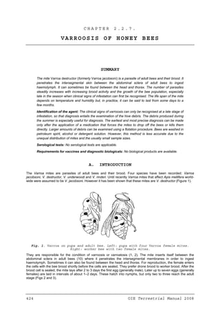
- 1. 424 OIE Terrestrial Manual 2008 C H A P T E R 2 . 2 . 7 . VARROOSIS OF HONEY BEES SUMMARY The mite Varroa destructor (formerly Varroa jacobsoni) is a parasite of adult bees and their brood. It penetrates the intersegmental skin between the abdominal sclera of adult bees to ingest haemolymph. It can sometimes be found between the head and thorax. The number of parasites steadily increases with increasing brood activity and the growth of the bee population, especially late in the season when clinical signs of infestation can first be recognised. The life span of the mite depends on temperature and humidity but, in practice, it can be said to last from some days to a few months. Identification of the agent: The clinical signs of varroosis can only be recognised at a late stage of infestation, so that diagnosis entails the examination of the hive debris. The debris produced during the summer is especially useful for diagnosis. The earliest and most precise diagnosis can be made only after the application of a medication that forces the mites to drop off the bees or kills them directly. Larger amounts of debris can be examined using a flotation procedure. Bees are washed in petroleum spirit, alcohol or detergent solution. However, this method is less accurate due to the unequal distribution of mites and the usually small sample sizes. Serological tests: No serological tests are applicable. Requirements for vaccines and diagnostic biologicals: No biological products are available. A. INTRODUCTION The Varroa mites are parasites of adult bees and their brood. Four species have been recorded: Varroa jacobsoni, V. destructor, V. underwoodi and V. rinderi. Until recently Varroa mites that affect Apis mellifera world- wide were assumed to be V. jacobsoni. However it has been shown that these mites are V. destructor (Figure 1). Fig. 1. Varroa on pupa and adult bee. Left: pupa with four Varroa female mites. Right: worker bee with two female mites. They are responsible for the condition of varroosis or varroatosis (1, 2). The mite inserts itself between the abdominal sclera in adult bees (10) where it penetrates the intersegmental membranes in order to ingest haemolymph. Sometimes it can also be found between the head and thorax. For reproduction, the female enters the cells with the bee brood shortly before the cells are sealed. They prefer drone brood to worker brood. After the brood cell is sealed, the mite lays after 2 to 3 days the first egg (generally male). Later up to seven eggs (generally females) are laid in intervals of about 1–2 days. These hatch into nymphs, but only two to three reach the adult stage (Figs 2 and 3).
- 2. Chapter 2.2.7. -- Varroosis of honey bees OIE Terrestrial Manual 2008 425 Fig. 2. Oviposition and development of Varroa in brood cells of worker bee (until about 9th day unsealed brood, until about 21st day sealed brood). Fig. 3. Development of Varroa: E = Egg, L = Larva, P = Protonymph, D = Deutonymph, A = Adult (Sex of eggs, larvae and protonymphs can only be distinguished by examining the chromosomes).
- 3. Chapter 2.2.7. -- Varroosis of honey bees 426 OIE Terrestrial Manual 2008 The number of mites usually increases slowly at the beginning of the season. Clinical signs may be seen at any time during the active season, although usually maximum numbers are reached late in the season (Figure 4), when the first clinical signs of infestation can be recognised. The course of this parasitism is usually lethal, except in some areas, such as tropical Latin America (6, 12). The life span of mites on larval or adult bees depends on temperature and humidity. Under practical conditions, the life span may vary from some days to a few months. Fig. 4. Graph of populations of bees and mites over 1 year in a temperate Northern Hemisphere climate: brood numbers (solid line); mite numbers (broken line). In heavily infested bee colonies, clinical signs of varroosis can often first be seen in the latter part of the season when the brood is reduced (12). Heavy infestations are usually reached 3–4 years after the primary invasion, but can occur within weeks if infested by bees from nearby colonies that are collapsing. Essentially, the brood is damaged by the parasitic mites. Bees and their offspring that have been infected during the brood phase by only one parasitic mite show various ill effects, such as a shortened life span, changes in behaviour and an increased disease susceptibility (8). The parasitism is critical if more than one mite enters the brood cell for reproduction. Only in the lethal stage immediately before the collapse of the colonies do clinical signs, such as shrunken wings and shortened abdomen, appear (Figure 5). This is due to an increased susceptibility to deformed wing and acute paralysis virus, as well as to the infection of wounds and loss of haemolymph (3, 4). If the brood dies shortly before or after sealing, clinical signs of European foulbrood appear without the presence of the specific agent Melissococcus pluton. If the brood survives, the emerging bees show various behavioural changes and their life span is considerably shortened (7, 11). Fig. 5. Effect of Varroa on bee morphology. Left: normal bee appearance. Right: bee heavily attacked by mites. This newly emerged bee has a deformed wing and reduced abdominal volume. B. DIAGNOSTIC TECHNIQUES 1. Identification of the agent
- 4. Chapter 2.2.7. -- Varroosis of honey bees OIE Terrestrial Manual 2008 427 The female mite is a dark reddish/brown colour and has a flat, oval-shaped body approximately 1.1 mm × 1.5 mm. It is the only common parasite of honey bees that can be seen with the naked eye (13).
- 5. Chapter 2.2.7. -- Varroosis of honey bees 428 OIE Terrestrial Manual 2008 a) Debris examination An easy method of diagnosis of varroosis is by the examination of the debris generated by bees themselves. An insert covered with a screen mesh is placed on the floor of the hive. Unless this insert is covered with such a gauze, or smeared with grease, the bees will dispose of the mites outside the hive. The debris produced within a few days in the late season usually contains little other than visible mites (9, 11). The debris collected in winter, however, must be examined in the laboratory. An insert is placed in the hive as before, but an effective medication is used to cause the mites to fall off the bees, so that after a given time, a number of mites may be observed on the floor insert. Some countries demand the diagnostic application of certain medications for proving the absence of mites. Large amounts of debris can be examined in the laboratory using a flotation procedure (5). o Test procedure i) Dry the debris for 24 hours. ii) Flood the debris with industrial alcohol. iii) Stir continuously for around 1 minute or, if debris contains wax or propolis particles, stir for 10– 20 minutes. iv) Identify and observe the mites that float to the surface. b) Brood examination For the second method, drone brood is examined, if available, otherwise worker brood is examined. When a large number of samples are examined, a rough determination of the degree of infection can be obtained. o Test procedure i) Remove the cappings of the brood cells with a knife. ii) Wash the brood cells directly into a sieve system with warm water from a hand-held shower. iii) Collect the mites in the lower fine sieve (mesh width 1 mm) while the brood is gathered in the upper coarse sieve (mesh width 2–3 mm). iv) Place the contents of the sieve on a bright plate, where the mites can be easily identified and counted. When a smaller number of samples are being studied, the individual cells are examined using an appropriate source of light. After removing the cappings and the bee brood, infected cells can be identified by the presence of small white spots – the faeces of the mite – found on the cell wall. The mites themselves should be sought for confirmation, by examining the bottom of the cell and the bee brood for attached mites. c) Bee examination In a third method, approximately 200–250 bees are removed from unsealed brood combs. Samples should be taken from both sides of at lest three uncapped brood combs. To determine an apiary’s percentage of infestation, it is necessary to collect and analyse individual samples from at least 10% of the beehives, and to determine later the average infestation rate based on these individual results. o Test procedure i) Kill the bees in a special container by submersion in alcohol. ii) Stir the container for 10 minutes. iii) Separate the bees from the mites by means of a sieve with a mesh size of approximately 2–3 mm. Under some circumstances, the Varroa mite may be confused with the bee louse, Braula coeca (Figure 6). The latter is round, not oval, and being an insect, has only three pairs of legs. A number of different species of mite may be associated with Varroa mites on bees, but these are easily distinguished. In addition, other parasitic mites, such as those of the Tropilaelaps spp., are known to cause similar damage to bee colonies as the Varroa mites. 2. Serological tests No serological tests are available for routine laboratory diagnosis.
- 6. Chapter 2.2.7. -- Varroosis of honey bees OIE Terrestrial Manual 2008 429 Fig. 6. Diagram of Varroa destructor (formerly Varroa jacobsoni Oudemans) (female). a) Dorsal aspect ⎫ b) Anterior aspect ⎬ Note the flat shell-like back and four pairs of legs. c) Ventral aspect ⎭ d) The bee louse (Braula coeca, female). Note the lack of shell-like back and only three pairs of legs. C. REQUIREMENTS FOR VACCINES AND DIAGNOSTIC BIOLOGICALS There are no biological products and vaccines available. Several medicaments and substances like Formic acid, oxalic acid, lactic acid and thymol can be used to control Varroa mites (http://www.apis.admin.ch/english/Themes/Varroa.htm). Some hygienic honeybee strains are less susceptible to Varroa parasites. o Acknowledgement Illustrations by Karl Weiss, extracted from Bienen-Pathologie, 1984. Reproduced with the kind permission of the author and Ehrenwirth-Verlag, Munich (Germany). REFERENCES 1. ANDERSON D.L. (2000). Variation in the parasitic bee mite Varroa jacobsoni Oud. Apidologie, 31, 281–292. 2. ANDERSON D.L. & TRUEMAN J.W.H. (2000). Varroa jacobsoni (Acari: Varroidae) is more than one species. Exp. Appl. Acarol., 24, 165–189. 3. BAILEY L. (1981). Honey Bee Pathology. Academic Press, London, UK. 4. BALL B.V. (1985). Acute paralysis virus isolated from honey bee colonies infested with Varroa jacobsoni. J. Apic. Res., 24, 115–119. 5. BREM S. (1980). Laboruntersuchungen von Wintergemull. In: Diagnose und Therapie der Varroatose. Apimondia Publishing House, Bucharest, Romania, 116–117. 6. DE JONG D. (1997). Varroa and other parasites of brood. In: Pests, Predators and Diseases of Honey Bees, Third Edition, Morse R.A., ed. A. I. Root, Medina, Ohio, USA, 231–279. 7. DE JONG D. & DE JONG P.H. (1983). Longevity of Africanized honey bees (Hymenoptera Apidae) infested by Varroa jacobsoni (Parasitiformes Varroidae). J. Econ. Entomol., 76, 766–768.
- 7. Chapter 2.2.7. -- Varroosis of honey bees 430 OIE Terrestrial Manual 2008 8. DE JONG P.H. & GONCALVES L.S. (1982). Weight loss and other damage to developing worker honey bees from infestation with Varroa jacobsoni. J. Apic. Res., 21, 165–167. 9. FRIES I., CAMAZINE S. & SNEYD J. (1994). Population dynamics of Varroa jacobsoni: a model and a review. Bee World, 75, 4–28. 10. RITTER W. (1980). Varroatosis: A new disease of honey bee Apis mellifera. Bee World, 6, 141–153. 11. RITTER W. (1996). Diagnostik und Bekämpfung der Bienenkrankheiten (Diagnosis and control of bee diseases). Gustav Fischer Verlag, Jena, Stuttgart, Germany. 12. RITTER W., LECLERQ E. & KOCH W. (1984). Observations des populations d’abeilles et de Varroa dans les colonies à différents niveaux d’infestation. Apidologie, 14, 389–400. 13. SHIMANUKI H. & KNOX D.A. (1991). United States Department of Agriculture (USDA) Handbook No. 690. 53p. * * * NB: There are OIE Reference Laboratories for Bee diseases (see Table in Part 3 of this Terrestrial Manual or consult the OIE Web site for the most up-to-date list: www.oie.int).