SlideShare uma empresa Scribd logo
1 de 11
Baixar para ler offline
the american college of obstetricians and gynecologists
women ’ s health care physicians

P R AC T I C E
BUL L E T I N
clinical management guidelines for obstetrician – gynecologists

Number 122, August 2011	

Replaces Practice Bulletin Number 42, April 2003

Breast Cancer Screening
Breast cancer is the most commonly diagnosed noncutaneous cancer in women in the United States, and the second
leading cause of death from cancer in American women—second only to lung cancer (1). Breast cancer mortality can
be effectively reduced through screening. The purpose of this Practice Bulletin is to review breast cancer screening
guidelines and the evidence used to support the recommendations and highlight new screening modalities and controversies surrounding screening.

Background
Incidence
Breast cancer accounts for 27% of all new cases of cancer diagnosed in women (2). A woman’s lifetime risk of
developing breast cancer is 12.08%, or 1 in 8 (Table 1)
(2, 3). In 2010, approximately 207,090 new cases of
invasive breast cancer were diagnosed, and 39,840
deaths were attributable to breast cancer (2). In addition,
54,010 new cases of in situ breast cancer were reported
in 2010 (3). There was a 2% per year decrease in the
incidence of breast cancer between 1999 and 2006, possibly reflecting decreased use of menopausal hormone
therapy and decreased use of screening mammography
(3–5). Breast cancer-related mortality has been decreasing steadily since 1990, reflecting both earlier detection
of breast cancer and improved treatment (3).

Components of Breast Cancer Screening
and Current Guidelines
Breast cancer screening has traditionally included three
elements: 1) breast imaging (primarily mammography),
2) clinical breast examination, and 3) patient self-screen-

Table 1. Age-Specific Probabilities of Developing Invasive
Female Breast Cancer*
	
	
If Current Age Is…	

The Probability of 	
Developing Breast Cancer
in the Next 10 Years†	

Or 1 in:


20	

0.06%	

1,760


30	

0.44%	

229


40	

1.44%	

69


50	

2.39%	

42


60	

3.40%	

29


70	

3.73%	

27

12.08%	

8

	 Lifetime risk	

*Among those free of cancer at the beginning of the age interval. Based on cases
diagnosed 2004–2006. Percentages and “1 in” numbers may not be numerically
equivalent because of rounding.
†
Probability derived using NCI DevCan Software, Version 6.4.0.
Reprinted with permission from American Cancer Society. Breast Cancer Facts &
Figures 2009–2010. Atlanta: American Cancer Society, Inc.

ing (breast self-examination or breast self-awareness).
The relative value of each element and appropriate age of
initiation, cessation, and frequency of screening remain

Committee on Practice Bulletins—Gynecology. This Practice Bulletin was developed by the Committee on Practice Bulletins—Gynecology with the
assistance of Jennifer Griffin, MD, Mary Gemignani, MD, and Mark Pearlman, MD. The information is designed to aid practitioners in making decisions
about appropriate obstetric and gynecologic care. These guidelines should not be construed as dictating an exclusive course of treatment or procedure.
Variations in practice may be warranted based on the needs of the individual patient, resources, and limitations unique to the institution or type of practice.
controversial. Table 2 outlines the current screening
guidelines of several major medical organizations. The
American College of Obstetricians and Gynecologists
(the College) continues to endorse inclusion of all three
strategies in breast cancer screening.

Screening Mammography
Rationale for Mammographic Screening
Tumors detected at an early stage that are small and
confined to the breast are more likely to be successfully
treated, with a 98% 5-year survival for localized disease
(3). After 18 years of follow-up, one initial study found
that 89% of tumors measuring 1 cm or less were cured
by primary surgery (mastectomy and axillary dissection) (6). Other studies have confirmed these results,
with 90% of patients experiencing 10-year (or longer)
disease-free survival periods after tumors measuring
1 cm or less were detected by mammography, indicating
the likelihood that the tumors had not yet metastasized
before they were diagnosed and treated (7–11).
By mathematical estimation, a typical ductal adenocarcinoma with a constant mean doubling time of 100
days would have been present for more than 11 years
before it grew to a generally palpable size of 2 cm
(12–14). Mammography screening could potentially
identify a nonpalpable mass measuring approximately
1 mm to 1 cm during its preclinical phase, 3 years before
it becomes palpable (12–15). This concept is commonly
referred to as sojourn time, which is the time interval
when cancer may be detected by screening before it

becomes symptomatic. The sojourn time of an individual
type of cancer varies, with more biologically aggressive
tumors typically having shorter sojourn times. The greatest predictor of sojourn time in breast cancer appears to
be age. Estimates of mean sojourn time for breast cancer
in women increase with age: for ages 40–49 years, mean
sojourn time is 2–2.4 years; 50–59 years, 2.5–3.7 years;
60–69 years, 3.5–4.2 years; and 70–74 years, 4–4.1
years (16).
The mean sojourn time has implications for breast
cancer screening because it is desirable to detect tumors
during this sojourn period. Individuals who are likely
to have types of cancer with shorter sojourn times are
more likely to benefit from more frequent screening
when compared with those with slow-growing tumors
that have a larger preclinical window. Screening strategies should be designed to maximize the likelihood
of detecting the cancer during the preclinical window,
when treatment options may be greater and outcomes
may be improved.

Evaluation of Mammography
A recent meta-analysis conducted for the United States
Preventive Services Task Force reviewed eight randomized controlled trials of mammographic screening conducted between 1986 and 2006 (17). Despite some study
limitations (all studies were rated of “fair” quality by
the United States Preventive Services Task Force), this
review reaffirmed a reduction in breast cancer-related
mortality for women aged 39–69 years who were invited

Table 2. Breast Cancer Screening Recommendations
		
	
Mammography	

Clinical Breast 	
Examination	

Breast Self-Examination 	
Instruction	

Breast SelfAwareness

American College of 	
Obstetricians and 	
Gynecologists	
	

Age 40 years and 	
older annually	


Age 20–39 years 	
every 1–3 years	
Age 40 years and	

Consider for high-risk	
patients	

Recommended	
	

American Cancer Society	
	

Age 40 years and 	
older annually	

Age 20–39 years	
every 1–3 years 	

Optional for age 20 years	
and older	

Recommended	


Age 40 years and	
older annually	


National Comprehensive 	
Cancer Network	

Age 40 years and	
older annually	

Age 20–39 years	
every 1–3 years


Age 40 years and	
older annually	


National Cancer Institute	
	

Age 40 years and	
older every 1–2 years	

Recommended	
	

Not recommended	
	

—	

U.S. Preventive Services 	
Task Force	

	
Age 50–74 years biennially	

	
Insufficient evidence	

	
Not recommended	

	
—

2	

older annually

Recommended	

Recommended	

Practice Bulletin No. 122
for screening. This is consistent with the mortality reductions demonstrated in previous meta-analyses (18–22).
Relative risk of breast cancer mortality in women
invited for screening was 0.85 for women aged 39–49
years; 0.86 for women aged 50–59 years; and 0.68 for
women aged 60–69 years. Evidence was insufficient
to demonstrate a mortality reduction for women aged
70 years and older (Table 3). However, because of
the increasing incidence of breast cancer as women
age, more young women (aged 39–49 years) had to be
invited for screening to prevent one breast cancer-related
death when compared with older women (Table 3). The
number needed to invite for screening (over several
rounds of screening and at least 11 years of follow-up)
to prevent one breast cancer death in women aged 39–49
years was 1,904, compared with 1,339 in women aged
50–59 years. This difference in number needed to invite
for screening to prevent one death was a key reason the
United States Preventive Services Task Force elected not
to recommend routine screening for women aged 40–49
years, despite similar mortality reductions (23).
Because of differences in study design, with most
randomized clinical trials evaluating biennial mammographic screening, these randomized controlled trials do
not provide meaningful comparisons between different
screening strategies in terms of age at initiation, cessation, and frequency. The lack of clear evidence on this
issue led the United States Preventive Services Task
Force to commission the creation of screening models
that could compare different strategies to help guide
national screening recommendations. These six models
were created by independent investigative groups that
are a part of the National Cancer Institute’s Cancer

Intervention and Surveillance Modeling Network. These
models were created using a common set of age-specific
variables, with the goal of identifying efficient screening strategies (ie, those that produce a gain in mortality
reduction or a gain in life years per additional screening
mammogram).
Biennial screening was used in seven of eight
screening strategies found to be efficient in reducing
mortality, and in six of these eight strategies, screening began at age 50 years. A review of the strategies
concluded that more lives could be saved by extending
screening to women older than 69 years, rather than
extending screening to women aged 40–49 years (24).
However, when defining benefit as number of life years
gained through screening, screening was started at age
40 years in four of eight strategies. Although annual
screening was an efficient strategy for reducing breast
cancer mortality and increasing life years gained, it was
noted by the authors to be more resource intensive (24).

Other Imaging Techniques
Ultrasonography is an established adjunct to mammography in the imaging evaluation. It is useful in evaluating inconclusive mammographic findings, in evaluating young patients and other women with dense breast
tissue, in guiding tissue core-needle biopsy and other
biopsy techniques, and in differentiating a cyst from
a solid mass. It is not recommended as a screening
modality for women at average risk of developing breast
cancer. Ultrasonography may be an option for additional
screening in women at high risk who are candidates for
magnetic resonance imaging (MRI) screening but can-

Table 3. Pooled Relative Risks for Breast Cancer Mortality From Mammography
Screening Trials for All Ages
			
		
RR for Breast Cancer 	
Age 	
Trials Included, n	
Mortality (95% CrI)	

NNI to Prevent 1
Breast Cancer Death
(95% CrI)

39–49 y	

8*	

0.85 (0.75–0.96)	

1904 (929–6378)

50–59 y	

†

6	

0.86 (0.75–0.99)	

1339 (322–7455)

60–69 y	

2‡	

0.68 (0.54–0.87)	

377 (230–1050)

70–74 y	

1	

1.12 (0.73–1.72)	

Not available

§

Abbreviations: CrI, credible interval; NNI, number needed to invite to screening; RR, relative risk; y, years.
*Health Insurance Plan of Greater New York, Canadian National Breast Screening Study-1, Stockholm, Malmö,
Swedish Two-County trial (2 trials), Gothenburg trial, and Age trial.
†
Canadian National Breast Screening Study-1, Stockholm, Malmö, Swedish Two-County trial (2 trials), and
Gothenburg trial.
‡
Malmö and Swedish Two-County trial (Östergötland).
§
Swedish Two-County trial (Östergötland).
Reprinted with permission from Nelson HD, Tyne K, Naik A, Bougatsos C, Chan BK, Humphrey L. Screening for
breast cancer: An update for the U.S. Preventive Services Task Force. Ann Intern Med 2009;151:727–37.

Practice Bul­e­in No. 122	
l t

3
not receive MRI because of gadolinium contrast allergy,
claustrophobia, or other barriers.
Magnetic resonance imaging can be a useful adjunct
to diagnostic mammography, but cost, duration of the
examination, and injection of contrast material prohibit
its use as a routine, population-based screening technique.
The American Cancer Society has issued guidelines
regarding the use of MRI for screening in high-risk
women. Based on expert panel review of the evidence, the
American Cancer Society recommends MRI screening for
women with a 20% or greater lifetime risk of developing
breast cancer, including women with the following:
•	 Have a known BRCA1 or BRCA2 gene mutation
•	 Have a first-degree relative with a BRCA1 or BRCA2
gene mutation and have not had any testing themselves
•	 A lifetime risk of breast cancer of 20% or greater,
according to risk assessment tools that are based
mainly on family history
•	 A history of radiation therapy to the chest between
the ages of 10 years and 30 years
•	 Other genetic syndromes, including Li–Fraumeni
syndrome, Cowden syndrome, or Bannayan–Riley–
Ruvalcaba syndrome or one of these syndromes in a
first-degree relative
The panel concluded that there was insufficient evidence
to recommend for or against MRI screening for women
with a personal history of breast cancer, carcinoma in
situ, atypical hyperplasia, and extremely dense breasts
on mammography (25). Breast MRI is not recommended
for screening women at average risk of developing
breast cancer.
Color Doppler ultrasonography, computer-aided
detection, positron emission tomography, scintimammography, and digital breast tomosynthesis have shown
promise in selected clinical situations or as adjuncts to
mammography for breast cancer diagnosis. However,
these technologies are not considered alternatives to routine mammography.

Digital Mammography
In 2005, the results of the Digital Mammographic
Imaging Screening Trial were reported (26). During the
2-year study period, 49,528 women were recruited to the
study from 33 sites in the United States and Canada. All
participants underwent both digital mammography and
film mammography in random order. The diagnostic
accuracy of digital mammography and film mammography was similar across the entire population. Digital
mammography was superior to film mammography in
women younger than 50 years, women with heteroge-

4	

neously dense or extremely dense breasts, and premenopausal and perimenopausal women.
In 2007, the follow-up and final results of the Oslo II
study were reported. This was a randomized trial of
screen-film versus full-field digital mammography in
a population-based screening program of women aged
45–69 years. In this trial, women aged 45–69 years were
assigned to undergo film mammography (n=16,985) or
digital mammography (n=6,944). The group of women
aged 45–49 years was monitored for 1.5 years and the
group of women aged 50–69 years was monitored for
2 years. There was a significant difference in the cancer
detection rate between the digital mammography (0.59%)
and film mammography (0.38%) (P=0.02) groups (27).
A recent meta-analysis of data from eight large
randomized studies found that, overall, digital mammography demonstrated a slightly higher detection rate
than film mammography, particularly for women aged
60 years or younger (28).

Clinical Considerations and
Recommendations
	 What is the difference between breast selfexamination and breast self-awareness, and
are these screening methods effective?
Breast self-examination is the performance of an examination of the breasts in a consistent, systematic way
by the individual on a regular basis, typically monthly.
Historically, physicians have been encouraged to educate their patients on how to perform these examinations, and public awareness campaigns have focused on
this intervention. It still may be appropriate for certain
high-risk populations and for other women who choose
to follow this approach.
Currently, there is an evolution away from teaching
breast self-examination toward the concept of breast selfawareness. The College, the American Cancer Society,
and the National Comprehensive Cancer Network endorse
breast self-awareness, which is defined as women’s awareness of the normal appearance and feel of their breasts.
This concept has arisen because approximately one half
of all cases of breast cancer in women 50 years and older
and more than 70% of cases of cancer in women younger
than 50 years are detected by women themselves, frequently as an incidental finding (29, 30). In addition, the
effectiveness of self-examination was at odds with what
was anticipated based on the aforementioned statistics.
Breast self-awareness should be encouraged and
can include breast self-examination. Women who desire
to perform self-examination as a part of this breast self-

Practice Bulletin No. 122
awareness strategy may be instructed in the appropriate
technique, although emphasis is not on examination
techniques. Women should report any changes in their
breasts to their health care providers. Although this
patient education strategy has not been studied to date,
breast awareness may be of particular importance as
part of a screening strategy because some women may
falsely assume that negative mammography or clinical
breast examination results definitively exclude the presence of breast cancer. New cases of cancer can arise
during screening intervals, and breast self-awareness
may prompt women not to delay in reporting breast
changes based on false reassurance of recent negative
screening result. Breast self-awareness aims to capture
the importance of self-detection and prompt evaluation
of symptoms because it relates to overall breast cancer
morbidity and mortality. However, the effect of breast
self-awareness education has not been studied.
The United States Preventive Services Task Force has
recommended against teaching breast self-examination
based on a lack of evidence to show benefit and potential
harm resulting from evaluation of false-positive findings
(23). A prospective study of 604 patients with breast cancer revealed that only 7.6% of the 448 women who practiced regular breast self-examination had detected their
own cancer, and those who did showed no survival advantage (31). The Shanghai breast self-examination trial of
266,000 women aged 39–72 years randomized to receive
either breast self-examination instruction plus follow-up
or no information on breast cancer screening reported
essentially no difference in breast cancer-related deaths
between the two groups (135 versus 131, respectively)
after 10–11 years of follow-up (32). However, women in
the instruction group were more likely to undergo breast
biopsy for benign lesions (32).
A Cochrane systematic review published in 2003
included the Shanghai breast trial (32) and another
large population-based study from Russia that compared breast self-examination with no intervention. In
both trials, almost twice as many biopsies with benign
results were performed in the screening group compared with the control group (33). The Canadian Task
Force on Preventive Health Care also recommended
against teaching breast self-examination based on fair
evidence that breast self-examination had no benefit and
good evidence that it was harmful. Their review cited
additional evidence that patients experience increased
worry, anxiety, and depression associated with breast
self-examination (34).
Because other screening methods (mammography
and clinical breast examination) can have false-negative
results and cases of breast cancer can occur in unscreened
women, there are clearly situations in which women

Practice Bul­e­in No. 122	
l t

will detect cancer themselves. As such, the College, the
American Cancer Society, and the National Comprehensive Cancer Network endorse educating women aged
20 years and older regarding breast self-awareness, as a
means to play a role in earlier detection (Table 2).

	 Is clinical (ie, health care provider performed)
breast examination effective for breast cancer
screening? If so, how frequently should it be
performed?
A study using data from the National Breast and Cervical
Cancer Early Detection Program, where 752,081 clinical
breast examinations were performed in the community
setting in women aged 40 years and older, concluded
that clinical breast examination alone has a sensitivity for cancer detection of 58.8%, with a specificity of
93.4%. In this population, five cases of cancer were
detected per 1,000 clinical breast examinations performed. When the clinical breast examination finding
was abnormal and the mammogram was normal, 7.4
cases of cancer were detected per 1,000 screenings. The
authors concluded that the addition of clinical breast
examination “modestly improved” early detection (35).
Multiple reviews have supported the combination
of clinical breast examination and mammography for
breast cancer screening (36–41). A recent study demonstrated improved sensitivity for breast cancer detection
(94.6% versus 88.6%) when comparing mammography
plus clinical breast examination with mammography
alone. However, the false-positive rate was also higher
in the group receiving clinical breast examination compared with the mammography-alone group (12.4%
versus 7.4%) (41). Based on available evidence, the
College, the American Cancer Society, and the National
Comprehensive Cancer Network recommend that clinical breast examination should be performed annually
for women aged 40 years and older. Although the value
of screening clinical breast examination for women
with a low prevalence of breast cancer (ie, women aged
20–39 years) is not clear, the College, the American
Cancer Society, and the National Comprehensive Cancer
Network continue to recommend clinical breast examination for these women every 1–3 years.

	 When should mammography begin, and
how frequently should mammography be
performed?
Based on the incidence of breast cancer, the sojourn time
for breast cancer growth, and the potential for reduction
in breast cancer mortality, the College recommends that
women aged 40 years and older be offered screening
mammography annually. However, as with any screen-

5
ing test, women should be educated on the predictive
value of the test and the potential for false-positive results
and false-negative results. Women should be informed
of the potential for additional imaging or biopsies that
may be recommended based on screening results. The
physician should work with the patient to determine the
best screening strategy based on individual risk and values. In some women, biennial screening may be a more
appropriate or acceptable strategy. Some average-risk
women may prefer biennial screening, which maintains
most of the benefits of screening while minimizing both
the frequency of screening and the potential for additional
testing, whereas other women prefer annual screening
because it maximizes cancer detection.
Various groups have offered recommendations on
the timing of initiation and frequency of mammography screening. Each group places different values on
competing considerations, such as published evidence,
cost-effectiveness, efficiency, accuracy, adverse consequences, specificity, sensitivity, false-positive results,
false-negative results, positive predictive value, patient
adherence, availability of health care resources, conflicting health care needs, and opinions of experts and
advocates. A summary of the recommendations can be
found in Table 2.
The American Cancer Society and the National
Comprehensive Cancer Network recommend annual
screening mammography beginning at age 40 years (42,
43). In contrast, the United States Preventive Services
Task Force recently changed their guidelines to recommend biennial mammography in women aged 50–74
years. Although routine screening in women aged 40–49
years was not recommended, the Task Force advised
that screening in women younger than 50 years should
be individualized based on “patient values regarding
specific benefits and harms” (23).
The American Cancer Society, the National
Comprehensive Cancer Network, and the United States
Preventive Services Task Force agree that screening
mammography reduces breast cancer-related mortality in women aged 40 years and older based on metaanalyses of available randomized controlled trials on
mammographic screening (see Table 3). However, the
United States Preventive Services Task Force arrived
at a different conclusion regarding routine screening for
women aged 40–49 years by placing greater weight on
the lower prevalence of disease in this population, which
reduces the positive predictive value of mammography
and increases the number needed to screen to prevent
one breast cancer-related death. In addition, the United
States Preventive Services Task Force argues that all
women who participate in breast cancer screening may
potentially experience a false-positive mammogram (as

6	

high as 49% of women after 10 years of screening), with
resultant additional imaging, biopsies, and psychologic
distress (44). The burden of these additional tests may
be perceived as greater on women aged 40–49 years
because they are less likely to experience breast cancer
and, therefore, less likely to benefit from screening (23).
In response to this argument, experts who support routine screening for women in their 40s note that
although the incidence of cancer is less in the 40–49
year age group than in all women older than 50 years,
the incidence of cancer in women in their 50s (1 in 38,
or 2.60%) is not much higher than in women in their 40s
(1 in 69, or 1.44%) (2). In addition, women who undergo
routine screening mammography in their 40s have comparable mortality reduction compared with women who
undergo routine mammography screening in their 50s
(16% reduction versus 15% reduction, respectively) (17).
This is of particular importance because each year in
the United States breast cancer is diagnosed in approximately 50,000 women younger than 50 years.
Regarding the frequency of screening mammography, the United States Preventive Services Task Force
also diverged from what has become common practice in
the United States by recommending biennial rather than
annual screening for women aged 50 years and older. This
decision was based on the National Cancer Institute’s
Cancer Intervention and Surveillance Modeling Network
screening models, which predicted that 81% of the benefits of screening (ie, mortality reduction) could be maintained by screening every other year (24). This strategy
is less resource intensive and is predicted to reduce falsepositive results by 50% (23). However, these models
demonstrate a greater reduction in mortality and increased
number of life years gained by screening annually; therefore, accepting a biennial strategy reduces some (19%) of
the potential benefits of screening.

	 What are the potential adverse consequences
of screening mammography?
Potential adverse outcomes of breast cancer screening mammography include false-positive mammograms, false-negative mammograms, and overdiagnosis.
Concerns about the risk of radiation exposure (eg, induction of breast cancer from radiation exposure) have
largely been decreased by improvements in mammography technique, technology, and clinical experience (45).
False-positive mammograms (ie, those with perceived abnormalities requiring further evaluation to
verify that the lesion is not cancer) are a continuing
concern (44, 46). False-positive screening mammograms
require diagnostic mammography with supplementary
views, ultrasonography, and even biopsy in 20–30% of

Practice Bulletin No. 122
cases in an attempt to reach an accurate diagnosis (44,
46). Psychosocial consequences of screening mammography, such as anxiety and distress, have been identified,
reviewed, and are generally short-lived and not severe
(47–50). Studies evaluating the effect of false-positive
results suggest that women in the United States are
highly tolerant of false-positive mammograms, and that
women who experience a false-positive mammogram
are more likely than women with a normal result to
adhere to routine screening in the future. Women with
false-positive results were more likely to have anxiety
about developing breast cancer, but not at a demonstrably
pathologic level (50, 51).

	 What are the factors that increase a woman’s
relative risk of breast cancer?
Most women in whom invasive breast cancer is diagnosed do not have unique identifiable risk factors;
however, women with certain characteristics do have an
increased lifetime prevalence of breast cancer compared
with the general population (52, 53). The incidence of
breast cancer increases with advancing age (2). Because
women at high risk need to be appropriately counseled
regarding increased surveillance or breast cancer risk
reduction, physicians should periodically assess breast
cancer risk. The goal of this risk assessment is to categorize the woman’s risk level as average for her age versus
elevated or high risk. Risk assessment should not be used
to consider a woman ineligible for screening appropriate
for her age, but rather to identify those who may qualify
for enhanced screening, such as addition of MRI screening or more frequent clinical breast examinations and
risk reduction strategies.

Reproductive Risk Factors
Certain reproductive factors also influence breast cancer risk, including age at menarche, age at first birth,
breastfeeding, parity, and age at menopause (Table 4).
A greater number of reproductive years, later age at first
birth, and lower parity or nulliparity generally increase
breast cancer risk.

Familial Risk Factors
Another important consideration in breast cancer risk
assessment is family history to identify cases of breast
cancer, ovarian cancer, prostate cancer, and other types
of cancer in first-degree relatives (parents, sibling, child),
second-degree relatives, and third-degree relatives, including the age of onset for the family members with cancer.
Based on this family history, women may be eligible for
testing for BRCA gene mutations or for referral to a genetic
counselor for further evaluation and consideration of test-

Practice Bul­e­in No. 122	
l t

Table 4. Factors That Increase the Relative Risk of Breast
Cancer in Women
Relative Risk	

Factor

>4.0	


Female
Age (65+ vs <65 years, although risk increases
across all ages until age 80)
Certain inherited genetic mutations for breast 	
cancer (BRCA1 and/or BRCA2)
Two or more first-degree relatives with breast 	
cancer diagnosed at an early age
Personal history of breast cancer
High breast tissue density
Biopsy-confirmed atypical hyperplasia

2.1–4.0	


One first-degree relative with breast cancer
High-dose radiation to chest
High bone density (postmenopausal)

1.1–2.0	
Factors that affect 	
circulating hormones


Late age at first full-term pregnancy (>30 years)
Early menarche (<12 years)
Late menopause (>55 years)
No full-term pregnancies
Never breastfed a child
Recent oral contraceptive use
Recent and long-term use of estrogen and	
progestin
Obesity (postmenopausal)

Other factors	


Personal history of endometrial or ovarian	
cancer
Alcohol consumption
Height (tall)
High socioeconomic status
Ashkenazi Jewish heritage

Reprinted with permission from American Cancer Society. Breast Cancer Facts
& Figures 2009-2010. Atlanta: American Cancer Society, Inc. Adapted with
permission from Hulka BS, Moorman PG. Breast cancer: hormones and other risk
factors. Maturitas. Feb 28 2001;38(1):103–113; discussion 113–106.

ing (see Practice Bulletin No.103, Hereditary Breast and
Ovarian Cancer Syndrome, April 2009).

Combined Factors
The Gail Model is a risk assessment tool that uses patient
age, some reproductive factors, limited family history
(breast cancer in first-degree female relatives), and a
history of breast biopsies to estimate 5-year and lifetime
breast cancer risks. This model has been validated in
Caucasian women and studied extensively in African
American women. The assessment can be completed in
less than a minute, and is available at www.cancer.gov/
bcrisktool. Women estimated to have a 5-year breast
cancer risk of 1.7% or greater (equivalent to the average

7
risk for a women aged 60 years) or a lifetime risk of 20%
or greater may be offered enhanced screening, including
a clinical breast examination every 6–12 months, yearly
mammography, and instruction in breast self-examination. The Gail model is not a good risk assessment tool
when there is a strong family history of breast cancer in
women other than the mother and sisters of the patient
or a family history of cancer other than breast cancer
(eg, ovarian cancer). Breast MRI is not typically recommended based on the Gail Model.

	 What screening is appropriate for women at
high risk?
For women who test positive for BRCA1 or BRCA2 mutations, enhanced screening should be recommended and
risk reduction methods discussed. Enhanced screening for
these women includes twice-yearly clinical breast examinations, annual mammography, annual breast MRI, and
instruction in breast self-examination. (Details regarding
the management of women with BRCA mutations are
reviewed in Practice Bulletin No.103, Hereditary Breast
and Ovarian Cancer Syndrome, April 2009.) Women
who have first-degree relatives with these mutations but
who are untested are generally managed as if they carry
these mutations until their BRCA status is known.
Women who are estimated to have a lifetime risk
of breast cancer of 20% or greater, based on risk models
that rely largely on family history (such as BRCAPRO,
BODACEA, or Claus), but who are either untested or
test negative for BRCA gene mutations, can be offered
enhanced screening as described previously for carriers
of the BRCA mutation. Risk-reduction strategies also
may be considered for these women.
Other women considered at high risk of development of future cases of breast cancer are those who
received thoracic irradiation (typically as a treatment
for lymphoma) between the ages 10 years and 30 years.
These women should be advised to receive annual mammography, annual MRI, and screening clinical breast
examination every 6–12 months beginning 8–10 years
after they received treatment or at age 25 years, whichever occurs last.
Women with a personal history of high-risk breast
biopsy results, including atypical hyperplasia and lobular
carcinoma in situ are at increased risk of future breast
cancer. These women should receive enhanced screening, including annual mammography, clinical breast
examination every 6–12 months, and instruction in
breast self-examination. Yearly breast MRI also has been
recommended for women with a history of lobular carcinoma in situ by some organizations (43). Women with a
personal history of ductal carcinoma in situ or invasive

8	

breast cancer should be monitored similarly, although
MRI is not routinely recommended in this population.

	 Is there an upper age range at which the risks
of mammography outweigh the benefits?
A consensus of recommendations on this issue does not
exist. Medical comorbidity and life expectancy should
be considered in a breast cancer screening program for
women aged 75 years or older because the benefit of
screening mammography decreases compared with the
harms of overtreatment with advancing age. Women
aged 75 years or older should, in consultation with their
physicians, decide whether or not to continue mammographic screening (17).
Figure 1 illustrates the progressively increasing incidence and mortality rates of invasive breast cancer by
race and age up to 84 years (3). Most of the screening
mammography clinical trials had an upper age limit
criteria ranging from 64 years to 74 years. However, a
meta-analysis concluded that screening mammography
in women aged 70–79 years is moderately cost-effective
and yields a small increase in life expectancy (54).

Summary of
Recommendations and
Conclusions
The following recommendations are based on limited and inconsistent scientific evidence (Level B):
	 Based on the incidence of breast cancer, the sojourn
time for breast cancer growth, and the potential
reduction in breast cancer mortality, the College
recommends that women aged 40 years and older be
offered screening mammography annually.

The following recommendations are based primarily on consensus and expert opinion (Level C):
	 Clinical breast examination should be performed
annually for women aged 40 years and older.

	 For women aged 20–39 years, clinical breast examinations are recommended every 1–3 years.

	 Breast self-awareness should be encouraged and can
include breast self-examination. Women should
report any changes in their breasts to their health
care providers.

	 Women should be educated on the predictive value
of screening mammography and the potential for
false-positive results and false-negative results.
Women should be informed of the potential for

Practice Bulletin No. 122
600	
500	
Incidence: White
Rate per 100,000

400	
Incidence: African American

300	
200	
Mortality: African American
100	

Mortality: White
0	

20–24	

25–29	 30–34	 35–39	

40–44	 45–49	 50–54	

55–59	 60–64	 65–69	 70–74	 75–79	 80–84	

85+

Age
Fig. 1. Female Breast Cancer—Incidence and Mortality Rates by Age and Race, United States, 2002–2006. Data from Incidence—North
American Association of Central Cancer Registries, 2009. Mortality––National Center for Health Statistics, Centers for Disease Control
and Prevention, 2009. Reprinted with permission from American Cancer Society. Breast Cancer Facts & Figures 2009–2010. Atlanta:
American Cancer Society, Inc.

additional imaging or biopsies that may be recommended based on screening results.

	 Women who are estimated to have a lifetime risk of
breast cancer of 20% or greater, based on risk models that rely largely on family history (such as
BRCAPRO, BODACEA, or Claus), but who are
either untested or test negative for BRCA gene
mutations, can be offered enhanced screening.

	 Breast MRI is not recommended for screening
women at average risk of developing breast cancer.

	 For women who test positive for BRCA1 and BRCA2
mutations, enhanced screening should be recommended and risk reduction methods discussed.

References
	 1.	 Jemal A, Siegel R, Ward E, Hao Y, Xu J, Thun MJ.
Cancer statistics, 2009. CA Cancer J Clin 2009;59:225–
49. (Level II-3)
	 2.	 Altekruse SF, Kosary CL, Krapcho M, Neyman N,
Aminou R, Waldron W et al, editors. SEER cancer statistics review, 1975-2007. Bethesda (MD): National Cancer
Institute; 2011. Available at: http://seer.cancer.gov/csr/
1975_2007. Retrieved February 17, 2011. (Level II-3)
	 3.	 American Cancer Society. Breast cancer facts & figures:
2009-2010. Atlanta (GA): ACS; 2009. Available at: http://
www.cancer.org/acs/groups/content/@nho/documents/
document/f861009final90809pdf.pdf. Retrieved February
17, 2011. (Level II-3)

Practice Bul­e­in No. 122	
l t

	 4.	 Ravdin PM, Cronin KA, Howlader N, Berg CD,
Chlebowski RT, Feuer EJ, et al. The decrease in breastcancer incidence in 2003 in the United States. N Engl J
Med 2007;356:1670–4. (Level III)
	 5.	 Glass AG, Lacey JV Jr, Carreon JD, Hoover RN. Breast
cancer incidence, 1980-2006: combined roles of menopausal hormone therapy, screening mammography, and
estrogen receptor status. J Natl Cancer Inst 2007;99:
1152–61. (Level II-3)
	 6.	 Rosen PP, Groshen S, Kinne DW. Survival and prognostic factors in node-negative breast cancer: results of
long-term follow-up studies. J Natl Cancer Inst Monogr
1992;(11):159–62. (Level III)
	 7.	 Tabar L, Chen HH, Duffy SW, Yen MF, Chiang CF, Dean PB,
et al. A novel method for prediction of long-term outcome
of women with T1a, T1b, and 10-14 mm invasive breast
cancers: a prospective study [published erratum appears
in Lancet 2000;355:1372]. Lancet 2000;355:429–33.
(Level II-3)
	 8.	 Tabar L, Dean PB, Kaufman CS, Duffy SW, Chen HH. A
new era in the diagnosis of breast cancer. Surg Oncol Clin
N Am 2000;9:233–77. (Level III)
	 9.	 Joensuu H, Pylkkanen L, Toikkanen S. Late mortality from pT1N0M0 breast carcinoma. Cancer 1999;85:
2183–9. (Level II-3)
1
	 0.	 Lopez MJ, Smart CR. Twenty-year follow-up of minimal breast cancer from the Breast Cancer Detection
Demonstration Project. Surg Oncol Clin N Am 1997;6:
393–401. (Level II-3)
11.	 Arnesson LG, Smeds S, Fagerberg G. Recurrence-free
survival in patients with small breast cancer. An analysis of cancers 10 mm or less detected clinically and by
screening. Eur J Surg 1994;160:271–6. (Level I)

9
12.	 Macdonald I. The natural history of mammary carcinoma.
Am J Surg 1966;111:435–42. (Level III)
13.	 Gullino PM. Natural history of breast cancer. Progression
from hyperplasia to neoplasia as predicted by angiogenesis. Cancer 1977;39:2697–703. (Level III)
14.	 Wertheimer MD, Costanza ME, Dodson TF, D’Orsi C,
Pastides H, Zapka JG. Increasing the effort toward breast
cancer detection. JAMA 1986;255:1311–5. (Level III)
15.	 Walter SD, Day NE. Estimation of the duration of a
pre-clinical disease state using screening data. Am J
Epidemiol 1983;118:865–86. (Level III)
16.	 Smith RA, Duffy SW, Gabe R, Tabar L, Yen AM, Chen TH.
The randomized trials of breast cancer screening: what
have we learned? Radiol Clin North Am 2004;42:793–
806; v. (Level III)
17.	 Nelson HD, Tyne K, Naik A, Bougatsos C, Chan BK,
Humphrey L. Screening for breast cancer: an update for
the U.S. Preventive Services Task Force. U.S. Preventive
Services Task Force. Ann Intern Med 2009;151:727-37;
W237–42. (Level III)
18.	 Humphrey LL, Helfand M, Chan BK, Woolf SH. Breast
cancer screening: a summary of the evidence for the
U.S. Preventive Services Task Force. Ann Intern Med
2002;137:347–60. (Level III)
19.	 Kerlikowske K, Grady D, Rubin SM, Sandrock C, Ernster
VL. Efficacy of screening mammography. A meta-analysis. JAMA 1995;273:149–54. (Meta-analysis)
20.	 Hendrick RE, Smith RA, Rutledge JH, 3rd, Smart CR.
Benefit of screening mammography in women aged 40-49:
a new meta-analysis of randomized controlled trials.J Natl
Cancer Inst Monogr 1997;(22):87–92. (Meta-analysis)
2
	 1.	 Olsen O, Gotzsche PC. Cochrane review on screening
for breast cancer with mammography [published erratum appears in Lancet 2006;367:474]. Lancet 2001;358:
1340–2. (Level III)
22.	 Nystrom L, Andersson I, Bjurstam N, Frisell J,
Nordenskjold B, Rutqvist LE. Long-term effects of mammography screening: updated overview of the Swedish
randomised trials [published erratum appears in Lancet
2002;360:724]. Lancet 2002;359:909–19. (Level III)
23.	 Screening for breast cancer: U.S. Preventive Services Task
Force recommendation statement. US Preventive Services
Task Force [published errata appear in Ann Intern Med
2010;152:199–200; Ann Intern Med 2010;152:688]. Ann
Intern Med 2009;151:716–26;W-236. (Level III)
24.	 Mandelblatt J, Saha S, Teutsch S, Hoerger T, Siu AL,
Atkins D, et al. The cost-effectiveness of screening mammography beyond age 65 years: a systematic review for
the U.S. Preventive Services Task Force. Cost Work
Group of the U.S. Preventive Services Task Force. Ann
Intern Med 2003;139:835–42. (Level III)
25.	 Saslow D, Boetes C, Burke W, Harms S, Leach MO,
Lehman CD, et al. American Cancer Society guidelines
for breast screening with MRI as an adjunct to mammography. American Cancer Society Breast Cancer Advisory
Group [published erratum appears in CA Cancer J Clin
2007;57:185]. CA Cancer J Clin 2007;57:75–89. (Level III)
26.	 Pisano ED, Gatsonis C, Hendrick E, Yaffe M, Baum JK,
Acharyya S, et al. Diagnostic performance of digital

10	

versus film mammography for breast-cancer screening. Digital Mammographic Imaging Screening Trial
(DMIST) Investigators Group [published erratum appears
in N Engl J Med 2006;355:1840]. N Engl J Med 2005;
353:1773–83. (Level II-3)
27.	 Skaane P, Hofvind S, Skjennald A. Randomized trial of
screen-film versus full-field digital mammography with
soft-copy reading in population-based screening program:
follow-up and final results of Oslo II study. Radiology
2007;244:708–17. (Level I)
28.	 Vinnicombe S, Pinto Pereira SM, McCormack VA, Shiel S,
Perry N, Dos Santos Silva IM. Full-field digital versus
screen-film mammography: comparison within the UK
breast screening program and systematic review of published data. Radiology 2009;251:347–58. (Level III)
29.	 Coates RJ, Uhler RJ, Brogan DJ, Gammon MD, Malone
KE, Swanson CA, et al. Patterns and predictors of the
breast cancer detection methods in women under age 45
years of age (United States). CCC cancer causes & control
2001;12(5):431–42. (Level II-2)
30.	 Newcomer L, Newcomb P, Trentham-Dietz A, Storer B,
Yasui Y, Daling J, et al. Detection method and breast carcinoma histology. Cancer 2002;95(3):470–7. (Level II-3)
31.	 Auvinen A, Elovainio L, Hakama M. Breast self-examination and survival from breast cancer: a prospective
follow-up study. Breast Cancer Res Treat 1996;38:161–8.
(Level II-2)
32.	 Thomas DB, Gao DL, Ray RM, Wang WW, Allison CJ,
Chen FL, et al. Randomized trial of breast self-examination in Shanghai: final results. J Natl Cancer Inst 2002;
94:1445–57. (Level I)
33.	 Kosters JP, Gotzsche PC. Regular self-examination or
clinical examination for early detection of breast cancer. Cochrane Database of Systematic Reviews 2003,
Issue 2. Art. No.: CD003373. DOI: 10.1002/14651858.
CD003373. (Meta-analysis)
34.	 Baxter N. Preventive health care, 2001 update: should
women be routinely taught breast self-examination to screen
for breast cancer? Canadian Task Force on Preventive
Health Care. CMAJ 2001;164:1837–46. (Level III)
35.	 Bobo JK, Lee NC, Thames SF. Findings from 752,081
clinical breast examinations reported to a national screening program from 1995 through 1998. J Natl Cancer Inst
2000;92:971–6. (Level II-3)
36.	 Shen Y, Zelen M. Screening sensitivity and sojourn
time from breast cancer early detection clinical trials:
mammograms and physical examinations. J Clin Oncol
2001;19:3490–9. (Level III)
37.	 Jatoi I. Breast cancer screening. Am J Surg 1999;177:
518–24. (Level III)
38.	 Primic-Zakelj M. Screening mammography for early
detection of breast cancer. Ann Oncol 1999;10(suppl
6):121–7. (Level III)
39.	 Fletcher SW, Black W, Harris R, Rimer BK, Shapiro S.
Report of the International Workshop on Screening for
Breast Cancer. J Natl Cancer Inst 1993;85:1644–56.
(Level III)
40.	 Miller AB, To T, Baines CJ, Wall C. Canadian national
breast screening study-2: 13-year results of a random-

Practice Bulletin No. 122
ized trial in women aged 50–59 years. J Natl Cancer Inst
2000;92:1490–9. (Level I)
41.	 Chiarelli AM, Majpruz V, Brown P, Theriault M, Shumak R,
Mai V. The contribution of clinical breast examination to
the accuracy of breast screening. J Natl Cancer Inst 2009;
101:1236–43. (Level II-2)
42.	 Smith RA, Saslow D, Sawyer KA, Burke W, Costanza ME,
Evans WP 3rd, et al. American Cancer Society guidelines
for breast cancer screening: update 2003. American Cancer
Society High-Risk Work Group; American Cancer Society
Screening Older Women Work Group; American Cancer
Society Mammography Work Group; American Cancer
Society Physical Examination Work Group; American
Cancer Society New Technologies Work Group; American
Cancer Society Breast Cancer Advisory Group. CA Cancer
J Clin 2003;53:141–69. (Level III)
43.	 National Comprehensive Cancer Network. NCCN
Clinical Practice Guidelines in OncologyTM: breast cancer. Fort Washington (PA): NCCN; 2011. (Level III)
44.	 Elmore JG, Barton MB, Moceri VM, Polk S, Arena PJ,
Fletcher SW. Ten-year risk of false positive screening
mammograms and clinical breast examinations. N Engl J
Med 1998;338:1089–96. (Level II-3)
45.	 Armstrong K, Moye E, Williams S, Berlin JA, Reynolds
EE. Screening mammography in women 40 to 49 years
of age: a systematic review for the American College of
Physicians. Ann Intern Med 2007;146:516–26. (Level III)
46.	 Harris R. Variation of benefits and harms of breast
cancer screening with age. J Natl Cancer Inst Monogr
1997;(22):139–43. (Level III)
47.	 Rimer BK, Bluman LG. The psychosocial consequences of mammography. J Natl Cancer Inst Monogr
1997;(22):131–8. (Level III)
48.	 Lerman C, Trock B, Rimer BK, Boyce A, Jepson C,
Engstrom PF. Psychological and behavioral implications of abnormal mammograms. Ann Intern Med 1991;
114:657–61. (Level III)
49.	 Brett J, Bankhead C, Henderson B, Watson E, Austoker J.
The psychological impact of mammographic screening.
A systematic review. Psychooncology 2005;14:917–38.
(Level III)
50.	 Brewer NT, Salz T, Lillie SE. Systematic review: the
long-term effects of false-positive mammograms. Ann
Intern Med 2007;146:502–10. (Meta-analysis)
51.	 Schwartz LM, Woloshin S, Sox HC, Fischhoff B, Welch HG.
US women’s attitudes to false positive mammography
results and detection of ductal carcinoma in situ: cross
sectional survey. BMJ 2000;320:1635–40. (Level III)
52.	 Overmoyer B. Breast cancer screening. Med Clin North
Am 1999;83:1443–66;vi–vii. (Level III)
53.	 Agency for Healthcare Research and Quality. Diagnosis
and management of specific breast abnormalities. Evidence
Report/Technology Assessment 33. Rockville (MD):
AHRQ; 2001. AHRQ publication no. 01-E046. (Level III)
54.	 Kerlikowske K, Salzmann P, Phillips KA, Cauley JA,
Cummings SR. Continuing screening mammography in
women aged 70 to 79 years: impact on life expectancy and
cost-effectiveness. JAMA 1999;282:2156–63. (Level III)

Practice Bul­e­in No. 122	
l t

The MEDLINE database, the Cochrane Library, and the
American College of Obstetricians and Gynecologists’
own internal resources and documents were used to con­
duct a lit­ r­ ­ure search to lo­ ate rel­ ­ ant ar­i­ les pub­ished
e at
c
ev
tc
l
be­ween January 1990–February 2011. The search was
t
re­ trict­ d to ar­i­ les pub­ished in the English lan­ uage.
s
e
tc
l
g
Pri­ r­­y was given to articles re­ ort­ng results of orig­­ al
o it
p i
in
re­ earch, although re­ iew ar­i­ les and com­ en­ar­es also
s
v
tc
m t i
were consulted. Ab­ tracts of re­ earch pre­ ent­ d at sym­ o­
s
s
s e
p
sia and sci­ n­if­c con­ er­ nc­ s were not con­ id­ red adequate
e t i
f e e
s e
for in­ lu­ ion in this doc­ ­ ent. Guide­ines pub­ished by
c s
um
l
l
or­ a­ i­ a­ions or in­ ti­u­ions such as the Na­ion­ l In­ ti­utes
g nz t
s t t
t a s t
of Health and the Amer­­ an Col­ege of Ob­ te­ri­ ians and
ic
l
s t c
Gy­ e­ ol­ ­ ists were re­ iewed, and ad­ i­ion­ l studies were
n c og
v
dt a
located by re­ iew­ng bib­iographies of identified articles.
v i
l
When re­i­ ble research was not available, expert opinions
la
from ob­ te­ri­ ian–gynecologists were used.
s t c
Studies were reviewed and evaluated for qual­ty ac­ ord­ng
i­
c i
to the method outlined by the U.S. Pre­ en­ive Services
v t
Task Force:
I	
Evidence obtained from at least one prop­ r­ y
el
de­ igned randomized controlled trial.
s
II-1	 Evidence obtained from well-designed con­rolled
t
tri­ ls without randomization.
a
II-2	 Evidence obtained from well-designed co­ ort or
h
case–control analytic studies, pref­ r­ ­ ly from more
e ab
than one center or research group.
II-3	 Evidence obtained from multiple time series with or
with­ ut the intervention. Dra­ at­c re­ ults in un­ on­
o
m i s
c
trolled ex­ er­­ ents also could be regarded as this
p im
type of ev­­ ence.
id
III	 Opinions of respected authorities, based on clin­­ al
ic
ex­ e­ i­ nce, descriptive stud­es, or re­ orts of ex­ ert
p re
i
p
p
committees.
Based on the highest level of evidence found in the data,
recommendations are provided and grad­ d ac­ ord­ng to the
e
c i
following categories:
Level A—Recommendations are based on good and con­
sis­ent sci­ n­if­c evidence.
t
e t i
Level B—Recommendations are based on limited or in­ on­
c
sis­ent scientific evidence.
t
Level C—Recommendations are based primarily on con­
sen­ us and expert opinion.
s
Copyright August 2011 by the American College of Ob­ te­s t
ri­ ians and Gynecologists. All rights reserved. No part of this
c
publication may be reproduced, stored in a re­riev­ l sys­em,
t
a
t
posted on the Internet, or transmitted, in any form or by any
means, elec­ron­c, me­ han­­ al, photocopying, recording, or
t i
c ic
oth­ r­ ise, without prior written permission from the publisher.
ew
Requests for authorization to make photocopies should be
directed to Copyright Clearance Center, 222 Rosewood Drive,
Danvers, MA 01923, (978) 750-8400.
ISSN 1099-3630
The American College of Obstetricians and Gynecologists
409 12th Street, SW, PO Box 96920, Washington, DC 20090-6920
Breast cancer screening. Practice Bulletin No. 122. American College
of Obstetricians and Gynecologists. Obstet Gynecol 2011;118:
372–82.

11

Mais conteúdo relacionado

Mais procurados

Breast cancer
Breast cancerBreast cancer
Breast cancerM_Gregory
 
Oncotype dx presentation
Oncotype dx presentationOncotype dx presentation
Oncotype dx presentationahmed mjali
 
Breast cancer screening, prevention and genetic counselling
Breast cancer screening, prevention and genetic counsellingBreast cancer screening, prevention and genetic counselling
Breast cancer screening, prevention and genetic counsellingDrAyush Garg
 
Genetic Connections to Breast Cancer - February 14, 2023
Genetic Connections to Breast Cancer - February 14, 2023Genetic Connections to Breast Cancer - February 14, 2023
Genetic Connections to Breast Cancer - February 14, 2023CHC Connecticut
 
EBRT IN CARCINOMA CERVIX
EBRT IN CARCINOMA CERVIXEBRT IN CARCINOMA CERVIX
EBRT IN CARCINOMA CERVIXIsha Jaiswal
 
Breast cancer screening
Breast cancer screeningBreast cancer screening
Breast cancer screeningshams atrash
 
Breast cancer screening and prevention
Breast cancer screening and prevention Breast cancer screening and prevention
Breast cancer screening and prevention RekhaYogsrivas
 
WOMEN CANCER AWARENESS
WOMEN CANCER AWARENESSWOMEN CANCER AWARENESS
WOMEN CANCER AWARENESSKanhu Charan
 
RADIOTHERAPY IN CARCINOMA OVARY
RADIOTHERAPY IN CARCINOMA OVARYRADIOTHERAPY IN CARCINOMA OVARY
RADIOTHERAPY IN CARCINOMA OVARYDR DEBASHIS PANDA
 
Breast cancer awareness
Breast cancer awarenessBreast cancer awareness
Breast cancer awarenessYana L'Fiana
 
Breast Cancer Awareness Conversation Starters Series by iStudentNurse
Breast Cancer Awareness Conversation Starters Series by iStudentNurseBreast Cancer Awareness Conversation Starters Series by iStudentNurse
Breast Cancer Awareness Conversation Starters Series by iStudentNurseiStudentNurse.com
 
What’s new in prostate cancer part 2, 2021
What’s new in prostate cancer part 2, 2021What’s new in prostate cancer part 2, 2021
What’s new in prostate cancer part 2, 2021Robert J Miller MD
 

Mais procurados (20)

Cancer Screening
Cancer ScreeningCancer Screening
Cancer Screening
 
Breast screening pallavi
Breast screening pallaviBreast screening pallavi
Breast screening pallavi
 
Breast cancer
Breast cancerBreast cancer
Breast cancer
 
Oncotype dx presentation
Oncotype dx presentationOncotype dx presentation
Oncotype dx presentation
 
Genetics of Breast Cancer
Genetics of Breast CancerGenetics of Breast Cancer
Genetics of Breast Cancer
 
Breast cancer screening, prevention and genetic counselling
Breast cancer screening, prevention and genetic counsellingBreast cancer screening, prevention and genetic counselling
Breast cancer screening, prevention and genetic counselling
 
Genetic Connections to Breast Cancer - February 14, 2023
Genetic Connections to Breast Cancer - February 14, 2023Genetic Connections to Breast Cancer - February 14, 2023
Genetic Connections to Breast Cancer - February 14, 2023
 
EBRT IN CARCINOMA CERVIX
EBRT IN CARCINOMA CERVIXEBRT IN CARCINOMA CERVIX
EBRT IN CARCINOMA CERVIX
 
Breast cancer screening
Breast cancer screeningBreast cancer screening
Breast cancer screening
 
Breast cancer screening and prevention
Breast cancer screening and prevention Breast cancer screening and prevention
Breast cancer screening and prevention
 
WOMEN CANCER AWARENESS
WOMEN CANCER AWARENESSWOMEN CANCER AWARENESS
WOMEN CANCER AWARENESS
 
Breast Cancer Screening Presentation - PiPP
Breast Cancer Screening Presentation - PiPPBreast Cancer Screening Presentation - PiPP
Breast Cancer Screening Presentation - PiPP
 
RADIOTHERAPY IN CARCINOMA OVARY
RADIOTHERAPY IN CARCINOMA OVARYRADIOTHERAPY IN CARCINOMA OVARY
RADIOTHERAPY IN CARCINOMA OVARY
 
Breast cancer awareness
Breast cancer awarenessBreast cancer awareness
Breast cancer awareness
 
Breast cancer awareness
Breast cancer awarenessBreast cancer awareness
Breast cancer awareness
 
Cancer awareness
Cancer awarenessCancer awareness
Cancer awareness
 
MANAGEMENT OF TRIPLE NEGATIVE BREAST CANCER.pptx
MANAGEMENT OF TRIPLE NEGATIVE BREAST CANCER.pptxMANAGEMENT OF TRIPLE NEGATIVE BREAST CANCER.pptx
MANAGEMENT OF TRIPLE NEGATIVE BREAST CANCER.pptx
 
Breast screening
Breast screeningBreast screening
Breast screening
 
Breast Cancer Awareness Conversation Starters Series by iStudentNurse
Breast Cancer Awareness Conversation Starters Series by iStudentNurseBreast Cancer Awareness Conversation Starters Series by iStudentNurse
Breast Cancer Awareness Conversation Starters Series by iStudentNurse
 
What’s new in prostate cancer part 2, 2021
What’s new in prostate cancer part 2, 2021What’s new in prostate cancer part 2, 2021
What’s new in prostate cancer part 2, 2021
 

Destaque

HPV infection, cervical abnormalities, and cancer in HIV-infected women in Mu...
HPV infection, cervical abnormalities, and cancer in HIV-infected women in Mu...HPV infection, cervical abnormalities, and cancer in HIV-infected women in Mu...
HPV infection, cervical abnormalities, and cancer in HIV-infected women in Mu...Dr.Samsuddin Khan
 
Mammography Screening
Mammography ScreeningMammography Screening
Mammography ScreeningDES Daughter
 
Infectious and non infectious diseases
Infectious and non infectious diseasesInfectious and non infectious diseases
Infectious and non infectious diseasesyr82011
 
Brain Waves Surfing - (In)security in EEG (Electroencephalography) Technologies
Brain Waves Surfing - (In)security in EEG (Electroencephalography) TechnologiesBrain Waves Surfing - (In)security in EEG (Electroencephalography) Technologies
Brain Waves Surfing - (In)security in EEG (Electroencephalography) TechnologiesAlejandro Hernández
 
Infectious and non infectious disease (AS and A Level Biology Chapter 10)
Infectious and non infectious disease (AS and A Level Biology Chapter 10)Infectious and non infectious disease (AS and A Level Biology Chapter 10)
Infectious and non infectious disease (AS and A Level Biology Chapter 10)shafiyyakaryadi
 
Infectious And Noninfectious Diseases Spring 2006
Infectious And Noninfectious Diseases Spring 2006Infectious And Noninfectious Diseases Spring 2006
Infectious And Noninfectious Diseases Spring 2006Patty Melody
 
Lifestyle modification and prevention of breast cancer
Lifestyle modification and prevention of breast cancerLifestyle modification and prevention of breast cancer
Lifestyle modification and prevention of breast cancerAbdul Basit
 
electromagnetic spectrum & its uses
 electromagnetic spectrum & its uses  electromagnetic spectrum & its uses
electromagnetic spectrum & its uses Tarun Joshi
 

Destaque (12)

HPV infection, cervical abnormalities, and cancer in HIV-infected women in Mu...
HPV infection, cervical abnormalities, and cancer in HIV-infected women in Mu...HPV infection, cervical abnormalities, and cancer in HIV-infected women in Mu...
HPV infection, cervical abnormalities, and cancer in HIV-infected women in Mu...
 
Mammography Screening
Mammography ScreeningMammography Screening
Mammography Screening
 
Cancer screening
Cancer screeningCancer screening
Cancer screening
 
Infectious and non infectious diseases
Infectious and non infectious diseasesInfectious and non infectious diseases
Infectious and non infectious diseases
 
Cancer Screening
Cancer ScreeningCancer Screening
Cancer Screening
 
Brain Waves Surfing - (In)security in EEG (Electroencephalography) Technologies
Brain Waves Surfing - (In)security in EEG (Electroencephalography) TechnologiesBrain Waves Surfing - (In)security in EEG (Electroencephalography) Technologies
Brain Waves Surfing - (In)security in EEG (Electroencephalography) Technologies
 
Infectious and non infectious disease (AS and A Level Biology Chapter 10)
Infectious and non infectious disease (AS and A Level Biology Chapter 10)Infectious and non infectious disease (AS and A Level Biology Chapter 10)
Infectious and non infectious disease (AS and A Level Biology Chapter 10)
 
Non infectious diseases of fish
Non infectious diseases of fishNon infectious diseases of fish
Non infectious diseases of fish
 
Infectious And Noninfectious Diseases Spring 2006
Infectious And Noninfectious Diseases Spring 2006Infectious And Noninfectious Diseases Spring 2006
Infectious And Noninfectious Diseases Spring 2006
 
Lifestyle modification and prevention of breast cancer
Lifestyle modification and prevention of breast cancerLifestyle modification and prevention of breast cancer
Lifestyle modification and prevention of breast cancer
 
electromagnetic spectrum & its uses
 electromagnetic spectrum & its uses  electromagnetic spectrum & its uses
electromagnetic spectrum & its uses
 
What's Hot in Breast Cancer Treatment
What's Hot in Breast Cancer TreatmentWhat's Hot in Breast Cancer Treatment
What's Hot in Breast Cancer Treatment
 

Semelhante a Breast Cancer Screening

Twenty five year follow up for breast cancer incidence and mortality of the c...
Twenty five year follow up for breast cancer incidence and mortality of the c...Twenty five year follow up for breast cancer incidence and mortality of the c...
Twenty five year follow up for breast cancer incidence and mortality of the c...Jaime dehais
 
Twenty five year follow up for breast cancer incidence 12-feb2014
Twenty five year follow up for breast cancer incidence 12-feb2014Twenty five year follow up for breast cancer incidence 12-feb2014
Twenty five year follow up for breast cancer incidence 12-feb2014Miguel Pizzanelli
 
Original StudyType of Breast Cancer Diagnosis, Screening,a.docx
Original StudyType of Breast Cancer Diagnosis, Screening,a.docxOriginal StudyType of Breast Cancer Diagnosis, Screening,a.docx
Original StudyType of Breast Cancer Diagnosis, Screening,a.docxvannagoforth
 
Screening for carcinoma prostate
Screening for carcinoma prostateScreening for carcinoma prostate
Screening for carcinoma prostatePrakash Hs
 
Breast cancer epidemiology
Breast cancer epidemiology Breast cancer epidemiology
Breast cancer epidemiology abdulaziz muslim
 
Effect of three decades of screening mammography on breast cancer incidence
Effect of three decades of screening mammography on breast cancer incidenceEffect of three decades of screening mammography on breast cancer incidence
Effect of three decades of screening mammography on breast cancer incidenceDave Chase
 
Knowledge Discovery from Breast Cancer Database
Knowledge Discovery from Breast Cancer DatabaseKnowledge Discovery from Breast Cancer Database
Knowledge Discovery from Breast Cancer Databaseiosrjce
 
I need a response for the 2 peers belowMany disorders, es
I need a response for the 2 peers belowMany disorders, esI need a response for the 2 peers belowMany disorders, es
I need a response for the 2 peers belowMany disorders, eskarinorchard1
 
Screening in carcinoma breast
Screening in carcinoma breast Screening in carcinoma breast
Screening in carcinoma breast pgclubrcc
 
Psa guideline exercise
Psa guideline exercisePsa guideline exercise
Psa guideline exerciseJohn Voss
 
Principles of Cancer Screening
Principles of Cancer ScreeningPrinciples of Cancer Screening
Principles of Cancer Screeningdaranisaha
 
Principles of Cancer Screening
Principles of Cancer ScreeningPrinciples of Cancer Screening
Principles of Cancer Screeningsemualkaira
 
Principles of Cancer Screening
Principles of Cancer ScreeningPrinciples of Cancer Screening
Principles of Cancer Screeningsemualkaira
 
Evolving recommendations in prostate cancer screening
Evolving recommendations in prostate cancer screeningEvolving recommendations in prostate cancer screening
Evolving recommendations in prostate cancer screeningsummer elmorshidy
 
Principles of Cancer Screening
Principles of Cancer ScreeningPrinciples of Cancer Screening
Principles of Cancer ScreeningJohnJulie1
 

Semelhante a Breast Cancer Screening (20)

Cancer screening for seniors
Cancer screening for seniorsCancer screening for seniors
Cancer screening for seniors
 
WomensHealth_ITN0515
WomensHealth_ITN0515WomensHealth_ITN0515
WomensHealth_ITN0515
 
Twenty five year follow up for breast cancer incidence and mortality of the c...
Twenty five year follow up for breast cancer incidence and mortality of the c...Twenty five year follow up for breast cancer incidence and mortality of the c...
Twenty five year follow up for breast cancer incidence and mortality of the c...
 
Twenty five year follow up for breast cancer incidence 12-feb2014
Twenty five year follow up for breast cancer incidence 12-feb2014Twenty five year follow up for breast cancer incidence 12-feb2014
Twenty five year follow up for breast cancer incidence 12-feb2014
 
Canadian cx cancer 2012
Canadian cx cancer 2012Canadian cx cancer 2012
Canadian cx cancer 2012
 
Original StudyType of Breast Cancer Diagnosis, Screening,a.docx
Original StudyType of Breast Cancer Diagnosis, Screening,a.docxOriginal StudyType of Breast Cancer Diagnosis, Screening,a.docx
Original StudyType of Breast Cancer Diagnosis, Screening,a.docx
 
Screening for carcinoma prostate
Screening for carcinoma prostateScreening for carcinoma prostate
Screening for carcinoma prostate
 
Breast cancer epidemiology
Breast cancer epidemiology Breast cancer epidemiology
Breast cancer epidemiology
 
Beyond.mammography
Beyond.mammographyBeyond.mammography
Beyond.mammography
 
CANCER PREVENTION & SCREENING IN INDIA.ppt
CANCER PREVENTION & SCREENING IN INDIA.pptCANCER PREVENTION & SCREENING IN INDIA.ppt
CANCER PREVENTION & SCREENING IN INDIA.ppt
 
Effect of three decades of screening mammography on breast cancer incidence
Effect of three decades of screening mammography on breast cancer incidenceEffect of three decades of screening mammography on breast cancer incidence
Effect of three decades of screening mammography on breast cancer incidence
 
Knowledge Discovery from Breast Cancer Database
Knowledge Discovery from Breast Cancer DatabaseKnowledge Discovery from Breast Cancer Database
Knowledge Discovery from Breast Cancer Database
 
I need a response for the 2 peers belowMany disorders, es
I need a response for the 2 peers belowMany disorders, esI need a response for the 2 peers belowMany disorders, es
I need a response for the 2 peers belowMany disorders, es
 
Screening in carcinoma breast
Screening in carcinoma breast Screening in carcinoma breast
Screening in carcinoma breast
 
Psa guideline exercise
Psa guideline exercisePsa guideline exercise
Psa guideline exercise
 
Principles of Cancer Screening
Principles of Cancer ScreeningPrinciples of Cancer Screening
Principles of Cancer Screening
 
Principles of Cancer Screening
Principles of Cancer ScreeningPrinciples of Cancer Screening
Principles of Cancer Screening
 
Principles of Cancer Screening
Principles of Cancer ScreeningPrinciples of Cancer Screening
Principles of Cancer Screening
 
Evolving recommendations in prostate cancer screening
Evolving recommendations in prostate cancer screeningEvolving recommendations in prostate cancer screening
Evolving recommendations in prostate cancer screening
 
Principles of Cancer Screening
Principles of Cancer ScreeningPrinciples of Cancer Screening
Principles of Cancer Screening
 

Mais de Asha Reddy

LIKELIHOOD RATIOS -Agathokleous 2013 Meta-analysis 2nd trimester T 21.
LIKELIHOOD RATIOS -Agathokleous 2013 Meta-analysis 2nd trimester T 21.LIKELIHOOD RATIOS -Agathokleous 2013 Meta-analysis 2nd trimester T 21.
LIKELIHOOD RATIOS -Agathokleous 2013 Meta-analysis 2nd trimester T 21.Asha Reddy
 
Dindigul district cervical screening study, india acceptability, effectivenes...
Dindigul district cervical screening study, india acceptability, effectivenes...Dindigul district cervical screening study, india acceptability, effectivenes...
Dindigul district cervical screening study, india acceptability, effectivenes...Asha Reddy
 
Visual inspection with Lugol’s iodine
 Visual inspection with Lugol’s iodine Visual inspection with Lugol’s iodine
Visual inspection with Lugol’s iodineAsha Reddy
 
cfDNA Screening for Fetal Aneuploidy- Positive predictive value
cfDNA Screening for Fetal Aneuploidy- Positive predictive valuecfDNA Screening for Fetal Aneuploidy- Positive predictive value
cfDNA Screening for Fetal Aneuploidy- Positive predictive valueAsha Reddy
 
SMFM : clarification of recommendations for cfDNA screening
SMFM : clarification of recommendations for cfDNA screeningSMFM : clarification of recommendations for cfDNA screening
SMFM : clarification of recommendations for cfDNA screeningAsha Reddy
 
Discordant noninvasive prenatal testing & cytogenetic results: 109 consecutiv...
Discordant noninvasive prenatal testing & cytogenetic results: 109 consecutiv...Discordant noninvasive prenatal testing & cytogenetic results: 109 consecutiv...
Discordant noninvasive prenatal testing & cytogenetic results: 109 consecutiv...Asha Reddy
 
Repeated failed non-invasive prenatal testing in autoimmune disease owing to...
Repeated failed non-invasive prenatal testing in  autoimmune disease owing to...Repeated failed non-invasive prenatal testing in  autoimmune disease owing to...
Repeated failed non-invasive prenatal testing in autoimmune disease owing to...Asha Reddy
 
A case of repeated failed non-invasive prenatal testing in a woman with immun...
A case of repeated failed non-invasive prenatal testing in a woman with immun...A case of repeated failed non-invasive prenatal testing in a woman with immun...
A case of repeated failed non-invasive prenatal testing in a woman with immun...Asha Reddy
 
Recurrent Third-Trimester Fetal Loss and Maternal Mosaicism for Long-QT Syndrome
Recurrent Third-Trimester Fetal Loss and Maternal Mosaicism for Long-QT SyndromeRecurrent Third-Trimester Fetal Loss and Maternal Mosaicism for Long-QT Syndrome
Recurrent Third-Trimester Fetal Loss and Maternal Mosaicism for Long-QT SyndromeAsha Reddy
 
Fetal orbital diameter (mm)- gestational age
Fetal orbital diameter (mm)- gestational ageFetal orbital diameter (mm)- gestational age
Fetal orbital diameter (mm)- gestational ageAsha Reddy
 
Recurrent NIHF Hydrops Investigations
Recurrent NIHF Hydrops InvestigationsRecurrent NIHF Hydrops Investigations
Recurrent NIHF Hydrops InvestigationsAsha Reddy
 
POF/ Primary Ovarian Insufficiency/ POI -Causes
POF/ Primary Ovarian Insufficiency/ POI -CausesPOF/ Primary Ovarian Insufficiency/ POI -Causes
POF/ Primary Ovarian Insufficiency/ POI -CausesAsha Reddy
 
Antiphospholipid syndrome - ACOG 2015 Recommendations for Heparin
Antiphospholipid syndrome - ACOG 2015 Recommendations for HeparinAntiphospholipid syndrome - ACOG 2015 Recommendations for Heparin
Antiphospholipid syndrome - ACOG 2015 Recommendations for HeparinAsha Reddy
 
Recommended Thromboprophylaxis for Pregnancies Complicated by Inherited Throm...
Recommended Thromboprophylaxis for Pregnancies Complicated by Inherited Throm...Recommended Thromboprophylaxis for Pregnancies Complicated by Inherited Throm...
Recommended Thromboprophylaxis for Pregnancies Complicated by Inherited Throm...Asha Reddy
 
Markers for endometriosis
Markers for endometriosisMarkers for endometriosis
Markers for endometriosisAsha Reddy
 
Conventional Units to SI units conversion table
Conventional Units to SI units conversion  table Conventional Units to SI units conversion  table
Conventional Units to SI units conversion table Asha Reddy
 
Expressed breast milk on refrigerator storage
Expressed breast milk on refrigerator storageExpressed breast milk on refrigerator storage
Expressed breast milk on refrigerator storageAsha Reddy
 
Human Milk Banking Guidelines -INDIAN ACADEMY OF PEDIATRICS
Human Milk Banking Guidelines  -INDIAN ACADEMY OF PEDIATRICSHuman Milk Banking Guidelines  -INDIAN ACADEMY OF PEDIATRICS
Human Milk Banking Guidelines -INDIAN ACADEMY OF PEDIATRICSAsha Reddy
 
Suture ligation of supernumerary digits and tags :An outmoded practice
Suture ligation of supernumerary digits and tags :An outmoded practiceSuture ligation of supernumerary digits and tags :An outmoded practice
Suture ligation of supernumerary digits and tags :An outmoded practiceAsha Reddy
 

Mais de Asha Reddy (20)

LIKELIHOOD RATIOS -Agathokleous 2013 Meta-analysis 2nd trimester T 21.
LIKELIHOOD RATIOS -Agathokleous 2013 Meta-analysis 2nd trimester T 21.LIKELIHOOD RATIOS -Agathokleous 2013 Meta-analysis 2nd trimester T 21.
LIKELIHOOD RATIOS -Agathokleous 2013 Meta-analysis 2nd trimester T 21.
 
Dindigul district cervical screening study, india acceptability, effectivenes...
Dindigul district cervical screening study, india acceptability, effectivenes...Dindigul district cervical screening study, india acceptability, effectivenes...
Dindigul district cervical screening study, india acceptability, effectivenes...
 
Visual inspection with Lugol’s iodine
 Visual inspection with Lugol’s iodine Visual inspection with Lugol’s iodine
Visual inspection with Lugol’s iodine
 
cfDNA Screening for Fetal Aneuploidy- Positive predictive value
cfDNA Screening for Fetal Aneuploidy- Positive predictive valuecfDNA Screening for Fetal Aneuploidy- Positive predictive value
cfDNA Screening for Fetal Aneuploidy- Positive predictive value
 
SMFM : clarification of recommendations for cfDNA screening
SMFM : clarification of recommendations for cfDNA screeningSMFM : clarification of recommendations for cfDNA screening
SMFM : clarification of recommendations for cfDNA screening
 
Discordant noninvasive prenatal testing & cytogenetic results: 109 consecutiv...
Discordant noninvasive prenatal testing & cytogenetic results: 109 consecutiv...Discordant noninvasive prenatal testing & cytogenetic results: 109 consecutiv...
Discordant noninvasive prenatal testing & cytogenetic results: 109 consecutiv...
 
Repeated failed non-invasive prenatal testing in autoimmune disease owing to...
Repeated failed non-invasive prenatal testing in  autoimmune disease owing to...Repeated failed non-invasive prenatal testing in  autoimmune disease owing to...
Repeated failed non-invasive prenatal testing in autoimmune disease owing to...
 
A case of repeated failed non-invasive prenatal testing in a woman with immun...
A case of repeated failed non-invasive prenatal testing in a woman with immun...A case of repeated failed non-invasive prenatal testing in a woman with immun...
A case of repeated failed non-invasive prenatal testing in a woman with immun...
 
Recurrent Third-Trimester Fetal Loss and Maternal Mosaicism for Long-QT Syndrome
Recurrent Third-Trimester Fetal Loss and Maternal Mosaicism for Long-QT SyndromeRecurrent Third-Trimester Fetal Loss and Maternal Mosaicism for Long-QT Syndrome
Recurrent Third-Trimester Fetal Loss and Maternal Mosaicism for Long-QT Syndrome
 
Fetal orbital diameter (mm)- gestational age
Fetal orbital diameter (mm)- gestational ageFetal orbital diameter (mm)- gestational age
Fetal orbital diameter (mm)- gestational age
 
Recurrent NIHF Hydrops Investigations
Recurrent NIHF Hydrops InvestigationsRecurrent NIHF Hydrops Investigations
Recurrent NIHF Hydrops Investigations
 
POF/ Primary Ovarian Insufficiency/ POI -Causes
POF/ Primary Ovarian Insufficiency/ POI -CausesPOF/ Primary Ovarian Insufficiency/ POI -Causes
POF/ Primary Ovarian Insufficiency/ POI -Causes
 
Antiphospholipid syndrome - ACOG 2015 Recommendations for Heparin
Antiphospholipid syndrome - ACOG 2015 Recommendations for HeparinAntiphospholipid syndrome - ACOG 2015 Recommendations for Heparin
Antiphospholipid syndrome - ACOG 2015 Recommendations for Heparin
 
Recommended Thromboprophylaxis for Pregnancies Complicated by Inherited Throm...
Recommended Thromboprophylaxis for Pregnancies Complicated by Inherited Throm...Recommended Thromboprophylaxis for Pregnancies Complicated by Inherited Throm...
Recommended Thromboprophylaxis for Pregnancies Complicated by Inherited Throm...
 
Markers for endometriosis
Markers for endometriosisMarkers for endometriosis
Markers for endometriosis
 
Conventional Units to SI units conversion table
Conventional Units to SI units conversion  table Conventional Units to SI units conversion  table
Conventional Units to SI units conversion table
 
Nifty
NiftyNifty
Nifty
 
Expressed breast milk on refrigerator storage
Expressed breast milk on refrigerator storageExpressed breast milk on refrigerator storage
Expressed breast milk on refrigerator storage
 
Human Milk Banking Guidelines -INDIAN ACADEMY OF PEDIATRICS
Human Milk Banking Guidelines  -INDIAN ACADEMY OF PEDIATRICSHuman Milk Banking Guidelines  -INDIAN ACADEMY OF PEDIATRICS
Human Milk Banking Guidelines -INDIAN ACADEMY OF PEDIATRICS
 
Suture ligation of supernumerary digits and tags :An outmoded practice
Suture ligation of supernumerary digits and tags :An outmoded practiceSuture ligation of supernumerary digits and tags :An outmoded practice
Suture ligation of supernumerary digits and tags :An outmoded practice
 

Breast Cancer Screening

  • 1. the american college of obstetricians and gynecologists women ’ s health care physicians P R AC T I C E BUL L E T I N clinical management guidelines for obstetrician – gynecologists Number 122, August 2011 Replaces Practice Bulletin Number 42, April 2003 Breast Cancer Screening Breast cancer is the most commonly diagnosed noncutaneous cancer in women in the United States, and the second leading cause of death from cancer in American women—second only to lung cancer (1). Breast cancer mortality can be effectively reduced through screening. The purpose of this Practice Bulletin is to review breast cancer screening guidelines and the evidence used to support the recommendations and highlight new screening modalities and controversies surrounding screening. Background Incidence Breast cancer accounts for 27% of all new cases of cancer diagnosed in women (2). A woman’s lifetime risk of developing breast cancer is 12.08%, or 1 in 8 (Table 1) (2, 3). In 2010, approximately 207,090 new cases of invasive breast cancer were diagnosed, and 39,840 deaths were attributable to breast cancer (2). In addition, 54,010 new cases of in situ breast cancer were reported in 2010 (3). There was a 2% per year decrease in the incidence of breast cancer between 1999 and 2006, possibly reflecting decreased use of menopausal hormone therapy and decreased use of screening mammography (3–5). Breast cancer-related mortality has been decreasing steadily since 1990, reflecting both earlier detection of breast cancer and improved treatment (3). Components of Breast Cancer Screening and Current Guidelines Breast cancer screening has traditionally included three elements: 1) breast imaging (primarily mammography), 2) clinical breast examination, and 3) patient self-screen- Table 1. Age-Specific Probabilities of Developing Invasive Female Breast Cancer* If Current Age Is… The Probability of Developing Breast Cancer in the Next 10 Years† Or 1 in: 20 0.06% 1,760 30 0.44% 229 40 1.44% 69 50 2.39% 42 60 3.40% 29 70 3.73% 27 12.08% 8 Lifetime risk *Among those free of cancer at the beginning of the age interval. Based on cases diagnosed 2004–2006. Percentages and “1 in” numbers may not be numerically equivalent because of rounding. † Probability derived using NCI DevCan Software, Version 6.4.0. Reprinted with permission from American Cancer Society. Breast Cancer Facts & Figures 2009–2010. Atlanta: American Cancer Society, Inc. ing (breast self-examination or breast self-awareness). The relative value of each element and appropriate age of initiation, cessation, and frequency of screening remain Committee on Practice Bulletins—Gynecology. This Practice Bulletin was developed by the Committee on Practice Bulletins—Gynecology with the assistance of Jennifer Griffin, MD, Mary Gemignani, MD, and Mark Pearlman, MD. The information is designed to aid practitioners in making decisions about appropriate obstetric and gynecologic care. These guidelines should not be construed as dictating an exclusive course of treatment or procedure. Variations in practice may be warranted based on the needs of the individual patient, resources, and limitations unique to the institution or type of practice.
  • 2. controversial. Table 2 outlines the current screening guidelines of several major medical organizations. The American College of Obstetricians and Gynecologists (the College) continues to endorse inclusion of all three strategies in breast cancer screening. Screening Mammography Rationale for Mammographic Screening Tumors detected at an early stage that are small and confined to the breast are more likely to be successfully treated, with a 98% 5-year survival for localized disease (3). After 18 years of follow-up, one initial study found that 89% of tumors measuring 1 cm or less were cured by primary surgery (mastectomy and axillary dissection) (6). Other studies have confirmed these results, with 90% of patients experiencing 10-year (or longer) disease-free survival periods after tumors measuring 1 cm or less were detected by mammography, indicating the likelihood that the tumors had not yet metastasized before they were diagnosed and treated (7–11). By mathematical estimation, a typical ductal adenocarcinoma with a constant mean doubling time of 100 days would have been present for more than 11 years before it grew to a generally palpable size of 2 cm (12–14). Mammography screening could potentially identify a nonpalpable mass measuring approximately 1 mm to 1 cm during its preclinical phase, 3 years before it becomes palpable (12–15). This concept is commonly referred to as sojourn time, which is the time interval when cancer may be detected by screening before it becomes symptomatic. The sojourn time of an individual type of cancer varies, with more biologically aggressive tumors typically having shorter sojourn times. The greatest predictor of sojourn time in breast cancer appears to be age. Estimates of mean sojourn time for breast cancer in women increase with age: for ages 40–49 years, mean sojourn time is 2–2.4 years; 50–59 years, 2.5–3.7 years; 60–69 years, 3.5–4.2 years; and 70–74 years, 4–4.1 years (16). The mean sojourn time has implications for breast cancer screening because it is desirable to detect tumors during this sojourn period. Individuals who are likely to have types of cancer with shorter sojourn times are more likely to benefit from more frequent screening when compared with those with slow-growing tumors that have a larger preclinical window. Screening strategies should be designed to maximize the likelihood of detecting the cancer during the preclinical window, when treatment options may be greater and outcomes may be improved. Evaluation of Mammography A recent meta-analysis conducted for the United States Preventive Services Task Force reviewed eight randomized controlled trials of mammographic screening conducted between 1986 and 2006 (17). Despite some study limitations (all studies were rated of “fair” quality by the United States Preventive Services Task Force), this review reaffirmed a reduction in breast cancer-related mortality for women aged 39–69 years who were invited Table 2. Breast Cancer Screening Recommendations Mammography Clinical Breast Examination Breast Self-Examination Instruction Breast SelfAwareness American College of Obstetricians and Gynecologists Age 40 years and older annually Age 20–39 years every 1–3 years Age 40 years and Consider for high-risk patients Recommended American Cancer Society Age 40 years and older annually Age 20–39 years every 1–3 years Optional for age 20 years and older Recommended Age 40 years and older annually National Comprehensive Cancer Network Age 40 years and older annually Age 20–39 years every 1–3 years Age 40 years and older annually National Cancer Institute Age 40 years and older every 1–2 years Recommended Not recommended — U.S. Preventive Services Task Force Age 50–74 years biennially Insufficient evidence Not recommended — 2 older annually Recommended Recommended Practice Bulletin No. 122
  • 3. for screening. This is consistent with the mortality reductions demonstrated in previous meta-analyses (18–22). Relative risk of breast cancer mortality in women invited for screening was 0.85 for women aged 39–49 years; 0.86 for women aged 50–59 years; and 0.68 for women aged 60–69 years. Evidence was insufficient to demonstrate a mortality reduction for women aged 70 years and older (Table 3). However, because of the increasing incidence of breast cancer as women age, more young women (aged 39–49 years) had to be invited for screening to prevent one breast cancer-related death when compared with older women (Table 3). The number needed to invite for screening (over several rounds of screening and at least 11 years of follow-up) to prevent one breast cancer death in women aged 39–49 years was 1,904, compared with 1,339 in women aged 50–59 years. This difference in number needed to invite for screening to prevent one death was a key reason the United States Preventive Services Task Force elected not to recommend routine screening for women aged 40–49 years, despite similar mortality reductions (23). Because of differences in study design, with most randomized clinical trials evaluating biennial mammographic screening, these randomized controlled trials do not provide meaningful comparisons between different screening strategies in terms of age at initiation, cessation, and frequency. The lack of clear evidence on this issue led the United States Preventive Services Task Force to commission the creation of screening models that could compare different strategies to help guide national screening recommendations. These six models were created by independent investigative groups that are a part of the National Cancer Institute’s Cancer Intervention and Surveillance Modeling Network. These models were created using a common set of age-specific variables, with the goal of identifying efficient screening strategies (ie, those that produce a gain in mortality reduction or a gain in life years per additional screening mammogram). Biennial screening was used in seven of eight screening strategies found to be efficient in reducing mortality, and in six of these eight strategies, screening began at age 50 years. A review of the strategies concluded that more lives could be saved by extending screening to women older than 69 years, rather than extending screening to women aged 40–49 years (24). However, when defining benefit as number of life years gained through screening, screening was started at age 40 years in four of eight strategies. Although annual screening was an efficient strategy for reducing breast cancer mortality and increasing life years gained, it was noted by the authors to be more resource intensive (24). Other Imaging Techniques Ultrasonography is an established adjunct to mammography in the imaging evaluation. It is useful in evaluating inconclusive mammographic findings, in evaluating young patients and other women with dense breast tissue, in guiding tissue core-needle biopsy and other biopsy techniques, and in differentiating a cyst from a solid mass. It is not recommended as a screening modality for women at average risk of developing breast cancer. Ultrasonography may be an option for additional screening in women at high risk who are candidates for magnetic resonance imaging (MRI) screening but can- Table 3. Pooled Relative Risks for Breast Cancer Mortality From Mammography Screening Trials for All Ages RR for Breast Cancer Age Trials Included, n Mortality (95% CrI) NNI to Prevent 1 Breast Cancer Death (95% CrI) 39–49 y 8* 0.85 (0.75–0.96) 1904 (929–6378) 50–59 y † 6 0.86 (0.75–0.99) 1339 (322–7455) 60–69 y 2‡ 0.68 (0.54–0.87) 377 (230–1050) 70–74 y 1 1.12 (0.73–1.72) Not available § Abbreviations: CrI, credible interval; NNI, number needed to invite to screening; RR, relative risk; y, years. *Health Insurance Plan of Greater New York, Canadian National Breast Screening Study-1, Stockholm, Malmö, Swedish Two-County trial (2 trials), Gothenburg trial, and Age trial. † Canadian National Breast Screening Study-1, Stockholm, Malmö, Swedish Two-County trial (2 trials), and Gothenburg trial. ‡ Malmö and Swedish Two-County trial (Östergötland). § Swedish Two-County trial (Östergötland). Reprinted with permission from Nelson HD, Tyne K, Naik A, Bougatsos C, Chan BK, Humphrey L. Screening for breast cancer: An update for the U.S. Preventive Services Task Force. Ann Intern Med 2009;151:727–37. Practice Bul­e­in No. 122 l t 3
  • 4. not receive MRI because of gadolinium contrast allergy, claustrophobia, or other barriers. Magnetic resonance imaging can be a useful adjunct to diagnostic mammography, but cost, duration of the examination, and injection of contrast material prohibit its use as a routine, population-based screening technique. The American Cancer Society has issued guidelines regarding the use of MRI for screening in high-risk women. Based on expert panel review of the evidence, the American Cancer Society recommends MRI screening for women with a 20% or greater lifetime risk of developing breast cancer, including women with the following: • Have a known BRCA1 or BRCA2 gene mutation • Have a first-degree relative with a BRCA1 or BRCA2 gene mutation and have not had any testing themselves • A lifetime risk of breast cancer of 20% or greater, according to risk assessment tools that are based mainly on family history • A history of radiation therapy to the chest between the ages of 10 years and 30 years • Other genetic syndromes, including Li–Fraumeni syndrome, Cowden syndrome, or Bannayan–Riley– Ruvalcaba syndrome or one of these syndromes in a first-degree relative The panel concluded that there was insufficient evidence to recommend for or against MRI screening for women with a personal history of breast cancer, carcinoma in situ, atypical hyperplasia, and extremely dense breasts on mammography (25). Breast MRI is not recommended for screening women at average risk of developing breast cancer. Color Doppler ultrasonography, computer-aided detection, positron emission tomography, scintimammography, and digital breast tomosynthesis have shown promise in selected clinical situations or as adjuncts to mammography for breast cancer diagnosis. However, these technologies are not considered alternatives to routine mammography. Digital Mammography In 2005, the results of the Digital Mammographic Imaging Screening Trial were reported (26). During the 2-year study period, 49,528 women were recruited to the study from 33 sites in the United States and Canada. All participants underwent both digital mammography and film mammography in random order. The diagnostic accuracy of digital mammography and film mammography was similar across the entire population. Digital mammography was superior to film mammography in women younger than 50 years, women with heteroge- 4 neously dense or extremely dense breasts, and premenopausal and perimenopausal women. In 2007, the follow-up and final results of the Oslo II study were reported. This was a randomized trial of screen-film versus full-field digital mammography in a population-based screening program of women aged 45–69 years. In this trial, women aged 45–69 years were assigned to undergo film mammography (n=16,985) or digital mammography (n=6,944). The group of women aged 45–49 years was monitored for 1.5 years and the group of women aged 50–69 years was monitored for 2 years. There was a significant difference in the cancer detection rate between the digital mammography (0.59%) and film mammography (0.38%) (P=0.02) groups (27). A recent meta-analysis of data from eight large randomized studies found that, overall, digital mammography demonstrated a slightly higher detection rate than film mammography, particularly for women aged 60 years or younger (28). Clinical Considerations and Recommendations What is the difference between breast selfexamination and breast self-awareness, and are these screening methods effective? Breast self-examination is the performance of an examination of the breasts in a consistent, systematic way by the individual on a regular basis, typically monthly. Historically, physicians have been encouraged to educate their patients on how to perform these examinations, and public awareness campaigns have focused on this intervention. It still may be appropriate for certain high-risk populations and for other women who choose to follow this approach. Currently, there is an evolution away from teaching breast self-examination toward the concept of breast selfawareness. The College, the American Cancer Society, and the National Comprehensive Cancer Network endorse breast self-awareness, which is defined as women’s awareness of the normal appearance and feel of their breasts. This concept has arisen because approximately one half of all cases of breast cancer in women 50 years and older and more than 70% of cases of cancer in women younger than 50 years are detected by women themselves, frequently as an incidental finding (29, 30). In addition, the effectiveness of self-examination was at odds with what was anticipated based on the aforementioned statistics. Breast self-awareness should be encouraged and can include breast self-examination. Women who desire to perform self-examination as a part of this breast self- Practice Bulletin No. 122
  • 5. awareness strategy may be instructed in the appropriate technique, although emphasis is not on examination techniques. Women should report any changes in their breasts to their health care providers. Although this patient education strategy has not been studied to date, breast awareness may be of particular importance as part of a screening strategy because some women may falsely assume that negative mammography or clinical breast examination results definitively exclude the presence of breast cancer. New cases of cancer can arise during screening intervals, and breast self-awareness may prompt women not to delay in reporting breast changes based on false reassurance of recent negative screening result. Breast self-awareness aims to capture the importance of self-detection and prompt evaluation of symptoms because it relates to overall breast cancer morbidity and mortality. However, the effect of breast self-awareness education has not been studied. The United States Preventive Services Task Force has recommended against teaching breast self-examination based on a lack of evidence to show benefit and potential harm resulting from evaluation of false-positive findings (23). A prospective study of 604 patients with breast cancer revealed that only 7.6% of the 448 women who practiced regular breast self-examination had detected their own cancer, and those who did showed no survival advantage (31). The Shanghai breast self-examination trial of 266,000 women aged 39–72 years randomized to receive either breast self-examination instruction plus follow-up or no information on breast cancer screening reported essentially no difference in breast cancer-related deaths between the two groups (135 versus 131, respectively) after 10–11 years of follow-up (32). However, women in the instruction group were more likely to undergo breast biopsy for benign lesions (32). A Cochrane systematic review published in 2003 included the Shanghai breast trial (32) and another large population-based study from Russia that compared breast self-examination with no intervention. In both trials, almost twice as many biopsies with benign results were performed in the screening group compared with the control group (33). The Canadian Task Force on Preventive Health Care also recommended against teaching breast self-examination based on fair evidence that breast self-examination had no benefit and good evidence that it was harmful. Their review cited additional evidence that patients experience increased worry, anxiety, and depression associated with breast self-examination (34). Because other screening methods (mammography and clinical breast examination) can have false-negative results and cases of breast cancer can occur in unscreened women, there are clearly situations in which women Practice Bul­e­in No. 122 l t will detect cancer themselves. As such, the College, the American Cancer Society, and the National Comprehensive Cancer Network endorse educating women aged 20 years and older regarding breast self-awareness, as a means to play a role in earlier detection (Table 2). Is clinical (ie, health care provider performed) breast examination effective for breast cancer screening? If so, how frequently should it be performed? A study using data from the National Breast and Cervical Cancer Early Detection Program, where 752,081 clinical breast examinations were performed in the community setting in women aged 40 years and older, concluded that clinical breast examination alone has a sensitivity for cancer detection of 58.8%, with a specificity of 93.4%. In this population, five cases of cancer were detected per 1,000 clinical breast examinations performed. When the clinical breast examination finding was abnormal and the mammogram was normal, 7.4 cases of cancer were detected per 1,000 screenings. The authors concluded that the addition of clinical breast examination “modestly improved” early detection (35). Multiple reviews have supported the combination of clinical breast examination and mammography for breast cancer screening (36–41). A recent study demonstrated improved sensitivity for breast cancer detection (94.6% versus 88.6%) when comparing mammography plus clinical breast examination with mammography alone. However, the false-positive rate was also higher in the group receiving clinical breast examination compared with the mammography-alone group (12.4% versus 7.4%) (41). Based on available evidence, the College, the American Cancer Society, and the National Comprehensive Cancer Network recommend that clinical breast examination should be performed annually for women aged 40 years and older. Although the value of screening clinical breast examination for women with a low prevalence of breast cancer (ie, women aged 20–39 years) is not clear, the College, the American Cancer Society, and the National Comprehensive Cancer Network continue to recommend clinical breast examination for these women every 1–3 years. When should mammography begin, and how frequently should mammography be performed? Based on the incidence of breast cancer, the sojourn time for breast cancer growth, and the potential for reduction in breast cancer mortality, the College recommends that women aged 40 years and older be offered screening mammography annually. However, as with any screen- 5
  • 6. ing test, women should be educated on the predictive value of the test and the potential for false-positive results and false-negative results. Women should be informed of the potential for additional imaging or biopsies that may be recommended based on screening results. The physician should work with the patient to determine the best screening strategy based on individual risk and values. In some women, biennial screening may be a more appropriate or acceptable strategy. Some average-risk women may prefer biennial screening, which maintains most of the benefits of screening while minimizing both the frequency of screening and the potential for additional testing, whereas other women prefer annual screening because it maximizes cancer detection. Various groups have offered recommendations on the timing of initiation and frequency of mammography screening. Each group places different values on competing considerations, such as published evidence, cost-effectiveness, efficiency, accuracy, adverse consequences, specificity, sensitivity, false-positive results, false-negative results, positive predictive value, patient adherence, availability of health care resources, conflicting health care needs, and opinions of experts and advocates. A summary of the recommendations can be found in Table 2. The American Cancer Society and the National Comprehensive Cancer Network recommend annual screening mammography beginning at age 40 years (42, 43). In contrast, the United States Preventive Services Task Force recently changed their guidelines to recommend biennial mammography in women aged 50–74 years. Although routine screening in women aged 40–49 years was not recommended, the Task Force advised that screening in women younger than 50 years should be individualized based on “patient values regarding specific benefits and harms” (23). The American Cancer Society, the National Comprehensive Cancer Network, and the United States Preventive Services Task Force agree that screening mammography reduces breast cancer-related mortality in women aged 40 years and older based on metaanalyses of available randomized controlled trials on mammographic screening (see Table 3). However, the United States Preventive Services Task Force arrived at a different conclusion regarding routine screening for women aged 40–49 years by placing greater weight on the lower prevalence of disease in this population, which reduces the positive predictive value of mammography and increases the number needed to screen to prevent one breast cancer-related death. In addition, the United States Preventive Services Task Force argues that all women who participate in breast cancer screening may potentially experience a false-positive mammogram (as 6 high as 49% of women after 10 years of screening), with resultant additional imaging, biopsies, and psychologic distress (44). The burden of these additional tests may be perceived as greater on women aged 40–49 years because they are less likely to experience breast cancer and, therefore, less likely to benefit from screening (23). In response to this argument, experts who support routine screening for women in their 40s note that although the incidence of cancer is less in the 40–49 year age group than in all women older than 50 years, the incidence of cancer in women in their 50s (1 in 38, or 2.60%) is not much higher than in women in their 40s (1 in 69, or 1.44%) (2). In addition, women who undergo routine screening mammography in their 40s have comparable mortality reduction compared with women who undergo routine mammography screening in their 50s (16% reduction versus 15% reduction, respectively) (17). This is of particular importance because each year in the United States breast cancer is diagnosed in approximately 50,000 women younger than 50 years. Regarding the frequency of screening mammography, the United States Preventive Services Task Force also diverged from what has become common practice in the United States by recommending biennial rather than annual screening for women aged 50 years and older. This decision was based on the National Cancer Institute’s Cancer Intervention and Surveillance Modeling Network screening models, which predicted that 81% of the benefits of screening (ie, mortality reduction) could be maintained by screening every other year (24). This strategy is less resource intensive and is predicted to reduce falsepositive results by 50% (23). However, these models demonstrate a greater reduction in mortality and increased number of life years gained by screening annually; therefore, accepting a biennial strategy reduces some (19%) of the potential benefits of screening. What are the potential adverse consequences of screening mammography? Potential adverse outcomes of breast cancer screening mammography include false-positive mammograms, false-negative mammograms, and overdiagnosis. Concerns about the risk of radiation exposure (eg, induction of breast cancer from radiation exposure) have largely been decreased by improvements in mammography technique, technology, and clinical experience (45). False-positive mammograms (ie, those with perceived abnormalities requiring further evaluation to verify that the lesion is not cancer) are a continuing concern (44, 46). False-positive screening mammograms require diagnostic mammography with supplementary views, ultrasonography, and even biopsy in 20–30% of Practice Bulletin No. 122
  • 7. cases in an attempt to reach an accurate diagnosis (44, 46). Psychosocial consequences of screening mammography, such as anxiety and distress, have been identified, reviewed, and are generally short-lived and not severe (47–50). Studies evaluating the effect of false-positive results suggest that women in the United States are highly tolerant of false-positive mammograms, and that women who experience a false-positive mammogram are more likely than women with a normal result to adhere to routine screening in the future. Women with false-positive results were more likely to have anxiety about developing breast cancer, but not at a demonstrably pathologic level (50, 51). What are the factors that increase a woman’s relative risk of breast cancer? Most women in whom invasive breast cancer is diagnosed do not have unique identifiable risk factors; however, women with certain characteristics do have an increased lifetime prevalence of breast cancer compared with the general population (52, 53). The incidence of breast cancer increases with advancing age (2). Because women at high risk need to be appropriately counseled regarding increased surveillance or breast cancer risk reduction, physicians should periodically assess breast cancer risk. The goal of this risk assessment is to categorize the woman’s risk level as average for her age versus elevated or high risk. Risk assessment should not be used to consider a woman ineligible for screening appropriate for her age, but rather to identify those who may qualify for enhanced screening, such as addition of MRI screening or more frequent clinical breast examinations and risk reduction strategies. Reproductive Risk Factors Certain reproductive factors also influence breast cancer risk, including age at menarche, age at first birth, breastfeeding, parity, and age at menopause (Table 4). A greater number of reproductive years, later age at first birth, and lower parity or nulliparity generally increase breast cancer risk. Familial Risk Factors Another important consideration in breast cancer risk assessment is family history to identify cases of breast cancer, ovarian cancer, prostate cancer, and other types of cancer in first-degree relatives (parents, sibling, child), second-degree relatives, and third-degree relatives, including the age of onset for the family members with cancer. Based on this family history, women may be eligible for testing for BRCA gene mutations or for referral to a genetic counselor for further evaluation and consideration of test- Practice Bul­e­in No. 122 l t Table 4. Factors That Increase the Relative Risk of Breast Cancer in Women Relative Risk Factor >4.0 Female Age (65+ vs <65 years, although risk increases across all ages until age 80) Certain inherited genetic mutations for breast cancer (BRCA1 and/or BRCA2) Two or more first-degree relatives with breast cancer diagnosed at an early age Personal history of breast cancer High breast tissue density Biopsy-confirmed atypical hyperplasia 2.1–4.0 One first-degree relative with breast cancer High-dose radiation to chest High bone density (postmenopausal) 1.1–2.0 Factors that affect circulating hormones Late age at first full-term pregnancy (>30 years) Early menarche (<12 years) Late menopause (>55 years) No full-term pregnancies Never breastfed a child Recent oral contraceptive use Recent and long-term use of estrogen and progestin Obesity (postmenopausal) Other factors Personal history of endometrial or ovarian cancer Alcohol consumption Height (tall) High socioeconomic status Ashkenazi Jewish heritage Reprinted with permission from American Cancer Society. Breast Cancer Facts & Figures 2009-2010. Atlanta: American Cancer Society, Inc. Adapted with permission from Hulka BS, Moorman PG. Breast cancer: hormones and other risk factors. Maturitas. Feb 28 2001;38(1):103–113; discussion 113–106. ing (see Practice Bulletin No.103, Hereditary Breast and Ovarian Cancer Syndrome, April 2009). Combined Factors The Gail Model is a risk assessment tool that uses patient age, some reproductive factors, limited family history (breast cancer in first-degree female relatives), and a history of breast biopsies to estimate 5-year and lifetime breast cancer risks. This model has been validated in Caucasian women and studied extensively in African American women. The assessment can be completed in less than a minute, and is available at www.cancer.gov/ bcrisktool. Women estimated to have a 5-year breast cancer risk of 1.7% or greater (equivalent to the average 7
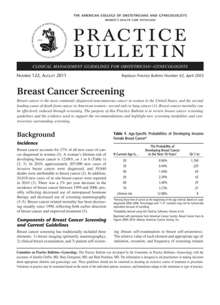
  • 8. risk for a women aged 60 years) or a lifetime risk of 20% or greater may be offered enhanced screening, including a clinical breast examination every 6–12 months, yearly mammography, and instruction in breast self-examination. The Gail model is not a good risk assessment tool when there is a strong family history of breast cancer in women other than the mother and sisters of the patient or a family history of cancer other than breast cancer (eg, ovarian cancer). Breast MRI is not typically recommended based on the Gail Model. What screening is appropriate for women at high risk? For women who test positive for BRCA1 or BRCA2 mutations, enhanced screening should be recommended and risk reduction methods discussed. Enhanced screening for these women includes twice-yearly clinical breast examinations, annual mammography, annual breast MRI, and instruction in breast self-examination. (Details regarding the management of women with BRCA mutations are reviewed in Practice Bulletin No.103, Hereditary Breast and Ovarian Cancer Syndrome, April 2009.) Women who have first-degree relatives with these mutations but who are untested are generally managed as if they carry these mutations until their BRCA status is known. Women who are estimated to have a lifetime risk of breast cancer of 20% or greater, based on risk models that rely largely on family history (such as BRCAPRO, BODACEA, or Claus), but who are either untested or test negative for BRCA gene mutations, can be offered enhanced screening as described previously for carriers of the BRCA mutation. Risk-reduction strategies also may be considered for these women. Other women considered at high risk of development of future cases of breast cancer are those who received thoracic irradiation (typically as a treatment for lymphoma) between the ages 10 years and 30 years. These women should be advised to receive annual mammography, annual MRI, and screening clinical breast examination every 6–12 months beginning 8–10 years after they received treatment or at age 25 years, whichever occurs last. Women with a personal history of high-risk breast biopsy results, including atypical hyperplasia and lobular carcinoma in situ are at increased risk of future breast cancer. These women should receive enhanced screening, including annual mammography, clinical breast examination every 6–12 months, and instruction in breast self-examination. Yearly breast MRI also has been recommended for women with a history of lobular carcinoma in situ by some organizations (43). Women with a personal history of ductal carcinoma in situ or invasive 8 breast cancer should be monitored similarly, although MRI is not routinely recommended in this population. Is there an upper age range at which the risks of mammography outweigh the benefits? A consensus of recommendations on this issue does not exist. Medical comorbidity and life expectancy should be considered in a breast cancer screening program for women aged 75 years or older because the benefit of screening mammography decreases compared with the harms of overtreatment with advancing age. Women aged 75 years or older should, in consultation with their physicians, decide whether or not to continue mammographic screening (17). Figure 1 illustrates the progressively increasing incidence and mortality rates of invasive breast cancer by race and age up to 84 years (3). Most of the screening mammography clinical trials had an upper age limit criteria ranging from 64 years to 74 years. However, a meta-analysis concluded that screening mammography in women aged 70–79 years is moderately cost-effective and yields a small increase in life expectancy (54). Summary of Recommendations and Conclusions The following recommendations are based on limited and inconsistent scientific evidence (Level B): Based on the incidence of breast cancer, the sojourn time for breast cancer growth, and the potential reduction in breast cancer mortality, the College recommends that women aged 40 years and older be offered screening mammography annually. The following recommendations are based primarily on consensus and expert opinion (Level C): Clinical breast examination should be performed annually for women aged 40 years and older. For women aged 20–39 years, clinical breast examinations are recommended every 1–3 years. Breast self-awareness should be encouraged and can include breast self-examination. Women should report any changes in their breasts to their health care providers. Women should be educated on the predictive value of screening mammography and the potential for false-positive results and false-negative results. Women should be informed of the potential for Practice Bulletin No. 122
  • 9. 600 500 Incidence: White Rate per 100,000 400 Incidence: African American 300 200 Mortality: African American 100 Mortality: White 0 20–24 25–29 30–34 35–39 40–44 45–49 50–54 55–59 60–64 65–69 70–74 75–79 80–84 85+ Age Fig. 1. Female Breast Cancer—Incidence and Mortality Rates by Age and Race, United States, 2002–2006. Data from Incidence—North American Association of Central Cancer Registries, 2009. Mortality––National Center for Health Statistics, Centers for Disease Control and Prevention, 2009. Reprinted with permission from American Cancer Society. Breast Cancer Facts & Figures 2009–2010. Atlanta: American Cancer Society, Inc. additional imaging or biopsies that may be recommended based on screening results. Women who are estimated to have a lifetime risk of breast cancer of 20% or greater, based on risk models that rely largely on family history (such as BRCAPRO, BODACEA, or Claus), but who are either untested or test negative for BRCA gene mutations, can be offered enhanced screening. Breast MRI is not recommended for screening women at average risk of developing breast cancer. For women who test positive for BRCA1 and BRCA2 mutations, enhanced screening should be recommended and risk reduction methods discussed. References 1. Jemal A, Siegel R, Ward E, Hao Y, Xu J, Thun MJ. Cancer statistics, 2009. CA Cancer J Clin 2009;59:225– 49. (Level II-3) 2. Altekruse SF, Kosary CL, Krapcho M, Neyman N, Aminou R, Waldron W et al, editors. SEER cancer statistics review, 1975-2007. Bethesda (MD): National Cancer Institute; 2011. Available at: http://seer.cancer.gov/csr/ 1975_2007. Retrieved February 17, 2011. (Level II-3) 3. American Cancer Society. Breast cancer facts & figures: 2009-2010. Atlanta (GA): ACS; 2009. Available at: http:// www.cancer.org/acs/groups/content/@nho/documents/ document/f861009final90809pdf.pdf. Retrieved February 17, 2011. (Level II-3) Practice Bul­e­in No. 122 l t 4. Ravdin PM, Cronin KA, Howlader N, Berg CD, Chlebowski RT, Feuer EJ, et al. The decrease in breastcancer incidence in 2003 in the United States. N Engl J Med 2007;356:1670–4. (Level III) 5. Glass AG, Lacey JV Jr, Carreon JD, Hoover RN. Breast cancer incidence, 1980-2006: combined roles of menopausal hormone therapy, screening mammography, and estrogen receptor status. J Natl Cancer Inst 2007;99: 1152–61. (Level II-3) 6. Rosen PP, Groshen S, Kinne DW. Survival and prognostic factors in node-negative breast cancer: results of long-term follow-up studies. J Natl Cancer Inst Monogr 1992;(11):159–62. (Level III) 7. Tabar L, Chen HH, Duffy SW, Yen MF, Chiang CF, Dean PB, et al. A novel method for prediction of long-term outcome of women with T1a, T1b, and 10-14 mm invasive breast cancers: a prospective study [published erratum appears in Lancet 2000;355:1372]. Lancet 2000;355:429–33. (Level II-3) 8. Tabar L, Dean PB, Kaufman CS, Duffy SW, Chen HH. A new era in the diagnosis of breast cancer. Surg Oncol Clin N Am 2000;9:233–77. (Level III) 9. Joensuu H, Pylkkanen L, Toikkanen S. Late mortality from pT1N0M0 breast carcinoma. Cancer 1999;85: 2183–9. (Level II-3) 1 0. Lopez MJ, Smart CR. Twenty-year follow-up of minimal breast cancer from the Breast Cancer Detection Demonstration Project. Surg Oncol Clin N Am 1997;6: 393–401. (Level II-3) 11. Arnesson LG, Smeds S, Fagerberg G. Recurrence-free survival in patients with small breast cancer. An analysis of cancers 10 mm or less detected clinically and by screening. Eur J Surg 1994;160:271–6. (Level I) 9
  • 10. 12. Macdonald I. The natural history of mammary carcinoma. Am J Surg 1966;111:435–42. (Level III) 13. Gullino PM. Natural history of breast cancer. Progression from hyperplasia to neoplasia as predicted by angiogenesis. Cancer 1977;39:2697–703. (Level III) 14. Wertheimer MD, Costanza ME, Dodson TF, D’Orsi C, Pastides H, Zapka JG. Increasing the effort toward breast cancer detection. JAMA 1986;255:1311–5. (Level III) 15. Walter SD, Day NE. Estimation of the duration of a pre-clinical disease state using screening data. Am J Epidemiol 1983;118:865–86. (Level III) 16. Smith RA, Duffy SW, Gabe R, Tabar L, Yen AM, Chen TH. The randomized trials of breast cancer screening: what have we learned? Radiol Clin North Am 2004;42:793– 806; v. (Level III) 17. Nelson HD, Tyne K, Naik A, Bougatsos C, Chan BK, Humphrey L. Screening for breast cancer: an update for the U.S. Preventive Services Task Force. U.S. Preventive Services Task Force. Ann Intern Med 2009;151:727-37; W237–42. (Level III) 18. Humphrey LL, Helfand M, Chan BK, Woolf SH. Breast cancer screening: a summary of the evidence for the U.S. Preventive Services Task Force. Ann Intern Med 2002;137:347–60. (Level III) 19. Kerlikowske K, Grady D, Rubin SM, Sandrock C, Ernster VL. Efficacy of screening mammography. A meta-analysis. JAMA 1995;273:149–54. (Meta-analysis) 20. Hendrick RE, Smith RA, Rutledge JH, 3rd, Smart CR. Benefit of screening mammography in women aged 40-49: a new meta-analysis of randomized controlled trials.J Natl Cancer Inst Monogr 1997;(22):87–92. (Meta-analysis) 2 1. Olsen O, Gotzsche PC. Cochrane review on screening for breast cancer with mammography [published erratum appears in Lancet 2006;367:474]. Lancet 2001;358: 1340–2. (Level III) 22. Nystrom L, Andersson I, Bjurstam N, Frisell J, Nordenskjold B, Rutqvist LE. Long-term effects of mammography screening: updated overview of the Swedish randomised trials [published erratum appears in Lancet 2002;360:724]. Lancet 2002;359:909–19. (Level III) 23. Screening for breast cancer: U.S. Preventive Services Task Force recommendation statement. US Preventive Services Task Force [published errata appear in Ann Intern Med 2010;152:199–200; Ann Intern Med 2010;152:688]. Ann Intern Med 2009;151:716–26;W-236. (Level III) 24. Mandelblatt J, Saha S, Teutsch S, Hoerger T, Siu AL, Atkins D, et al. The cost-effectiveness of screening mammography beyond age 65 years: a systematic review for the U.S. Preventive Services Task Force. Cost Work Group of the U.S. Preventive Services Task Force. Ann Intern Med 2003;139:835–42. (Level III) 25. Saslow D, Boetes C, Burke W, Harms S, Leach MO, Lehman CD, et al. American Cancer Society guidelines for breast screening with MRI as an adjunct to mammography. American Cancer Society Breast Cancer Advisory Group [published erratum appears in CA Cancer J Clin 2007;57:185]. CA Cancer J Clin 2007;57:75–89. (Level III) 26. Pisano ED, Gatsonis C, Hendrick E, Yaffe M, Baum JK, Acharyya S, et al. Diagnostic performance of digital 10 versus film mammography for breast-cancer screening. Digital Mammographic Imaging Screening Trial (DMIST) Investigators Group [published erratum appears in N Engl J Med 2006;355:1840]. N Engl J Med 2005; 353:1773–83. (Level II-3) 27. Skaane P, Hofvind S, Skjennald A. Randomized trial of screen-film versus full-field digital mammography with soft-copy reading in population-based screening program: follow-up and final results of Oslo II study. Radiology 2007;244:708–17. (Level I) 28. Vinnicombe S, Pinto Pereira SM, McCormack VA, Shiel S, Perry N, Dos Santos Silva IM. Full-field digital versus screen-film mammography: comparison within the UK breast screening program and systematic review of published data. Radiology 2009;251:347–58. (Level III) 29. Coates RJ, Uhler RJ, Brogan DJ, Gammon MD, Malone KE, Swanson CA, et al. Patterns and predictors of the breast cancer detection methods in women under age 45 years of age (United States). CCC cancer causes & control 2001;12(5):431–42. (Level II-2) 30. Newcomer L, Newcomb P, Trentham-Dietz A, Storer B, Yasui Y, Daling J, et al. Detection method and breast carcinoma histology. Cancer 2002;95(3):470–7. (Level II-3) 31. Auvinen A, Elovainio L, Hakama M. Breast self-examination and survival from breast cancer: a prospective follow-up study. Breast Cancer Res Treat 1996;38:161–8. (Level II-2) 32. Thomas DB, Gao DL, Ray RM, Wang WW, Allison CJ, Chen FL, et al. Randomized trial of breast self-examination in Shanghai: final results. J Natl Cancer Inst 2002; 94:1445–57. (Level I) 33. Kosters JP, Gotzsche PC. Regular self-examination or clinical examination for early detection of breast cancer. Cochrane Database of Systematic Reviews 2003, Issue 2. Art. No.: CD003373. DOI: 10.1002/14651858. CD003373. (Meta-analysis) 34. Baxter N. Preventive health care, 2001 update: should women be routinely taught breast self-examination to screen for breast cancer? Canadian Task Force on Preventive Health Care. CMAJ 2001;164:1837–46. (Level III) 35. Bobo JK, Lee NC, Thames SF. Findings from 752,081 clinical breast examinations reported to a national screening program from 1995 through 1998. J Natl Cancer Inst 2000;92:971–6. (Level II-3) 36. Shen Y, Zelen M. Screening sensitivity and sojourn time from breast cancer early detection clinical trials: mammograms and physical examinations. J Clin Oncol 2001;19:3490–9. (Level III) 37. Jatoi I. Breast cancer screening. Am J Surg 1999;177: 518–24. (Level III) 38. Primic-Zakelj M. Screening mammography for early detection of breast cancer. Ann Oncol 1999;10(suppl 6):121–7. (Level III) 39. Fletcher SW, Black W, Harris R, Rimer BK, Shapiro S. Report of the International Workshop on Screening for Breast Cancer. J Natl Cancer Inst 1993;85:1644–56. (Level III) 40. Miller AB, To T, Baines CJ, Wall C. Canadian national breast screening study-2: 13-year results of a random- Practice Bulletin No. 122
  • 11. ized trial in women aged 50–59 years. J Natl Cancer Inst 2000;92:1490–9. (Level I) 41. Chiarelli AM, Majpruz V, Brown P, Theriault M, Shumak R, Mai V. The contribution of clinical breast examination to the accuracy of breast screening. J Natl Cancer Inst 2009; 101:1236–43. (Level II-2) 42. Smith RA, Saslow D, Sawyer KA, Burke W, Costanza ME, Evans WP 3rd, et al. American Cancer Society guidelines for breast cancer screening: update 2003. American Cancer Society High-Risk Work Group; American Cancer Society Screening Older Women Work Group; American Cancer Society Mammography Work Group; American Cancer Society Physical Examination Work Group; American Cancer Society New Technologies Work Group; American Cancer Society Breast Cancer Advisory Group. CA Cancer J Clin 2003;53:141–69. (Level III) 43. National Comprehensive Cancer Network. NCCN Clinical Practice Guidelines in OncologyTM: breast cancer. Fort Washington (PA): NCCN; 2011. (Level III) 44. Elmore JG, Barton MB, Moceri VM, Polk S, Arena PJ, Fletcher SW. Ten-year risk of false positive screening mammograms and clinical breast examinations. N Engl J Med 1998;338:1089–96. (Level II-3) 45. Armstrong K, Moye E, Williams S, Berlin JA, Reynolds EE. Screening mammography in women 40 to 49 years of age: a systematic review for the American College of Physicians. Ann Intern Med 2007;146:516–26. (Level III) 46. Harris R. Variation of benefits and harms of breast cancer screening with age. J Natl Cancer Inst Monogr 1997;(22):139–43. (Level III) 47. Rimer BK, Bluman LG. The psychosocial consequences of mammography. J Natl Cancer Inst Monogr 1997;(22):131–8. (Level III) 48. Lerman C, Trock B, Rimer BK, Boyce A, Jepson C, Engstrom PF. Psychological and behavioral implications of abnormal mammograms. Ann Intern Med 1991; 114:657–61. (Level III) 49. Brett J, Bankhead C, Henderson B, Watson E, Austoker J. The psychological impact of mammographic screening. A systematic review. Psychooncology 2005;14:917–38. (Level III) 50. Brewer NT, Salz T, Lillie SE. Systematic review: the long-term effects of false-positive mammograms. Ann Intern Med 2007;146:502–10. (Meta-analysis) 51. Schwartz LM, Woloshin S, Sox HC, Fischhoff B, Welch HG. US women’s attitudes to false positive mammography results and detection of ductal carcinoma in situ: cross sectional survey. BMJ 2000;320:1635–40. (Level III) 52. Overmoyer B. Breast cancer screening. Med Clin North Am 1999;83:1443–66;vi–vii. (Level III) 53. Agency for Healthcare Research and Quality. Diagnosis and management of specific breast abnormalities. Evidence Report/Technology Assessment 33. Rockville (MD): AHRQ; 2001. AHRQ publication no. 01-E046. (Level III) 54. Kerlikowske K, Salzmann P, Phillips KA, Cauley JA, Cummings SR. Continuing screening mammography in women aged 70 to 79 years: impact on life expectancy and cost-effectiveness. JAMA 1999;282:2156–63. (Level III) Practice Bul­e­in No. 122 l t The MEDLINE database, the Cochrane Library, and the American College of Obstetricians and Gynecologists’ own internal resources and documents were used to con­ duct a lit­ r­ ­ure search to lo­ ate rel­ ­ ant ar­i­ les pub­ished e at c ev tc l be­ween January 1990–February 2011. The search was t re­ trict­ d to ar­i­ les pub­ished in the English lan­ uage. s e tc l g Pri­ r­­y was given to articles re­ ort­ng results of orig­­ al o it p i in re­ earch, although re­ iew ar­i­ les and com­ en­ar­es also s v tc m t i were consulted. Ab­ tracts of re­ earch pre­ ent­ d at sym­ o­ s s s e p sia and sci­ n­if­c con­ er­ nc­ s were not con­ id­ red adequate e t i f e e s e for in­ lu­ ion in this doc­ ­ ent. Guide­ines pub­ished by c s um l l or­ a­ i­ a­ions or in­ ti­u­ions such as the Na­ion­ l In­ ti­utes g nz t s t t t a s t of Health and the Amer­­ an Col­ege of Ob­ te­ri­ ians and ic l s t c Gy­ e­ ol­ ­ ists were re­ iewed, and ad­ i­ion­ l studies were n c og v dt a located by re­ iew­ng bib­iographies of identified articles. v i l When re­i­ ble research was not available, expert opinions la from ob­ te­ri­ ian–gynecologists were used. s t c Studies were reviewed and evaluated for qual­ty ac­ ord­ng i­ c i to the method outlined by the U.S. Pre­ en­ive Services v t Task Force: I Evidence obtained from at least one prop­ r­ y el de­ igned randomized controlled trial. s II-1 Evidence obtained from well-designed con­rolled t tri­ ls without randomization. a II-2 Evidence obtained from well-designed co­ ort or h case–control analytic studies, pref­ r­ ­ ly from more e ab than one center or research group. II-3 Evidence obtained from multiple time series with or with­ ut the intervention. Dra­ at­c re­ ults in un­ on­ o m i s c trolled ex­ er­­ ents also could be regarded as this p im type of ev­­ ence. id III Opinions of respected authorities, based on clin­­ al ic ex­ e­ i­ nce, descriptive stud­es, or re­ orts of ex­ ert p re i p p committees. Based on the highest level of evidence found in the data, recommendations are provided and grad­ d ac­ ord­ng to the e c i following categories: Level A—Recommendations are based on good and con­ sis­ent sci­ n­if­c evidence. t e t i Level B—Recommendations are based on limited or in­ on­ c sis­ent scientific evidence. t Level C—Recommendations are based primarily on con­ sen­ us and expert opinion. s Copyright August 2011 by the American College of Ob­ te­s t ri­ ians and Gynecologists. All rights reserved. No part of this c publication may be reproduced, stored in a re­riev­ l sys­em, t a t posted on the Internet, or transmitted, in any form or by any means, elec­ron­c, me­ han­­ al, photocopying, recording, or t i c ic oth­ r­ ise, without prior written permission from the publisher. ew Requests for authorization to make photocopies should be directed to Copyright Clearance Center, 222 Rosewood Drive, Danvers, MA 01923, (978) 750-8400. ISSN 1099-3630 The American College of Obstetricians and Gynecologists 409 12th Street, SW, PO Box 96920, Washington, DC 20090-6920 Breast cancer screening. Practice Bulletin No. 122. American College of Obstetricians and Gynecologists. Obstet Gynecol 2011;118: 372–82. 11