SlideShare uma empresa Scribd logo
1 de 61
SpirochetsSpirochets
ARUNI.I.S,
Reader
Dept of Microbiology
Noorul Islam Institute of Medical Science
Trivandum
Spirochetes are elongated, motile, flexibleSpirochetes are elongated, motile, flexible
bacteria twisted spirally alone the long axis.bacteria twisted spirally alone the long axis.
Motile withMotile with endoflagellaendoflagella..
Human pathogens are found in 3 genera:Human pathogens are found in 3 genera:
TreponemaTreponema
BorreliaBorrelia
LeptospiraLeptospira
SpirochetesSpirochetes
TreponemaTreponema
 Some of theSome of the Treponema speciesTreponema species are pathogensare pathogens
while others occur as commensals in thewhile others occur as commensals in the
mouth, intestine and genitalia.mouth, intestine and genitalia.
 Causes the following diseases in humans:Causes the following diseases in humans:
1.1. Venereal syphilis by T.pallidumVenereal syphilis by T.pallidum
2.Endemic syphilis T.pallidum(T.endemicum)2.Endemic syphilis T.pallidum(T.endemicum)
3.Yaws by T.pertenue3.Yaws by T.pertenue
4. Pinta by T.carateum4. Pinta by T.carateum
TreponemaTreponema
Treponema pallidumTreponema pallidum
 Causative agent ofCausative agent of SyphilisSyphilis..
 Name pallidum refers to it’s pale staining.Name pallidum refers to it’s pale staining.
 Delicate spirochete withDelicate spirochete with active motilityactive motility..
 Thin with tapering ends.10 mm long & 0.1-0.2 mm wide.Thin with tapering ends.10 mm long & 0.1-0.2 mm wide.
 Morphology and motility can be seen under theMorphology and motility can be seen under the darkdark
ground or phase contrast microscopeground or phase contrast microscope..
 Can be stained by Silver impregnation methodCan be stained by Silver impregnation method
 Very delicateVery delicate and highly susceptible to heat.and highly susceptible to heat.
 Do not grow in artificial culture media, but possible byDo not grow in artificial culture media, but possible by co-co-
cultivationcultivation with tissue culture cells.with tissue culture cells.
 Natural infection occur only inNatural infection occur only in human beingshuman beings..
 Antigenic structure:Antigenic structure:
 3 types of antigens are seen in3 types of antigens are seen in TreponemaTreponema::
a)a) Cardiolipin antigenCardiolipin antigen present inpresent in T.pallidumT.pallidum againstagainst
which reagin Abs are producedwhich reagin Abs are produced
b)b) AA group specific antigengroup specific antigen present inpresent in T.pallidumT.pallidum asas
well as non-pathogenic Reiter strain of Treponema.well as non-pathogenic Reiter strain of Treponema.
c)c) AA species specific polysaccharidespecies specific polysaccharide present inpresent in
pathogenic strain ofpathogenic strain of TreponemaTreponema..
SYPHILISSYPHILIS
Caused by Treponema pallidum
Can enter any mucous membrane or break in the skin,
can be spread by kissing and close contact
SYPHILISSYPHILIS
NONO
SYMPTOMSSYMPTOMS
Up to 50% of people don’t know they have it.
Symptoms may be mild or painless or
nearly invisible.
 Organism enters the body through cuts or abrasion on the skinOrganism enters the body through cuts or abrasion on the skin
or mucosa during sexual contact.or mucosa during sexual contact.
 IP is about a month(range 10-90 days)IP is about a month(range 10-90 days)
 3stages of venereal syphilis3stages of venereal syphilis
 Primary:Primary: chancre, disappears spontaneously.chancre, disappears spontaneously.
 Secondary:Secondary: penetrate mucus membranes, enter blood-stream,penetrate mucus membranes, enter blood-stream,
fever, rash.fever, rash.
 Tertiary:Tertiary: invade heart, musculoskeletal system, CNS, relativelyinvade heart, musculoskeletal system, CNS, relatively
noninfectious.noninfectious.
PathogenesisPathogenesis
11stst
Stage SYPHILIS(Primary)Stage SYPHILIS(Primary)
10 to 90 days after contact, painless, avascular, indurated
superficially ulcerated lesion may appear, called hard chancres.
It occurs at the site of entry of the bacteria – genital organs,
mouth , nipples- and heals spontaneously after 10-40 days.
They can appear ANYWHERE that the spirochetes
entered a mucous membrane or break in the skin.
penis
finger
tongue
cervix
vulva
 The chancre is covered by a thick ,glairyThe chancre is covered by a thick ,glairy
exudate ,rich in spirochetesexudate ,rich in spirochetes
 The regional lymph nodes are swollenThe regional lymph nodes are swollen
 Even before the chancre appears, theEven before the chancre appears, the
spirochetes spread from the site of entry in tospirochetes spread from the site of entry in to
lymph & blood streamlymph & blood stream
 So the patient may be infectious during the lateSo the patient may be infectious during the late
incubation periodincubation period
 The chancre heals in 10-40 days ,even with outThe chancre heals in 10-40 days ,even with out
treatment, leaving a thin scartreatment, leaving a thin scar
 Persistent or multiple chancre may be seen inPersistent or multiple chancre may be seen in
HIV & othe immunodeficient patiensHIV & othe immunodeficient patiens
22ndnd
Stage SYPHILIS(Secondary)Stage SYPHILIS(Secondary)
Symptoms appear 2 weeks to 6 months after primary lesion.
Characterized by papular skin rashes, mucous patches in
oropharynx and condylomata at the mucocutaneous junctions.
Patient is highly infectious and these signs disappear without
treatment.
• Rash on hands
• Rash over entire body • Lesions of tongue
• Rash on feet
•Lesions around mouth
ORAL MANIFESTATIONSORAL MANIFESTATIONS
The oral lesions are usuallyThe oral lesions are usually
multiple,painless. Greenish white plaguesmultiple,painless. Greenish white plagues
overlying an ulcerated surfaceoverlying an ulcerated surface
These are called mucous patchesThese are called mucous patches
They occur mostly on the tongue, buccalThey occur mostly on the tongue, buccal
mucosa or gingivamucosa or gingiva
These lesions are highly infectious as theyThese lesions are highly infectious as they
contain a large number of microorganismscontain a large number of microorganisms
Oral lesion in Secondary SyphilisOral lesion in Secondary Syphilis
 Spirocetes are abundant in the lesions & the patient isSpirocetes are abundant in the lesions & the patient is
most infectious during the secondary stage.most infectious during the secondary stage.
 The lesions undergo spontaneous healing ,in someThe lesions undergo spontaneous healing ,in some
instances taking as long as 4 or 5 yearsinstances taking as long as 4 or 5 years
 After the secondary lesion disappears, there is a periodAfter the secondary lesion disappears, there is a period
of quiescence known as latent syphilis.of quiescence known as latent syphilis.
 Diagnosis during this period is possible only by serologyDiagnosis during this period is possible only by serology
 In many cases this is followed by natural cure, but inIn many cases this is followed by natural cure, but in
others after several years, manifestations of Tertiaryothers after several years, manifestations of Tertiary
syphilis appearssyphilis appears
33rdrd
Stage SYPHILIS(Tertiary)Stage SYPHILIS(Tertiary)
The period after the healing of the 2ry lesions- latent syphilis.
Occur after several years and diagnose serologically.
Damages heart and aorta, causes
hardening, distortion, ulceration….can
lead to rupture, hemorrhaging and death.
Brain damage and infection, causes
headaches, dizziness, blurred vision,
paralysis, and can lead to insanity.
Skin Damage, causes “gummata”, large
open sores which are slow to heal.
Syphilis can also damage the bones.
CONGENITSAL SYPHILISCONGENITSAL SYPHILIS
Syphilis is transmitted from
the mother to the baby trough
the placenta.
The more recent the infection,
especially during the pregnancy, the
more damaging to the baby.
COMPLICATIONS:
Miscarriage Enlarged liver and spleen
Stillbirth Hydrocephalus
Meningitis Mental Retardation
Convulsions Sever Skin Rashes
Blindness Deafness
Deformities of face, nose, teeth, jaw, leg bones
Laboratory DiagnosisLaboratory Diagnosis
a)a) Microscopy:Microscopy: wet films of exudates examinedwet films of exudates examined
under the dark ground microscope.under the dark ground microscope.
b)b) Serology:Serology:
1)1) Reagin antibody test: tests for antibodiesReagin antibody test: tests for antibodies
reacting with cardiolipin antigen and arereacting with cardiolipin antigen and are
known asknown as standard test for syphilis (STS).standard test for syphilis (STS).
i.i. Wasserman complement fixation testWasserman complement fixation test
ii.ii. Khan flocculation testKhan flocculation test
iii.iii. VDRL / RPR testVDRL / RPR test
2)2) Group specific treponemal tests - Here cultivableGroup specific treponemal tests - Here cultivable
treponems (Reiter strain) used as antigen.treponems (Reiter strain) used as antigen.
Reiter Protein Complement Fixation Test(RPCF)Reiter Protein Complement Fixation Test(RPCF)
3)3) SpecificSpecific T.pallidumT.pallidum test - Here virulent Nichol’s strainstest - Here virulent Nichol’s strains
ofof T.pallidumT.pallidum is used as antigen.is used as antigen.
i.i. T.pallidumT.pallidum immobilization (TPI) testimmobilization (TPI) test
ii.ii. Fluorecent treponemal antibody (FTA) testFluorecent treponemal antibody (FTA) test
iii.iii. T.pallidumT.pallidum Haemagglutination (TPHA) testHaemagglutination (TPHA) test
 EIA for Treponemal antibody detectionEIA for Treponemal antibody detection
 CSF VDRL is used for neurosyphilisCSF VDRL is used for neurosyphilis
Prophylaxis & TreatmentProphylaxis & Treatment
Avoid sexual contact with an infectedAvoid sexual contact with an infected
individual.individual.
Use of physical barriers, antiseptics orUse of physical barriers, antiseptics or
antibiotics may minimise the risk.antibiotics may minimise the risk.
Chemoprophylaxis by penicillin.Chemoprophylaxis by penicillin.
No vaccine is available.No vaccine is available.
Treatment by Penicillin G, Erythromycin,Treatment by Penicillin G, Erythromycin,
Non-venereal TreponemesNon-venereal Treponemes
 Organisms closely resemblesOrganisms closely resembles T.pallidumT.pallidum but do not causesbut do not causes
VD.VD.
 Transmission occurs through direct body to body contactTransmission occurs through direct body to body contact
among people of unhygienic practices.among people of unhygienic practices.
 Important non-venereal infections include:Important non-venereal infections include:

Endemic syphilisEndemic syphilis

YawsYaws

PintaPinta
Endemic SyphilisEndemic Syphilis
 Causative agent isCausative agent is T.endemicum.T.endemicum.
 Common in children of poor personal hygiene.Common in children of poor personal hygiene.
 Infection can also be seen on the nipples of the mother infected byInfection can also be seen on the nipples of the mother infected by
her children.her children.
 Usually seen with the manifestations of secondary syphilis such asUsually seen with the manifestations of secondary syphilis such as
mucous patches & skin eruptionsmucous patches & skin eruptions
 The disease may progress to gummatous lesions on the skin, boneThe disease may progress to gummatous lesions on the skin, bone
and nasopharynyx.and nasopharynyx.
 Laboratory diagnosis and treatment are same as venereal syphilis.Laboratory diagnosis and treatment are same as venereal syphilis.
YawsYaws
In India, it has been reported from Orissa,In India, it has been reported from Orissa,
Andra Pradesh and Madhya Pradesh.Andra Pradesh and Madhya Pradesh.
Like in syphilis, the primary lesion isLike in syphilis, the primary lesion is
followed by secondary and tertiaryfollowed by secondary and tertiary
manifestations.manifestations.
 Causative agent isCausative agent is T.pertenue.T.pertenue.
 The primary lesion in yaws is anThe primary lesion in yaws is an
extra-genital papula which breaksextra-genital papula which breaks
down to form an ulceratingdown to form an ulcerating
granuloma.granuloma.
PintaPinta
 Causative agent isCausative agent is T.carateum.T.carateum.
 Disease is seen mainly in central and south America.Disease is seen mainly in central and south America.
 Infection occurs by direct person to person contact.Infection occurs by direct person to person contact.
 The primary lesion is an extra-genital papule.The primary lesion is an extra-genital papule.
 Hypopigmentation or hyperpigmentation is seen.Hypopigmentation or hyperpigmentation is seen.
 Laboratory diagnosis and treatment are similar asLaboratory diagnosis and treatment are similar as
T.pallidum.T.pallidum.
BorreliaBorrelia
BorreliaBorrelia
Microaerophilic-to-anaerobicMicroaerophilic-to-anaerobic
 Loose, irregular coilsLoose, irregular coils
 In vitroIn vitro cultivation is difficultcultivation is difficult
 Visualization byVisualization by
Giemsa stainGiemsa stain
Gram staining-Gram negativeGram staining-Gram negative
Silver stainingSilver staining
Dark field microscopyDark field microscopy
 Highly adapted to arthropod transmissionHighly adapted to arthropod transmission
 Antigenic variation
Borrelia undergoes antigenic variations in vivo
and is found to be the cause of relapses in the
diseases.
Recovery from the disease is believed to be due
to the development of immunity to theses
antigenic variants.
 Species of medical importance:Species of medical importance:
B.recurrentisB.recurrentis – Relapsing fever– Relapsing fever
B.vincentiB.vincenti – Fusospirochetosis(Vincent’s– Fusospirochetosis(Vincent’s
angina)angina)
B.burgdorferiB.burgdorferi – Lyme disease– Lyme disease
 This Acute infection is caused byThis Acute infection is caused by B.recurrentisB.recurrentis..
 Sudden onset with fever, septicemia, headache, muscle pain.Sudden onset with fever, septicemia, headache, muscle pain.
 After 3-5 days, fever subsides and during this afebrile period ofAfter 3-5 days, fever subsides and during this afebrile period of
4-19 days borreliae are not found in the blood.4-19 days borreliae are not found in the blood.
 After a afebrile period when another bout of fever sets in,After a afebrile period when another bout of fever sets in,
borreliae reappear in blood.borreliae reappear in blood.
 Patient returns to normal, after 4-10 relapses.Patient returns to normal, after 4-10 relapses.
 Sporadic in US, distributed worldwide.Sporadic in US, distributed worldwide.
Relapsing Fever
Transmitted animal-to-animal, animal-to-Transmitted animal-to-animal, animal-to-
human by ticks, human-to-human by bodyhuman by ticks, human-to-human by body
lice.lice.
Causative agents:Causative agents:
Louse borne diseaseLouse borne disease :: B. recurrentisB. recurrentis
Tick borne diseaseTick borne disease :: B.duttonii, B.hermsii,B.duttonii, B.hermsii,
B.parkeriB.parkeri etc.etc.
Relapsing fever- 2 types:Relapsing fever- 2 types:
Tickborne RFTickborne RF
Transmitted by soft ticks (genusTransmitted by soft ticks (genus
OrnithodorosOrnithodoros))
O. turicatae, O. parkeri, O. hermsiO. turicatae, O. parkeri, O. hermsi in US,in US,
15 others worldwide15 others worldwide
Louseborne RFLouseborne RF
Body / head / pubic lice (Body / head / pubic lice (Pediculus humanusPediculus humanus
corporis / capitis / pubiscorporis / capitis / pubis))
Also by bed bugs.Also by bed bugs.
 Laboratory diagnosis:Laboratory diagnosis:
 Microscopy:Microscopy:
Light microscopy- Giemsa / Leishman stain.Light microscopy- Giemsa / Leishman stain.
Silver stainingSilver staining
Dark ground microscopy.Dark ground microscopy.
 Culture:Culture: too difficult to grow.too difficult to grow.
 Animal inoculationAnimal inoculation: into white mouse.: into white mouse.
 Serology:Serology: not reliable.not reliable.
 It is an ulcerativeIt is an ulcerative gingivostomatitisgingivostomatitis oror oropharyngitisoropharyngitis
caused bycaused by Borrelia vincentiBorrelia vincenti..
 B.vincentiB.vincenti is a normal mouth commensal motileis a normal mouth commensal motile
spirochete, about 5-20spirochete, about 5-20 μμm wide, with 3-8 coils ofm wide, with 3-8 coils of
variable size.variable size.
 It is a normal mouth commensal ,but under certainIt is a normal mouth commensal ,but under certain
pre disposing condition(malnutrition ,viral infection)pre disposing condition(malnutrition ,viral infection)
give rise to ulcerative gingivo stomatitis orgive rise to ulcerative gingivo stomatitis or
orophangitis(Vincent’s angina)orophangitis(Vincent’s angina)
 In this infection,In this infection, B.vincentiB.vincenti is associated withis associated with
fusiform bacilli and this symbitic infection is knownfusiform bacilli and this symbitic infection is known
asas fusospirochetosisfusospirochetosis..
Vincent’s AnginaVincent’s Angina
DiagnosisDiagnosis may be made by demonstratingmay be made by demonstrating
spirochetes and fusiform bacilli in stainedspirochetes and fusiform bacilli in stained
smears of exudates form the lesions.smears of exudates form the lesions.
TreatmentTreatment-- Penicillin & metronidazolePenicillin & metronidazole
Lyme DiseaseLyme Disease
 First observed in 1975, in Old Lyme, USA.First observed in 1975, in Old Lyme, USA.
 Caused byCaused by Borrelia burgdorferiBorrelia burgdorferi..
 Incubation period: 7 - 14 days (range 3 - 30 days).Incubation period: 7 - 14 days (range 3 - 30 days).
 Infection may be sub-clinical, manifest onlyInfection may be sub-clinical, manifest only
nonspecific symptoms: fever, headache, myalgia.nonspecific symptoms: fever, headache, myalgia.
 Most prevalent human tick-borne disease in theMost prevalent human tick-borne disease in the
US, Europe, parts of Asia.US, Europe, parts of Asia.
EpidemiologyEpidemiology
Human encroachment into habitat of white-Human encroachment into habitat of white-
tailed deer.tailed deer.
Spread from deer, mice to humans by theSpread from deer, mice to humans by the bitebite
of soft ticksof soft ticks (genus(genus Ixodes)Ixodes)
 First stage:First stage: Erythema migransErythema migrans
 spirochetes multiply in skin, quickly enter circulationspirochetes multiply in skin, quickly enter circulation
 rashrash appears days-to-weeks after tick biteappears days-to-weeks after tick bite
 surround site of inoculation, expand insurround site of inoculation, expand in concentric ringsconcentric rings
 disappear spontaneously after several weeksdisappear spontaneously after several weeks
Second stage:Second stage: disseminated infectiondisseminated infection
associated with fever, headache, myalgia,associated with fever, headache, myalgia,
arthralgia and lymphadenopathy.arthralgia and lymphadenopathy.
Musculoskeletal signs (migratory joint, muscleMusculoskeletal signs (migratory joint, muscle
pains), CNS signs, cardiac damage may alsopains), CNS signs, cardiac damage may also
appear.appear.
Third stageThird stage of persistent infection,of persistent infection, occurringoccurring
months or years latermonths or years later..
Develops chronic arthritis, polyneuropathy,Develops chronic arthritis, polyneuropathy,
encephalopathy, etc.encephalopathy, etc.
Symptoms include arthritis, facial palsy andSymptoms include arthritis, facial palsy and
meningitis.meningitis.
45
Borrelia burgdorferiBorrelia burgdorferi
46
Ixodes scapularis, tick vector for Lyme disease.
47
Lyme DiseaseLyme Disease
erythematous rasherythematous rash
DiagnosisDiagnosis made be isolating the organismsmade be isolating the organisms
form skin lesions, CSF, blood or by serology.form skin lesions, CSF, blood or by serology.
Treatment:Treatment: Amoxycillin, doxycycline andAmoxycillin, doxycycline and
cefuroxime.cefuroxime.
Tick control important forTick control important for prevention.prevention.
LeptospirLeptospiraa
Leptospira are actively motile, delicateLeptospira are actively motile, delicate
spirochetes, which can causespirochetes, which can cause Leptospirosis orLeptospirosis or
Weil’s diseasesWeil’s diseases..
Leptospirosis is an acute zoonotic infection ofLeptospirosis is an acute zoonotic infection of
worldwide significance caused by spirochaeteworldwide significance caused by spirochaete
Leptospira interrogansLeptospira interrogans ..
Various factors influencing the animal activity,Various factors influencing the animal activity,
suitability of the environment for the survivalsuitability of the environment for the survival
of the organism and behavioral andof the organism and behavioral and
occupational habits of human beings can beoccupational habits of human beings can be
the determinants of incidence and prevalencethe determinants of incidence and prevalence
of the disease.of the disease.
 Leptospirosis, also known as canicola fever,Leptospirosis, also known as canicola fever,
hemorrhagic jaundice, , mud fever, spirochetalhemorrhagic jaundice, , mud fever, spirochetal
jaundice, swamp fever, swineherd's disease, caver'sjaundice, swamp fever, swineherd's disease, caver's
flu or sewerman's flu.flu or sewerman's flu.
 There is an acute form of human infection known asThere is an acute form of human infection known as
Weil's diseaseWeil's disease, where the patient suffers from, where the patient suffers from
jaundice.jaundice.
 This condition is usually caused byThis condition is usually caused by
L.L.icterohaemorrhagiaeicterohaemorrhagiae serogroup of L.serogroup of L.interrogansinterrogans
Leptospira interrogansLeptospira interrogans
 Leptospira appear tightly coiled thinLeptospira appear tightly coiled thin
flexible Spirochetes with 5 – 15flexible Spirochetes with 5 – 15
microns x 0.1 micron size.microns x 0.1 micron size.
 One end appears bent forms a hook.One end appears bent forms a hook.
 Actively motile.Actively motile.
 Seen best with dark fieldSeen best with dark field
Microscopy.Microscopy.
 13 serogroups, >200 serovars13 serogroups, >200 serovars
Cultivation of LeptospiraCultivation of Leptospira
 Leptospira grows best underLeptospira grows best under
aerobic conditions at 28aerobic conditions at 2800
to 30to 3000
cc
and pH 7.2 – 7.5.and pH 7.2 – 7.5.
 EMJHEMJH (Ellinghausen,(Ellinghausen,
McCullough, Johnson, Harris)McCullough, Johnson, Harris)
mediamedia is commonly used.is commonly used.
 Other MediaOther Media
 Fletchers MediaFletchers Media
..Optimal growth after 1 – 2 weeksOptimal growth after 1 – 2 weeks
 ..
എലിപ്പനിഎലിപ്പനി PathogenesisPathogenesis
 Rats, Mice, Wild Rodents, Dogs, Swine, Cattle areRats, Mice, Wild Rodents, Dogs, Swine, Cattle are
principleprinciple source of infectionsource of infection..
 The animals excrete Leptospira both in active infectionThe animals excrete Leptospira both in active infection
and Asymptomatic stage through urine.and Asymptomatic stage through urine.
 The Leptospira survive and remain viable for severalThe Leptospira survive and remain viable for several
weeks in stagnant water.weeks in stagnant water.
 Enter through cuts and abrasionsEnter through cuts and abrasions in the skin andin the skin and
mucous membranes or through mouth, nose ormucous membranes or through mouth, nose or
conjunctive.conjunctive.
 Man is the end host, and there is no evidence ofMan is the end host, and there is no evidence of
human to human infection.human to human infection.
 Incubation periodIncubation period 1 – 2 weeks1 – 2 weeks..
 Multiplication of leptospires inMultiplication of leptospires in blood streamblood stream
producesproduces feverfever..
 Establish organ involvement inEstablish organ involvement in Kidney and LiverKidney and Liver..
 May produceMay produce hemorrhage and necrosishemorrhage and necrosis in the tissuesin the tissues
and initiatesand initiates dysfunctiondysfunction of these organs.of these organs.
PP
aa
tt
hh
oo
gg
ee
nn
ee
ss
ii
ss
 Clinical Pictures:Clinical Pictures:
 JaundiceJaundice
 HemorrhageHemorrhage
 Nitrogen retentionNitrogen retention
 TheThe Illness is BiphasicIllness is Biphasic with initial temperaturewith initial temperature
when the second phase comes with raise of IgMwhen the second phase comes with raise of IgM
titers raise.titers raise.
 Aseptic meingitisAseptic meingitis – initial headache, stiffness of– initial headache, stiffness of
neck, pleocytosis of Cerebro spinal fluid.neck, pleocytosis of Cerebro spinal fluid.
 ComplicationsComplications
 NephritisNephritis
 HepatitisHepatitis
 Manifestations in eyeManifestations in eye
 Muscular lesionsMuscular lesions
 Many infections are mild and subclinicalMany infections are mild and subclinical
Weil’s SyndromeWeil’s Syndrome
 Weil's syndrome is a severe form of leptospirosis thatWeil's syndrome is a severe form of leptospirosis that
causes continuouscauses continuous fever, stuporfever, stupor & reduction in the& reduction in the
blood's ability to clot, which leads toblood's ability to clot, which leads to bleeding withinbleeding within
tissuestissues..
 Blood tests reveal anemia.Blood tests reveal anemia.
 By the third to sixth day, signs ofBy the third to sixth day, signs of kidney damagekidney damage andand
liver injuryliver injury appear.appear.
 Kidney abnormalities may causeKidney abnormalities may cause blood in the urineblood in the urine
and painful urinationand painful urination..
 Liver injury may be mild & usually heals completely.Liver injury may be mild & usually heals completely.
Hepatitis - LeptospirosisHepatitis - Leptospirosis
 Hepatitis is the frequent complication.Hepatitis is the frequent complication.
 Elevation of serum creatine phospholipaseElevation of serum creatine phospholipase
enzyme raise differentiates from Viral hepatitisenzyme raise differentiates from Viral hepatitis
where the enzyme is not raised.where the enzyme is not raised.
Nephritis - LeptospirosisNephritis - Leptospirosis
 Kidney involvement in animals produce chronicKidney involvement in animals produce chronic
disease of the kidney and the infected animaldisease of the kidney and the infected animal
startsstarts shedding large number of leptospirashedding large number of leptospira andand
main source of environmental contamination ofmain source of environmental contamination of
bacteria and results in human infections.bacteria and results in human infections.
 Human urine also contain Spirochetes in theHuman urine also contain Spirochetes in the
second and third week of infection.second and third week of infection.
 Laboratory diagnosis:Laboratory diagnosis:
 Specimen:Specimen: Blood, urine, CSF.Blood, urine, CSF.
 Blood examinationBlood examination is significant only in the early stage of theis significant only in the early stage of the
disease.disease.
 Urine examinationUrine examination is significant from second week onwardsis significant from second week onwards
up to 6 weeks.up to 6 weeks.
Microscopy:Microscopy: Blood film / urine sediments under dark groundBlood film / urine sediments under dark ground
microscope or by IF.microscope or by IF.
Culture:Culture: Blood culture in EMJH medium.Blood culture in EMJH medium.
Animal inoculation:Animal inoculation: Blood / urine into guinea pig.Blood / urine into guinea pig.
Serology:Serology: Detection of antibodies by ELISADetection of antibodies by ELISA..
Prophylaxis:Prophylaxis:
 Rodent control, disinfection of water andRodent control, disinfection of water and
wearing of protective clothing.wearing of protective clothing.
 Vaccination on trial basis.Vaccination on trial basis.
Treatment:Treatment:
 Penicillin, Tetracycline, Doxycycline.Penicillin, Tetracycline, Doxycycline.
Thank youThank you

Mais conteúdo relacionado

Mais procurados (20)

Corynebacterium diptheriae
Corynebacterium diptheriaeCorynebacterium diptheriae
Corynebacterium diptheriae
 
Candida
CandidaCandida
Candida
 
12. mycobacterium leprae
12. mycobacterium leprae12. mycobacterium leprae
12. mycobacterium leprae
 
Bacteriology
BacteriologyBacteriology
Bacteriology
 
Laboratory diagnosis of viral infection
Laboratory diagnosis of viral infectionLaboratory diagnosis of viral infection
Laboratory diagnosis of viral infection
 
Spirochaetes
SpirochaetesSpirochaetes
Spirochaetes
 
Treponema pallidum
Treponema pallidumTreponema pallidum
Treponema pallidum
 
Mycetoma
MycetomaMycetoma
Mycetoma
 
Ziehl Neelsen staining
Ziehl Neelsen stainingZiehl Neelsen staining
Ziehl Neelsen staining
 
Gram Stain Practical Briefing
Gram Stain Practical BriefingGram Stain Practical Briefing
Gram Stain Practical Briefing
 
Camp test
Camp testCamp test
Camp test
 
Mycobacterium leprae
Mycobacterium lepraeMycobacterium leprae
Mycobacterium leprae
 
Staphylococcus
StaphylococcusStaphylococcus
Staphylococcus
 
Staphylococci
StaphylococciStaphylococci
Staphylococci
 
Gram negative rods
Gram negative rodsGram negative rods
Gram negative rods
 
gram staining
gram staininggram staining
gram staining
 
Mycobacteria
MycobacteriaMycobacteria
Mycobacteria
 
Mycobacterium
MycobacteriumMycobacterium
Mycobacterium
 
Bacillus
Bacillus Bacillus
Bacillus
 
Spore forming bacteria - bacillus and clostridia
Spore forming bacteria - bacillus and clostridiaSpore forming bacteria - bacillus and clostridia
Spore forming bacteria - bacillus and clostridia
 

Destaque

Spirochetes for undergraduate students. treponema, leptospira, borrelia
Spirochetes for undergraduate students. treponema, leptospira, borreliaSpirochetes for undergraduate students. treponema, leptospira, borrelia
Spirochetes for undergraduate students. treponema, leptospira, borreliaRiyaz Sheriff
 
common oral lesions by ravindra daggupati
common oral lesions by ravindra daggupaticommon oral lesions by ravindra daggupati
common oral lesions by ravindra daggupatiRavindra Daggupati
 
Cholera update ddhs group agm 05 sep2014 dr sarkodie
Cholera update ddhs group agm 05 sep2014 dr sarkodieCholera update ddhs group agm 05 sep2014 dr sarkodie
Cholera update ddhs group agm 05 sep2014 dr sarkodieGeorge Akowuah
 
Presentation On Buruli Ulcer
Presentation On Buruli UlcerPresentation On Buruli Ulcer
Presentation On Buruli UlcerABURA Foundation
 
Cancrum oris or noma
Cancrum oris or nomaCancrum oris or noma
Cancrum oris or nomaAndre_M626
 
Mycobacterium leprae
Mycobacterium lepraeMycobacterium leprae
Mycobacterium lepraeKimzzy_Tyne
 
Proton pump inhibitors- ome, esome, lanso, pant
Proton pump  inhibitors- ome, esome, lanso, pantProton pump  inhibitors- ome, esome, lanso, pant
Proton pump inhibitors- ome, esome, lanso, pantSudha Tallapaneni
 
proton pump inhibitors discovery and development
proton pump inhibitors discovery and developmentproton pump inhibitors discovery and development
proton pump inhibitors discovery and developmentAli Hussein
 
Mycobacterium leprae - Lab diagnosis
Mycobacterium leprae - Lab diagnosisMycobacterium leprae - Lab diagnosis
Mycobacterium leprae - Lab diagnosissana_arman
 
Mycobacteria slides for lecture
Mycobacteria slides for lectureMycobacteria slides for lecture
Mycobacteria slides for lectureBruno Mmassy
 
acute gingival infections
 acute gingival infections   acute gingival infections
acute gingival infections neeti shinde
 
Proton pump inhibitor
Proton pump inhibitorProton pump inhibitor
Proton pump inhibitorAsiful alam
 

Destaque (20)

Spirochetes for undergraduate students. treponema, leptospira, borrelia
Spirochetes for undergraduate students. treponema, leptospira, borreliaSpirochetes for undergraduate students. treponema, leptospira, borrelia
Spirochetes for undergraduate students. treponema, leptospira, borrelia
 
Spirochetes
SpirochetesSpirochetes
Spirochetes
 
Spirochaetes
SpirochaetesSpirochaetes
Spirochaetes
 
Spirochaetes
SpirochaetesSpirochaetes
Spirochaetes
 
Spirochetes
SpirochetesSpirochetes
Spirochetes
 
common oral lesions by ravindra daggupati
common oral lesions by ravindra daggupaticommon oral lesions by ravindra daggupati
common oral lesions by ravindra daggupati
 
Spirochaetes treponema, borrelia & leptospira
Spirochaetes  treponema, borrelia & leptospiraSpirochaetes  treponema, borrelia & leptospira
Spirochaetes treponema, borrelia & leptospira
 
Cholera update ddhs group agm 05 sep2014 dr sarkodie
Cholera update ddhs group agm 05 sep2014 dr sarkodieCholera update ddhs group agm 05 sep2014 dr sarkodie
Cholera update ddhs group agm 05 sep2014 dr sarkodie
 
Noma
NomaNoma
Noma
 
Presentation On Buruli Ulcer
Presentation On Buruli UlcerPresentation On Buruli Ulcer
Presentation On Buruli Ulcer
 
Cancrum oris or noma
Cancrum oris or nomaCancrum oris or noma
Cancrum oris or noma
 
Mycobacterium leprae
Mycobacterium lepraeMycobacterium leprae
Mycobacterium leprae
 
Proton pump inhibitors- ome, esome, lanso, pant
Proton pump  inhibitors- ome, esome, lanso, pantProton pump  inhibitors- ome, esome, lanso, pant
Proton pump inhibitors- ome, esome, lanso, pant
 
proton pump inhibitors discovery and development
proton pump inhibitors discovery and developmentproton pump inhibitors discovery and development
proton pump inhibitors discovery and development
 
Proton Pump Inhibitors
Proton Pump InhibitorsProton Pump Inhibitors
Proton Pump Inhibitors
 
Mycobacterium leprae - Lab diagnosis
Mycobacterium leprae - Lab diagnosisMycobacterium leprae - Lab diagnosis
Mycobacterium leprae - Lab diagnosis
 
Mycobacteria slides for lecture
Mycobacteria slides for lectureMycobacteria slides for lecture
Mycobacteria slides for lecture
 
acute gingival infections
 acute gingival infections   acute gingival infections
acute gingival infections
 
Campylobacter
Campylobacter Campylobacter
Campylobacter
 
Proton pump inhibitor
Proton pump inhibitorProton pump inhibitor
Proton pump inhibitor
 

Semelhante a 11. spirochetes

Sexually transmitted diseases
 	Sexually transmitted diseases			 	Sexually transmitted diseases
Sexually transmitted diseases golden4host
 
د حاتم البيطار استشاري وجراح الفم والاسنان 01005684344 اتصل للحجز بالعيادة مح...
د حاتم البيطار استشاري وجراح الفم والاسنان 01005684344 اتصل للحجز بالعيادة مح...د حاتم البيطار استشاري وجراح الفم والاسنان 01005684344 اتصل للحجز بالعيادة مح...
د حاتم البيطار استشاري وجراح الفم والاسنان 01005684344 اتصل للحجز بالعيادة مح...د حاتم البيطار
 
SPIROCHAETES TREPONEMA K R .pptx
SPIROCHAETES TREPONEMA     K R     .pptxSPIROCHAETES TREPONEMA     K R     .pptx
SPIROCHAETES TREPONEMA K R .pptxKARTHIK REDDY C A
 
non conventional pathogens.ppt
non conventional pathogens.pptnon conventional pathogens.ppt
non conventional pathogens.pptabdalla ibrahim
 
leptospirosis-1216524917579477-9 (2).pdf
leptospirosis-1216524917579477-9 (2).pdfleptospirosis-1216524917579477-9 (2).pdf
leptospirosis-1216524917579477-9 (2).pdfJosephfleming17
 
Bohomolets Microbiology Lecture #22
Bohomolets Microbiology Lecture #22Bohomolets Microbiology Lecture #22
Bohomolets Microbiology Lecture #22Dr. Rubz
 
Intestinal nematoda summary for Medical Parasitology
Intestinal nematoda summary for Medical ParasitologyIntestinal nematoda summary for Medical Parasitology
Intestinal nematoda summary for Medical ParasitologyAbdullatif Al-Rashed
 
Anthrax ..
Anthrax ..Anthrax ..
Anthrax ..El_Omda
 

Semelhante a 11. spirochetes (20)

Gynecology 5th year, 2nd lecture (Dr. Sindus)
Gynecology 5th year, 2nd lecture (Dr. Sindus)Gynecology 5th year, 2nd lecture (Dr. Sindus)
Gynecology 5th year, 2nd lecture (Dr. Sindus)
 
Sexually transmitted diseases
 	Sexually transmitted diseases			 	Sexually transmitted diseases
Sexually transmitted diseases
 
د حاتم البيطار استشاري وجراح الفم والاسنان 01005684344 اتصل للحجز بالعيادة مح...
د حاتم البيطار استشاري وجراح الفم والاسنان 01005684344 اتصل للحجز بالعيادة مح...د حاتم البيطار استشاري وجراح الفم والاسنان 01005684344 اتصل للحجز بالعيادة مح...
د حاتم البيطار استشاري وجراح الفم والاسنان 01005684344 اتصل للحجز بالعيادة مح...
 
17 spirochetes
17  spirochetes17  spirochetes
17 spirochetes
 
Leprosy, Indian scenario
Leprosy, Indian scenarioLeprosy, Indian scenario
Leprosy, Indian scenario
 
Persistant pneumonia
Persistant pneumoniaPersistant pneumonia
Persistant pneumonia
 
SPIROCHAETES TREPONEMA K R .pptx
SPIROCHAETES TREPONEMA     K R     .pptxSPIROCHAETES TREPONEMA     K R     .pptx
SPIROCHAETES TREPONEMA K R .pptx
 
Gynecology 5th year, 1st lecture (Dr. Sindus)
Gynecology 5th year, 1st lecture (Dr. Sindus)Gynecology 5th year, 1st lecture (Dr. Sindus)
Gynecology 5th year, 1st lecture (Dr. Sindus)
 
non conventional pathogens.ppt
non conventional pathogens.pptnon conventional pathogens.ppt
non conventional pathogens.ppt
 
leptospirosis-1216524917579477-9 (2).pdf
leptospirosis-1216524917579477-9 (2).pdfleptospirosis-1216524917579477-9 (2).pdf
leptospirosis-1216524917579477-9 (2).pdf
 
Chlamydia
ChlamydiaChlamydia
Chlamydia
 
Bohomolets Microbiology Lecture #22
Bohomolets Microbiology Lecture #22Bohomolets Microbiology Lecture #22
Bohomolets Microbiology Lecture #22
 
Trichomonas jp
Trichomonas jpTrichomonas jp
Trichomonas jp
 
Measles
MeaslesMeasles
Measles
 
Trichomonas jp
Trichomonas jpTrichomonas jp
Trichomonas jp
 
Intestinal nematoda summary for Medical Parasitology
Intestinal nematoda summary for Medical ParasitologyIntestinal nematoda summary for Medical Parasitology
Intestinal nematoda summary for Medical Parasitology
 
Spirochataceae
SpirochataceaeSpirochataceae
Spirochataceae
 
Strongyloides stercoralis
Strongyloides stercoralisStrongyloides stercoralis
Strongyloides stercoralis
 
Anthrax ..
Anthrax ..Anthrax ..
Anthrax ..
 
Syphilis
SyphilisSyphilis
Syphilis
 

11. spirochetes

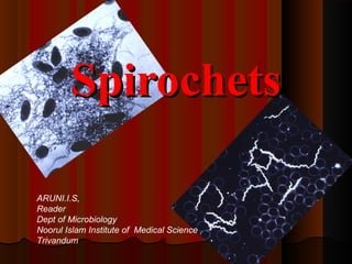
  • 1. SpirochetsSpirochets ARUNI.I.S, Reader Dept of Microbiology Noorul Islam Institute of Medical Science Trivandum
  • 2. Spirochetes are elongated, motile, flexibleSpirochetes are elongated, motile, flexible bacteria twisted spirally alone the long axis.bacteria twisted spirally alone the long axis. Motile withMotile with endoflagellaendoflagella.. Human pathogens are found in 3 genera:Human pathogens are found in 3 genera: TreponemaTreponema BorreliaBorrelia LeptospiraLeptospira SpirochetesSpirochetes
  • 4.  Some of theSome of the Treponema speciesTreponema species are pathogensare pathogens while others occur as commensals in thewhile others occur as commensals in the mouth, intestine and genitalia.mouth, intestine and genitalia.  Causes the following diseases in humans:Causes the following diseases in humans: 1.1. Venereal syphilis by T.pallidumVenereal syphilis by T.pallidum 2.Endemic syphilis T.pallidum(T.endemicum)2.Endemic syphilis T.pallidum(T.endemicum) 3.Yaws by T.pertenue3.Yaws by T.pertenue 4. Pinta by T.carateum4. Pinta by T.carateum TreponemaTreponema
  • 5. Treponema pallidumTreponema pallidum  Causative agent ofCausative agent of SyphilisSyphilis..  Name pallidum refers to it’s pale staining.Name pallidum refers to it’s pale staining.  Delicate spirochete withDelicate spirochete with active motilityactive motility..  Thin with tapering ends.10 mm long & 0.1-0.2 mm wide.Thin with tapering ends.10 mm long & 0.1-0.2 mm wide.  Morphology and motility can be seen under theMorphology and motility can be seen under the darkdark ground or phase contrast microscopeground or phase contrast microscope..  Can be stained by Silver impregnation methodCan be stained by Silver impregnation method  Very delicateVery delicate and highly susceptible to heat.and highly susceptible to heat.  Do not grow in artificial culture media, but possible byDo not grow in artificial culture media, but possible by co-co- cultivationcultivation with tissue culture cells.with tissue culture cells.  Natural infection occur only inNatural infection occur only in human beingshuman beings..
  • 6.  Antigenic structure:Antigenic structure:  3 types of antigens are seen in3 types of antigens are seen in TreponemaTreponema:: a)a) Cardiolipin antigenCardiolipin antigen present inpresent in T.pallidumT.pallidum againstagainst which reagin Abs are producedwhich reagin Abs are produced b)b) AA group specific antigengroup specific antigen present inpresent in T.pallidumT.pallidum asas well as non-pathogenic Reiter strain of Treponema.well as non-pathogenic Reiter strain of Treponema. c)c) AA species specific polysaccharidespecies specific polysaccharide present inpresent in pathogenic strain ofpathogenic strain of TreponemaTreponema..
  • 7. SYPHILISSYPHILIS Caused by Treponema pallidum Can enter any mucous membrane or break in the skin, can be spread by kissing and close contact
  • 8. SYPHILISSYPHILIS NONO SYMPTOMSSYMPTOMS Up to 50% of people don’t know they have it. Symptoms may be mild or painless or nearly invisible.
  • 9.  Organism enters the body through cuts or abrasion on the skinOrganism enters the body through cuts or abrasion on the skin or mucosa during sexual contact.or mucosa during sexual contact.  IP is about a month(range 10-90 days)IP is about a month(range 10-90 days)  3stages of venereal syphilis3stages of venereal syphilis  Primary:Primary: chancre, disappears spontaneously.chancre, disappears spontaneously.  Secondary:Secondary: penetrate mucus membranes, enter blood-stream,penetrate mucus membranes, enter blood-stream, fever, rash.fever, rash.  Tertiary:Tertiary: invade heart, musculoskeletal system, CNS, relativelyinvade heart, musculoskeletal system, CNS, relatively noninfectious.noninfectious. PathogenesisPathogenesis
  • 10. 11stst Stage SYPHILIS(Primary)Stage SYPHILIS(Primary) 10 to 90 days after contact, painless, avascular, indurated superficially ulcerated lesion may appear, called hard chancres. It occurs at the site of entry of the bacteria – genital organs, mouth , nipples- and heals spontaneously after 10-40 days. They can appear ANYWHERE that the spirochetes entered a mucous membrane or break in the skin. penis finger tongue cervix vulva
  • 11.  The chancre is covered by a thick ,glairyThe chancre is covered by a thick ,glairy exudate ,rich in spirochetesexudate ,rich in spirochetes  The regional lymph nodes are swollenThe regional lymph nodes are swollen  Even before the chancre appears, theEven before the chancre appears, the spirochetes spread from the site of entry in tospirochetes spread from the site of entry in to lymph & blood streamlymph & blood stream  So the patient may be infectious during the lateSo the patient may be infectious during the late incubation periodincubation period  The chancre heals in 10-40 days ,even with outThe chancre heals in 10-40 days ,even with out treatment, leaving a thin scartreatment, leaving a thin scar  Persistent or multiple chancre may be seen inPersistent or multiple chancre may be seen in HIV & othe immunodeficient patiensHIV & othe immunodeficient patiens
  • 12. 22ndnd Stage SYPHILIS(Secondary)Stage SYPHILIS(Secondary) Symptoms appear 2 weeks to 6 months after primary lesion. Characterized by papular skin rashes, mucous patches in oropharynx and condylomata at the mucocutaneous junctions. Patient is highly infectious and these signs disappear without treatment. • Rash on hands • Rash over entire body • Lesions of tongue • Rash on feet •Lesions around mouth
  • 13. ORAL MANIFESTATIONSORAL MANIFESTATIONS The oral lesions are usuallyThe oral lesions are usually multiple,painless. Greenish white plaguesmultiple,painless. Greenish white plagues overlying an ulcerated surfaceoverlying an ulcerated surface These are called mucous patchesThese are called mucous patches They occur mostly on the tongue, buccalThey occur mostly on the tongue, buccal mucosa or gingivamucosa or gingiva These lesions are highly infectious as theyThese lesions are highly infectious as they contain a large number of microorganismscontain a large number of microorganisms
  • 14. Oral lesion in Secondary SyphilisOral lesion in Secondary Syphilis
  • 15.  Spirocetes are abundant in the lesions & the patient isSpirocetes are abundant in the lesions & the patient is most infectious during the secondary stage.most infectious during the secondary stage.  The lesions undergo spontaneous healing ,in someThe lesions undergo spontaneous healing ,in some instances taking as long as 4 or 5 yearsinstances taking as long as 4 or 5 years  After the secondary lesion disappears, there is a periodAfter the secondary lesion disappears, there is a period of quiescence known as latent syphilis.of quiescence known as latent syphilis.  Diagnosis during this period is possible only by serologyDiagnosis during this period is possible only by serology  In many cases this is followed by natural cure, but inIn many cases this is followed by natural cure, but in others after several years, manifestations of Tertiaryothers after several years, manifestations of Tertiary syphilis appearssyphilis appears
  • 16. 33rdrd Stage SYPHILIS(Tertiary)Stage SYPHILIS(Tertiary) The period after the healing of the 2ry lesions- latent syphilis. Occur after several years and diagnose serologically. Damages heart and aorta, causes hardening, distortion, ulceration….can lead to rupture, hemorrhaging and death. Brain damage and infection, causes headaches, dizziness, blurred vision, paralysis, and can lead to insanity. Skin Damage, causes “gummata”, large open sores which are slow to heal. Syphilis can also damage the bones.
  • 17. CONGENITSAL SYPHILISCONGENITSAL SYPHILIS Syphilis is transmitted from the mother to the baby trough the placenta. The more recent the infection, especially during the pregnancy, the more damaging to the baby. COMPLICATIONS: Miscarriage Enlarged liver and spleen Stillbirth Hydrocephalus Meningitis Mental Retardation Convulsions Sever Skin Rashes Blindness Deafness Deformities of face, nose, teeth, jaw, leg bones
  • 18.
  • 19. Laboratory DiagnosisLaboratory Diagnosis a)a) Microscopy:Microscopy: wet films of exudates examinedwet films of exudates examined under the dark ground microscope.under the dark ground microscope. b)b) Serology:Serology: 1)1) Reagin antibody test: tests for antibodiesReagin antibody test: tests for antibodies reacting with cardiolipin antigen and arereacting with cardiolipin antigen and are known asknown as standard test for syphilis (STS).standard test for syphilis (STS). i.i. Wasserman complement fixation testWasserman complement fixation test ii.ii. Khan flocculation testKhan flocculation test iii.iii. VDRL / RPR testVDRL / RPR test
  • 20. 2)2) Group specific treponemal tests - Here cultivableGroup specific treponemal tests - Here cultivable treponems (Reiter strain) used as antigen.treponems (Reiter strain) used as antigen. Reiter Protein Complement Fixation Test(RPCF)Reiter Protein Complement Fixation Test(RPCF) 3)3) SpecificSpecific T.pallidumT.pallidum test - Here virulent Nichol’s strainstest - Here virulent Nichol’s strains ofof T.pallidumT.pallidum is used as antigen.is used as antigen. i.i. T.pallidumT.pallidum immobilization (TPI) testimmobilization (TPI) test ii.ii. Fluorecent treponemal antibody (FTA) testFluorecent treponemal antibody (FTA) test iii.iii. T.pallidumT.pallidum Haemagglutination (TPHA) testHaemagglutination (TPHA) test  EIA for Treponemal antibody detectionEIA for Treponemal antibody detection  CSF VDRL is used for neurosyphilisCSF VDRL is used for neurosyphilis
  • 21. Prophylaxis & TreatmentProphylaxis & Treatment Avoid sexual contact with an infectedAvoid sexual contact with an infected individual.individual. Use of physical barriers, antiseptics orUse of physical barriers, antiseptics or antibiotics may minimise the risk.antibiotics may minimise the risk. Chemoprophylaxis by penicillin.Chemoprophylaxis by penicillin. No vaccine is available.No vaccine is available. Treatment by Penicillin G, Erythromycin,Treatment by Penicillin G, Erythromycin,
  • 22. Non-venereal TreponemesNon-venereal Treponemes  Organisms closely resemblesOrganisms closely resembles T.pallidumT.pallidum but do not causesbut do not causes VD.VD.  Transmission occurs through direct body to body contactTransmission occurs through direct body to body contact among people of unhygienic practices.among people of unhygienic practices.  Important non-venereal infections include:Important non-venereal infections include:  Endemic syphilisEndemic syphilis  YawsYaws  PintaPinta
  • 23. Endemic SyphilisEndemic Syphilis  Causative agent isCausative agent is T.endemicum.T.endemicum.  Common in children of poor personal hygiene.Common in children of poor personal hygiene.  Infection can also be seen on the nipples of the mother infected byInfection can also be seen on the nipples of the mother infected by her children.her children.  Usually seen with the manifestations of secondary syphilis such asUsually seen with the manifestations of secondary syphilis such as mucous patches & skin eruptionsmucous patches & skin eruptions  The disease may progress to gummatous lesions on the skin, boneThe disease may progress to gummatous lesions on the skin, bone and nasopharynyx.and nasopharynyx.  Laboratory diagnosis and treatment are same as venereal syphilis.Laboratory diagnosis and treatment are same as venereal syphilis.
  • 24. YawsYaws In India, it has been reported from Orissa,In India, it has been reported from Orissa, Andra Pradesh and Madhya Pradesh.Andra Pradesh and Madhya Pradesh. Like in syphilis, the primary lesion isLike in syphilis, the primary lesion is followed by secondary and tertiaryfollowed by secondary and tertiary manifestations.manifestations.  Causative agent isCausative agent is T.pertenue.T.pertenue.  The primary lesion in yaws is anThe primary lesion in yaws is an extra-genital papula which breaksextra-genital papula which breaks down to form an ulceratingdown to form an ulcerating granuloma.granuloma.
  • 25. PintaPinta  Causative agent isCausative agent is T.carateum.T.carateum.  Disease is seen mainly in central and south America.Disease is seen mainly in central and south America.  Infection occurs by direct person to person contact.Infection occurs by direct person to person contact.  The primary lesion is an extra-genital papule.The primary lesion is an extra-genital papule.  Hypopigmentation or hyperpigmentation is seen.Hypopigmentation or hyperpigmentation is seen.  Laboratory diagnosis and treatment are similar asLaboratory diagnosis and treatment are similar as T.pallidum.T.pallidum.
  • 27. BorreliaBorrelia Microaerophilic-to-anaerobicMicroaerophilic-to-anaerobic  Loose, irregular coilsLoose, irregular coils  In vitroIn vitro cultivation is difficultcultivation is difficult  Visualization byVisualization by Giemsa stainGiemsa stain Gram staining-Gram negativeGram staining-Gram negative Silver stainingSilver staining Dark field microscopyDark field microscopy  Highly adapted to arthropod transmissionHighly adapted to arthropod transmission
  • 28.  Antigenic variation Borrelia undergoes antigenic variations in vivo and is found to be the cause of relapses in the diseases. Recovery from the disease is believed to be due to the development of immunity to theses antigenic variants.
  • 29.  Species of medical importance:Species of medical importance: B.recurrentisB.recurrentis – Relapsing fever– Relapsing fever B.vincentiB.vincenti – Fusospirochetosis(Vincent’s– Fusospirochetosis(Vincent’s angina)angina) B.burgdorferiB.burgdorferi – Lyme disease– Lyme disease
  • 30.  This Acute infection is caused byThis Acute infection is caused by B.recurrentisB.recurrentis..  Sudden onset with fever, septicemia, headache, muscle pain.Sudden onset with fever, septicemia, headache, muscle pain.  After 3-5 days, fever subsides and during this afebrile period ofAfter 3-5 days, fever subsides and during this afebrile period of 4-19 days borreliae are not found in the blood.4-19 days borreliae are not found in the blood.  After a afebrile period when another bout of fever sets in,After a afebrile period when another bout of fever sets in, borreliae reappear in blood.borreliae reappear in blood.  Patient returns to normal, after 4-10 relapses.Patient returns to normal, after 4-10 relapses.  Sporadic in US, distributed worldwide.Sporadic in US, distributed worldwide. Relapsing Fever
  • 31. Transmitted animal-to-animal, animal-to-Transmitted animal-to-animal, animal-to- human by ticks, human-to-human by bodyhuman by ticks, human-to-human by body lice.lice. Causative agents:Causative agents: Louse borne diseaseLouse borne disease :: B. recurrentisB. recurrentis Tick borne diseaseTick borne disease :: B.duttonii, B.hermsii,B.duttonii, B.hermsii, B.parkeriB.parkeri etc.etc.
  • 32. Relapsing fever- 2 types:Relapsing fever- 2 types: Tickborne RFTickborne RF Transmitted by soft ticks (genusTransmitted by soft ticks (genus OrnithodorosOrnithodoros)) O. turicatae, O. parkeri, O. hermsiO. turicatae, O. parkeri, O. hermsi in US,in US, 15 others worldwide15 others worldwide Louseborne RFLouseborne RF Body / head / pubic lice (Body / head / pubic lice (Pediculus humanusPediculus humanus corporis / capitis / pubiscorporis / capitis / pubis)) Also by bed bugs.Also by bed bugs.
  • 33.  Laboratory diagnosis:Laboratory diagnosis:  Microscopy:Microscopy: Light microscopy- Giemsa / Leishman stain.Light microscopy- Giemsa / Leishman stain. Silver stainingSilver staining Dark ground microscopy.Dark ground microscopy.  Culture:Culture: too difficult to grow.too difficult to grow.  Animal inoculationAnimal inoculation: into white mouse.: into white mouse.  Serology:Serology: not reliable.not reliable.
  • 34.  It is an ulcerativeIt is an ulcerative gingivostomatitisgingivostomatitis oror oropharyngitisoropharyngitis caused bycaused by Borrelia vincentiBorrelia vincenti..  B.vincentiB.vincenti is a normal mouth commensal motileis a normal mouth commensal motile spirochete, about 5-20spirochete, about 5-20 μμm wide, with 3-8 coils ofm wide, with 3-8 coils of variable size.variable size.  It is a normal mouth commensal ,but under certainIt is a normal mouth commensal ,but under certain pre disposing condition(malnutrition ,viral infection)pre disposing condition(malnutrition ,viral infection) give rise to ulcerative gingivo stomatitis orgive rise to ulcerative gingivo stomatitis or orophangitis(Vincent’s angina)orophangitis(Vincent’s angina)  In this infection,In this infection, B.vincentiB.vincenti is associated withis associated with fusiform bacilli and this symbitic infection is knownfusiform bacilli and this symbitic infection is known asas fusospirochetosisfusospirochetosis.. Vincent’s AnginaVincent’s Angina
  • 35. DiagnosisDiagnosis may be made by demonstratingmay be made by demonstrating spirochetes and fusiform bacilli in stainedspirochetes and fusiform bacilli in stained smears of exudates form the lesions.smears of exudates form the lesions. TreatmentTreatment-- Penicillin & metronidazolePenicillin & metronidazole
  • 36. Lyme DiseaseLyme Disease  First observed in 1975, in Old Lyme, USA.First observed in 1975, in Old Lyme, USA.  Caused byCaused by Borrelia burgdorferiBorrelia burgdorferi..  Incubation period: 7 - 14 days (range 3 - 30 days).Incubation period: 7 - 14 days (range 3 - 30 days).  Infection may be sub-clinical, manifest onlyInfection may be sub-clinical, manifest only nonspecific symptoms: fever, headache, myalgia.nonspecific symptoms: fever, headache, myalgia.  Most prevalent human tick-borne disease in theMost prevalent human tick-borne disease in the US, Europe, parts of Asia.US, Europe, parts of Asia.
  • 37. EpidemiologyEpidemiology Human encroachment into habitat of white-Human encroachment into habitat of white- tailed deer.tailed deer. Spread from deer, mice to humans by theSpread from deer, mice to humans by the bitebite of soft ticksof soft ticks (genus(genus Ixodes)Ixodes)
  • 38.  First stage:First stage: Erythema migransErythema migrans  spirochetes multiply in skin, quickly enter circulationspirochetes multiply in skin, quickly enter circulation  rashrash appears days-to-weeks after tick biteappears days-to-weeks after tick bite  surround site of inoculation, expand insurround site of inoculation, expand in concentric ringsconcentric rings  disappear spontaneously after several weeksdisappear spontaneously after several weeks
  • 39. Second stage:Second stage: disseminated infectiondisseminated infection associated with fever, headache, myalgia,associated with fever, headache, myalgia, arthralgia and lymphadenopathy.arthralgia and lymphadenopathy. Musculoskeletal signs (migratory joint, muscleMusculoskeletal signs (migratory joint, muscle pains), CNS signs, cardiac damage may alsopains), CNS signs, cardiac damage may also appear.appear.
  • 40. Third stageThird stage of persistent infection,of persistent infection, occurringoccurring months or years latermonths or years later.. Develops chronic arthritis, polyneuropathy,Develops chronic arthritis, polyneuropathy, encephalopathy, etc.encephalopathy, etc. Symptoms include arthritis, facial palsy andSymptoms include arthritis, facial palsy and meningitis.meningitis.
  • 42. 46 Ixodes scapularis, tick vector for Lyme disease.
  • 44. DiagnosisDiagnosis made be isolating the organismsmade be isolating the organisms form skin lesions, CSF, blood or by serology.form skin lesions, CSF, blood or by serology. Treatment:Treatment: Amoxycillin, doxycycline andAmoxycillin, doxycycline and cefuroxime.cefuroxime. Tick control important forTick control important for prevention.prevention.
  • 46. Leptospira are actively motile, delicateLeptospira are actively motile, delicate spirochetes, which can causespirochetes, which can cause Leptospirosis orLeptospirosis or Weil’s diseasesWeil’s diseases..
  • 47. Leptospirosis is an acute zoonotic infection ofLeptospirosis is an acute zoonotic infection of worldwide significance caused by spirochaeteworldwide significance caused by spirochaete Leptospira interrogansLeptospira interrogans .. Various factors influencing the animal activity,Various factors influencing the animal activity, suitability of the environment for the survivalsuitability of the environment for the survival of the organism and behavioral andof the organism and behavioral and occupational habits of human beings can beoccupational habits of human beings can be the determinants of incidence and prevalencethe determinants of incidence and prevalence of the disease.of the disease.
  • 48.  Leptospirosis, also known as canicola fever,Leptospirosis, also known as canicola fever, hemorrhagic jaundice, , mud fever, spirochetalhemorrhagic jaundice, , mud fever, spirochetal jaundice, swamp fever, swineherd's disease, caver'sjaundice, swamp fever, swineherd's disease, caver's flu or sewerman's flu.flu or sewerman's flu.  There is an acute form of human infection known asThere is an acute form of human infection known as Weil's diseaseWeil's disease, where the patient suffers from, where the patient suffers from jaundice.jaundice.  This condition is usually caused byThis condition is usually caused by L.L.icterohaemorrhagiaeicterohaemorrhagiae serogroup of L.serogroup of L.interrogansinterrogans
  • 49. Leptospira interrogansLeptospira interrogans  Leptospira appear tightly coiled thinLeptospira appear tightly coiled thin flexible Spirochetes with 5 – 15flexible Spirochetes with 5 – 15 microns x 0.1 micron size.microns x 0.1 micron size.  One end appears bent forms a hook.One end appears bent forms a hook.  Actively motile.Actively motile.  Seen best with dark fieldSeen best with dark field Microscopy.Microscopy.  13 serogroups, >200 serovars13 serogroups, >200 serovars
  • 50. Cultivation of LeptospiraCultivation of Leptospira  Leptospira grows best underLeptospira grows best under aerobic conditions at 28aerobic conditions at 2800 to 30to 3000 cc and pH 7.2 – 7.5.and pH 7.2 – 7.5.  EMJHEMJH (Ellinghausen,(Ellinghausen, McCullough, Johnson, Harris)McCullough, Johnson, Harris) mediamedia is commonly used.is commonly used.  Other MediaOther Media  Fletchers MediaFletchers Media ..Optimal growth after 1 – 2 weeksOptimal growth after 1 – 2 weeks  ..
  • 51. എലിപ്പനിഎലിപ്പനി PathogenesisPathogenesis  Rats, Mice, Wild Rodents, Dogs, Swine, Cattle areRats, Mice, Wild Rodents, Dogs, Swine, Cattle are principleprinciple source of infectionsource of infection..  The animals excrete Leptospira both in active infectionThe animals excrete Leptospira both in active infection and Asymptomatic stage through urine.and Asymptomatic stage through urine.  The Leptospira survive and remain viable for severalThe Leptospira survive and remain viable for several weeks in stagnant water.weeks in stagnant water.  Enter through cuts and abrasionsEnter through cuts and abrasions in the skin andin the skin and mucous membranes or through mouth, nose ormucous membranes or through mouth, nose or conjunctive.conjunctive.
  • 52.  Man is the end host, and there is no evidence ofMan is the end host, and there is no evidence of human to human infection.human to human infection.  Incubation periodIncubation period 1 – 2 weeks1 – 2 weeks..  Multiplication of leptospires inMultiplication of leptospires in blood streamblood stream producesproduces feverfever..  Establish organ involvement inEstablish organ involvement in Kidney and LiverKidney and Liver..  May produceMay produce hemorrhage and necrosishemorrhage and necrosis in the tissuesin the tissues and initiatesand initiates dysfunctiondysfunction of these organs.of these organs.
  • 54.  Clinical Pictures:Clinical Pictures:  JaundiceJaundice  HemorrhageHemorrhage  Nitrogen retentionNitrogen retention  TheThe Illness is BiphasicIllness is Biphasic with initial temperaturewith initial temperature when the second phase comes with raise of IgMwhen the second phase comes with raise of IgM titers raise.titers raise.  Aseptic meingitisAseptic meingitis – initial headache, stiffness of– initial headache, stiffness of neck, pleocytosis of Cerebro spinal fluid.neck, pleocytosis of Cerebro spinal fluid.
  • 55.  ComplicationsComplications  NephritisNephritis  HepatitisHepatitis  Manifestations in eyeManifestations in eye  Muscular lesionsMuscular lesions  Many infections are mild and subclinicalMany infections are mild and subclinical
  • 56. Weil’s SyndromeWeil’s Syndrome  Weil's syndrome is a severe form of leptospirosis thatWeil's syndrome is a severe form of leptospirosis that causes continuouscauses continuous fever, stuporfever, stupor & reduction in the& reduction in the blood's ability to clot, which leads toblood's ability to clot, which leads to bleeding withinbleeding within tissuestissues..  Blood tests reveal anemia.Blood tests reveal anemia.  By the third to sixth day, signs ofBy the third to sixth day, signs of kidney damagekidney damage andand liver injuryliver injury appear.appear.  Kidney abnormalities may causeKidney abnormalities may cause blood in the urineblood in the urine and painful urinationand painful urination..  Liver injury may be mild & usually heals completely.Liver injury may be mild & usually heals completely.
  • 57. Hepatitis - LeptospirosisHepatitis - Leptospirosis  Hepatitis is the frequent complication.Hepatitis is the frequent complication.  Elevation of serum creatine phospholipaseElevation of serum creatine phospholipase enzyme raise differentiates from Viral hepatitisenzyme raise differentiates from Viral hepatitis where the enzyme is not raised.where the enzyme is not raised.
  • 58. Nephritis - LeptospirosisNephritis - Leptospirosis  Kidney involvement in animals produce chronicKidney involvement in animals produce chronic disease of the kidney and the infected animaldisease of the kidney and the infected animal startsstarts shedding large number of leptospirashedding large number of leptospira andand main source of environmental contamination ofmain source of environmental contamination of bacteria and results in human infections.bacteria and results in human infections.  Human urine also contain Spirochetes in theHuman urine also contain Spirochetes in the second and third week of infection.second and third week of infection.
  • 59.  Laboratory diagnosis:Laboratory diagnosis:  Specimen:Specimen: Blood, urine, CSF.Blood, urine, CSF.  Blood examinationBlood examination is significant only in the early stage of theis significant only in the early stage of the disease.disease.  Urine examinationUrine examination is significant from second week onwardsis significant from second week onwards up to 6 weeks.up to 6 weeks. Microscopy:Microscopy: Blood film / urine sediments under dark groundBlood film / urine sediments under dark ground microscope or by IF.microscope or by IF. Culture:Culture: Blood culture in EMJH medium.Blood culture in EMJH medium. Animal inoculation:Animal inoculation: Blood / urine into guinea pig.Blood / urine into guinea pig. Serology:Serology: Detection of antibodies by ELISADetection of antibodies by ELISA..
  • 60. Prophylaxis:Prophylaxis:  Rodent control, disinfection of water andRodent control, disinfection of water and wearing of protective clothing.wearing of protective clothing.  Vaccination on trial basis.Vaccination on trial basis. Treatment:Treatment:  Penicillin, Tetracycline, Doxycycline.Penicillin, Tetracycline, Doxycycline.