

Recomendados
Mais conteúdo relacionado
Mais procurados
Mais procurados (20)
Destaque
Destaque (20)
Semelhante a Cardiovascular system
Semelhante a Cardiovascular system (20)
Mais de Ann Alcid
Mais de Ann Alcid (20)
Último
Último (20)
Cardiovascular system
- 2. CARDIOVASCULAR Comes from the Greek work “cardia”, meaning “heart”, and the Latin “vasculum”, meaning “small vessel” The system can be compared to a large muscular pump (heart) that sends a fluid (blood) through a series of large and small tubes (blood vessels)
- 3. HEART It is a hollow, cone-shaped muscular organ located behind and slightly to the left of the sternum or breastbone It is a muscular organ that rhythmically contracts forcing the blood through a system of vessels Septum divides the heart into a right portion and left portion. Intrinsic Conduction Activity – the hearts own nervous system that controls its beating activity. It is located within the heart tissue.
- 5. The heart is further subdivided into: a) Atrium – upper chambers. The thin walled atria (singular atrium) collect the blood flowing from the heat into the veins (RIGHT & LEFT ATRIUM) b) Ventricles – collects and pumps the blood into the arteries. (RIGHT & LEFT ARTERIES) The valves control the flow of blood through the heart: 1. TRICUSPID VALVE 2. MITRAL VALVE 3. PULMONARY SEMILUNAR VALVE 4. AORTIC VALVE
- 6. Pericardium thin tough membrane that protects the heart from rubbing against the lungs and walls of the chest Atrioventricular or AV valves located between the atria and ventricles. It produces the “lub” sound; the semilunar valves produce the “dup” sound. 2 Phases of cardiovascular: 1. Systole – contraction 2. Diastole – relaxation 1st organ to develop in a fetus Beats 2.5 billion times in an average life span Interstitial fluid immediate source of oxygen and nutrients
- 7. HEART DISORDERS RHEUMATIC HEART DISEASE – caused by streptococcal infections BACTERIAL ENDOCARDITIS – inflammation of the heart’s lining CORONARY HEART DISEASE – opening of the coronary artery is blocked by cholesterol deposits HYPERTENSIONS – high blood pressure
- 8. HEART MURMURS – are abnormal, extra heart sounds made by the blood moving through the heart and its valves CONGENITAL HEART DISEASE – is any defect in the heart or its main blood vessels that is present at birth
- 9. BLOOD VESSEL Form a closed transport system of tubes measuring about 60,000 miles (96,500 kilometers) in length – more than twice the distance. The entire blood vessel system can be thought of as a series of connected roads and highways Blood leaves the heart through large vessels (highways) that travel forth into the body.
- 11. At various points, these large vessels divide to become smaller vessels (secondary roads). In turn, these vessels continue to divide into smaller and smaller vessels (on-lane roads). And on its return trip, the blood travels vice- versa until it reaches the heart. In 1628,English physician William Harvey (1578-1657) published a new concept of blood circulation. He maintained that there was a continuing flow of blood through the body. He is the father of Modern Medicine. The valves are most numerous in the legs, where blood must flow against the force of gravity on its way back to the heart.
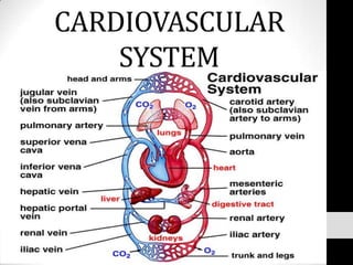
- 12. PULMONARYCIRCULATION– movementof bloodfrom the heart to the lungs SUPERIOR VENA CAVA INFERIOR VENA CAVA RIGHT ATRIUM TRICUSPID VALVE RIGHT VENTRICLE PULMONARY SEMILUNAR VALVE PULMONARY ARTERY LUNGS
- 13. SYSTEMATICCIRCULATION– movementof heart to the lungs to the bodysystems LUNGS LEFT ATRIUM MITRAL VALVE/BICUSPID VALVE LEFT VENTRICLE AORTIC VALVEAORTA BODY SYSTEMS
- 15. BLOOD Is the fluid pumped by the heart through the blood vessels to all parts of the body It is connective tissue; as its name, connects body parts, providing support, storage and protection. Non-living fluid matrix plasma and formed elements Dull red in color, depending on the amount of the oxygen carried Normal adult blood volume – 5 to 6 liters Agglutination – the blood cells that is clumped together and can block small vessels. It can be fatal to a person that is given the wrong blood type
- 16. Found everywhere in the body, connective tissue is the most abundant type of the 4 types of tissues (the other 3 are epithelial, muscle and nervous) Of all the tissues in the body, it is unique – it is the only one that is fluid It carries everything that must be transported from one place to another within the body It helps protect the body by clotting & by acting as a defense against foreign microorganisms Has a temperature of about 100.4⁰F (38⁰C) It makes up approximately 8% of a person’s body weight
- 17. Classification of Blood Cells/Formed Elements 1. Erythrocytes – red blood cells, for oxygen transport - 45% of total blood volume - Small biconcave disk shaped cell - Contains Hemoglobin – a protein pigment - 1 red blood cell = 250 million hemoglobin molecule - 1 hemoglobin molecule = 4 molecules of oxygen - Normal blood contains 12to 18 g hemoglobin/100ml of blood - 4 main blood types: A, B, O and AB
- 18. 2. Leukocytes – white blood cells - Body’s defense against diseases - 4,000 to 11,000 WBC/cubic volume - Forms a protective army that helps defend the body - Capable to slip in and out of the blood vessel by the process of diapedesis 3. Plasma – a watery, straw-colored fluid - Approximately 92% water - Over 100 diff. substances are dissolved in this - Albumins help to keep water in the bloodstream
- 19. 4. Platelets – or thrombocytes, are not truly cells like RBC and WBC - Small, disk-shaped fragments of extraordinarily large cells called megakaryocytes that are located in bone marrow - 300,000 per cubic mm of blood - Help to control bleeding in a complex process called homeostasis, or the stoppage of blood flow - Releases serotonin, a chemical that causes the blood vessel to spasm and narrow, decreasing the amount of blood flowing to the site of the injury
- 20. BLOOD DISEASES ANEMIA – deficiencies in the number of RBC or hemoglobin content; large quantity of blood loss thru bleeding or iron deficiency LEUKEMIA – uncontrolled overproduction of abnormal leukocytes HEMOPHILIA – delayed clotting time leading to profuse blood loss; hereditary disease carried by women but manifested only in men
- 21. ARTERIOSCLEROSIS – the walls of arteries become thickened and hard, interfering with the circulation of blood ATHEROSCLEROSIS – General term for hardening of the arteries. Fatty material accumulates on the interior walls of arteries making them narrower SICKLE CELL ANEMIA – inherited blood disorder in which RBC are sickle-shaped instead of round because of defective hemoglobin molecules