Mais conteúdo relacionado Semelhante a Chapter 50: Sensory and Motor Mechansims Semelhante a Chapter 50: Sensory and Motor Mechansims (20) 1. Copyright © 2005 Pearson Education, Inc. publishing as Benjamin Cummings
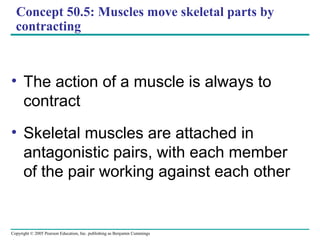
Concept 50.5: Muscles move skeletal parts by
contracting
• The action of a muscle is always to
contract
• Skeletal muscles are attached in
antagonistic pairs, with each member
of the pair working against each other
2. Copyright © 2005 Pearson Education, Inc. publishing as Benjamin Cummings
Concept 50.5: Muscles move skeletal parts by
contracting
• The action of a muscle is always to
contract
• Skeletal muscles are attached in
antagonistic pairs, with each member
of the pair working against each other
3. Copyright © 2005 Pearson Education, Inc. publishing as Benjamin Cummings
Biceps
contracts
Human
Triceps
relaxes
Forearm
flexes
Biceps
relaxes
Triceps
contracts
Forearm
extends
Extensor
muscle
relaxes
Flexor
muscle
contracts
Grasshopper
Extensor
muscle
contracts
Flexor
muscle
relaxes
Tibia
extends
Tibia
flexes
4. Copyright © 2005 Pearson Education, Inc. publishing as Benjamin Cummings
Vertebrate Skeletal Muscle
• Vertebrate skeletal muscle is characterized by a
hierarchy of smaller and smaller units
• A skeletal muscle consists of a bundle of long
fibers running parallel to the length of the muscle
• A muscle fiber is itself a bundle of smaller
myofibrils arranged longitudinally
5. Copyright © 2005 Pearson Education, Inc. publishing as Benjamin Cummings
• The myofibrils are composed to two kinds of
myofilaments:
– Thin filaments consist of two strands of actin
and one strand of regulatory protein
– Thick filaments are staggered arrays of myosin
molecules
6. Copyright © 2005 Pearson Education, Inc. publishing as Benjamin Cummings
• Skeletal muscle is also called striated muscle
because the regular arrangement of myofilaments
creates a pattern of light and dark bands
• Each unit is a sarcomere, bordered by Z lines
• Areas that contain the myofilments are the I band,
A band, and H zone
7. Copyright © 2005 Pearson Education, Inc. publishing as Benjamin Cummings
Bundle of
muscle fibers
Single muscle fiber
(cell)
Plasma membrane
Nuclei
Muscle
Myofibril
Dark band
Sarcomere
Z lineLight
band
I band
TEM
A band I band
0.5 µm
M line
Thick filaments
(myosin)
Sarcomere
H zoneZ line
Thin filaments
(actin)
Z line
8. Copyright © 2005 Pearson Education, Inc. publishing as Benjamin Cummings
The Sliding-Filament Model of Muscle Contraction
• According to the
sliding-filament
model, filaments
slide past each
other longitudinally,
producing more
overlap between
thin and thick
filaments
• As a result of
sliding, the I band
and H zone shrink
Sarcomere
0.5 µm
Z
H
A
Relaxed muscle fiber
I
Contracting muscle fiber
Fully contracted muscle fiber
9. Copyright © 2005 Pearson Education, Inc. publishing as Benjamin Cummings
• The sliding of filaments is based on interaction
between actin and myosin molecules of the thick
and thin filaments
• The “head” of a myosin molecule binds to an actin
filament, forming a cross-bridge and pulling the
thin filament toward the center of the sarcomere
10. Copyright © 2005 Pearson Education, Inc. publishing as Benjamin Cummings
PowerPoint Lectures for
Biology, Seventh Edition
Neil Campbell and Jane Reece
Lectures by Chris Romero
LE 49-30–4
Thin filaments
Thick filament
Thin filament
Thick
filament
Myosin head (low-energy
configuration)
Cross-bridge
binding site
Myosin head (high-
energy configuration)
Actin
Cross-bridge
Myosin head (low-
energy configuration)
Thin filament moves
toward center of sacomere.
11. Copyright © 2005 Pearson Education, Inc. publishing as Benjamin Cummings
The Role of Calcium and Regulatory Proteins
• A skeletal muscle fiber contracts only when
stimulated by a motor neuron
• When a muscle is at rest, myosin-binding sites on
the thin filament are blocked by the regulatory
protein tropomyosin
12. Copyright © 2005 Pearson Education, Inc. publishing as Benjamin Cummings
PowerPoint Lectures for
Biology, Seventh Edition
Neil Campbell and Jane Reece
Lectures by Chris Romero
LE 50-28
Myosin-binding sites blocked.
Myosin-binding sites exposed.
Tropomyosin Ca2+
-binding sites
Actin Troponin complex
Myosin-
binding site
Ca2+
13. Copyright © 2005 Pearson Education, Inc. publishing as Benjamin Cummings
• For a muscle fiber to contract, myosin-binding
sites must be uncovered
• This occurs when calcium ions (Ca2+
) bind to a set
of regulatory proteins, the troponin complex
• The stimulus leading to contraction of a muscle
fiber is an action potential in a motor neuron that
makes a synapse with the muscle fiber
14. Copyright © 2005 Pearson Education, Inc. publishing as Benjamin Cummings
LE 49-32
Ca2+
released
from sarcoplasmic
reticulum
Mitochondrion
Motor
neuron axon
Synaptic
terminal
T tubule
Sarcoplasmic
reticulum
Myofibril
Plasma membrane
of muscle fiber
Sarcomere
15. Copyright © 2005 Pearson Education, Inc. publishing as Benjamin Cummings
• The synaptic terminal of the motor neuron
releases the neurotransmitter acetylcholine
• Acetylcholine depolarizes the muscle, causing it to
produce an action potential
16. Copyright © 2005 Pearson Education, Inc. publishing as Benjamin Cummings
• Action potentials travel to the interior of the muscle
fiber along transverse (T) tubules
• The action potential along T tubules causes the
sarcoplasmic reticulum to release Ca2+
• The Ca2+
binds to the troponin-tropomyosin
complex on the thin filaments
• This binding exposes myosin-binding sites and
allows the cross-bridge cycle to proceed
17. Copyright © 2005 Pearson Education, Inc. publishing as Benjamin Cummings
LE 49-33
Ca2+
CYTOSOL
Ca2+
SR
PLASMA
MEMBRANET TUBULESynaptic cleft
Synaptic terminal
of motor neuron
ACh
18. Copyright © 2005 Pearson Education, Inc. publishing as Benjamin Cummings
Neural Control of Muscle Tension
• Contraction of a whole muscle is graded, which
means that the extent and strength of its
contraction can be voluntarily altered
• There are two basic mechanisms by which the
nervous system produces graded contractions:
– Varying the number of fibers that contract
– Varying the rate at which fibers are stimulated
19. Copyright © 2005 Pearson Education, Inc. publishing as Benjamin Cummings
• In a vertebrate skeletal muscle, each branched
muscle fiber is innervated by one motor neuron
• Each motor neuron may synapse with multiple
muscle fibers
20. Copyright © 2005 Pearson Education, Inc. publishing as Benjamin Cummings
PowerPoint Lectures for
Biology, Seventh Edition
Neil Campbell and Jane Reece
Lectures by Chris Romero
LE 49-34
Motor
unit 1
Motor
unit 2
Nerve
Synaptic terminals
Motor neuron
cell body
Spinal cord
Motor neuron
axon
Muscle
Tendon
Muscle fibers
21. Copyright © 2005 Pearson Education, Inc. publishing as Benjamin Cummings
• A motor unit consists of a single motor neuron and
all the muscle fibers it controls
• Recruitment of multiple motor neurons results in
stronger contractions
• A twitch results from a single action potential in a
motor neuron
• More rapidly delivered action potentials produce a
graded contraction by summation
22. Copyright © 2005 Pearson Education, Inc. publishing as Benjamin Cummings
PowerPoint Lectures for
Biology, Seventh Edition
Neil Campbell and Jane Reece
Lectures by Chris Romero
LE 49-35
Tetanus
Summation of
two twitches
Single
twitch
Tension
Action
potential Pair of
action
potentials
Time
Series of action
potentials at
high frequency
23. Copyright © 2005 Pearson Education, Inc. publishing as Benjamin Cummings
• Tetanus is a state of smooth and sustained
contraction produced when motor neurons deliver
a volley of action potentials
• The bacterial tetanus toxin blocks the release of
inhibitory neurotransmitters. When nervous
impulses cannot be checked by normal inhibitory
mechanisms, the generalized muscular spasms
characteristic of tetanus are produced.
24. Copyright © 2005 Pearson Education, Inc. publishing as Benjamin Cummings
Types of Muscle Fibers
• Skeletal muscle fibers are classified as slow
oxidative, fast oxidative, and fast glycolytic
• These categories are based on their contraction
speed and major pathway for producing ATP
26. Copyright © 2005 Pearson Education, Inc. publishing as Benjamin Cummings
Other Types of Muscle
• Cardiac muscle, found only in the heart, consists
of striated cells electrically connected by
intercalated discs
• Cardiac muscle can generate action potentials
without neural input
27. Copyright © 2005 Pearson Education, Inc. publishing as Benjamin Cummings
• In smooth muscle, found mainly in walls of hollow
organs, contractions are relatively slow and may
be initiated by the muscles themselves
• Contractions may also be caused by stimulation
from neurons in the autonomic nervous system
28. Copyright © 2005 Pearson Education, Inc. publishing as Benjamin Cummings
Biceps
contracts
Human
Triceps
relaxes
Forearm
flexes
Biceps
relaxes
Triceps
contracts
Forearm
extends
Extensor
muscle
relaxes
Flexor
muscle
contracts
Grasshopper
Extensor
muscle
contracts
Flexor
muscle
relaxes
Tibia
extends
Tibia
flexes
29. Copyright © 2005 Pearson Education, Inc. publishing as Benjamin Cummings
Vertebrate Skeletal Muscle
• Vertebrate skeletal muscle is characterized by a
hierarchy of smaller and smaller units
• A skeletal muscle consists of a bundle of long
fibers running parallel to the length of the muscle
• A muscle fiber is itself a bundle of smaller
myofibrils arranged longitudinally
30. Copyright © 2005 Pearson Education, Inc. publishing as Benjamin Cummings
• The myofibrils are composed to two kinds of
myofilaments:
– Thin filaments consist of two strands of actin
and one strand of regulatory protein
– Thick filaments are staggered arrays of myosin
molecules
31. Copyright © 2005 Pearson Education, Inc. publishing as Benjamin Cummings
• Skeletal muscle is also called striated muscle
because the regular arrangement of myofilaments
creates a pattern of light and dark bands
• Each unit is a sarcomere, bordered by Z lines
• Areas that contain the myofilments are the I band,
A band, and H zone
32. Copyright © 2005 Pearson Education, Inc. publishing as Benjamin Cummings
Bundle of
muscle fibers
Single muscle fiber
(cell)
Plasma membrane
Nuclei
Muscle
Myofibril
Dark band
Sarcomere
Z lineLight
band
I band
TEM
A band I band
0.5 µm
M line
Thick filaments
(myosin)
Sarcomere
H zoneZ line
Thin filaments
(actin)
Z line
33. Copyright © 2005 Pearson Education, Inc. publishing as Benjamin Cummings
The Sliding-Filament Model of Muscle Contraction
• According to the
sliding-filament
model, filaments
slide past each
other longitudinally,
producing more
overlap between
thin and thick
filaments
• As a result of
sliding, the I band
and H zone shrink
Sarcomere
0.5 µm
Z
H
A
Relaxed muscle fiber
I
Contracting muscle fiber
Fully contracted muscle fiber
34. Copyright © 2005 Pearson Education, Inc. publishing as Benjamin Cummings
• The sliding of filaments is based on interaction
between actin and myosin molecules of the thick
and thin filaments
• The “head” of a myosin molecule binds to an actin
filament, forming a cross-bridge and pulling the
thin filament toward the center of the sarcomere
35. Copyright © 2005 Pearson Education, Inc. publishing as Benjamin Cummings
PowerPoint Lectures for
Biology, Seventh Edition
Neil Campbell and Jane Reece
Lectures by Chris Romero
LE 49-30–4
Thin filaments
Thick filament
Thin filament
Thick
filament
Myosin head (low-energy
configuration)
Cross-bridge
binding site
Myosin head (high-
energy configuration)
Actin
Cross-bridge
Myosin head (low-
energy configuration)
Thin filament moves
toward center of sacomere.
36. Copyright © 2005 Pearson Education, Inc. publishing as Benjamin Cummings
The Role of Calcium and Regulatory Proteins
• A skeletal muscle fiber contracts only when
stimulated by a motor neuron
• When a muscle is at rest, myosin-binding sites on
the thin filament are blocked by the regulatory
protein tropomyosin
37. Copyright © 2005 Pearson Education, Inc. publishing as Benjamin Cummings
PowerPoint Lectures for
Biology, Seventh Edition
Neil Campbell and Jane Reece
Lectures by Chris Romero
LE 50-28
Myosin-binding sites blocked.
Myosin-binding sites exposed.
Tropomyosin Ca2+
-binding sites
Actin Troponin complex
Myosin-
binding site
Ca2+
38. Copyright © 2005 Pearson Education, Inc. publishing as Benjamin Cummings
• For a muscle fiber to contract, myosin-binding
sites must be uncovered
• This occurs when calcium ions (Ca2+
) bind to a set
of regulatory proteins, the troponin complex
• The stimulus leading to contraction of a muscle
fiber is an action potential in a motor neuron that
makes a synapse with the muscle fiber
39. Copyright © 2005 Pearson Education, Inc. publishing as Benjamin Cummings
LE 49-32
Ca2+
released
from sarcoplasmic
reticulum
Mitochondrion
Motor
neuron axon
Synaptic
terminal
T tubule
Sarcoplasmic
reticulum
Myofibril
Plasma membrane
of muscle fiber
Sarcomere
40. Copyright © 2005 Pearson Education, Inc. publishing as Benjamin Cummings
• The synaptic terminal of the motor neuron
releases the neurotransmitter acetylcholine
• Acetylcholine depolarizes the muscle, causing it to
produce an action potential
41. Copyright © 2005 Pearson Education, Inc. publishing as Benjamin Cummings
• Action potentials travel to the interior of the muscle
fiber along transverse (T) tubules
• The action potential along T tubules causes the
sarcoplasmic reticulum to release Ca2+
• The Ca2+
binds to the troponin-tropomyosin
complex on the thin filaments
• This binding exposes myosin-binding sites and
allows the cross-bridge cycle to proceed
42. Copyright © 2005 Pearson Education, Inc. publishing as Benjamin Cummings
LE 49-33
Ca2+
CYTOSOL
Ca2+
SR
PLASMA
MEMBRANET TUBULESynaptic cleft
Synaptic terminal
of motor neuron
ACh
43. Copyright © 2005 Pearson Education, Inc. publishing as Benjamin Cummings
Neural Control of Muscle Tension
• Contraction of a whole muscle is graded, which
means that the extent and strength of its
contraction can be voluntarily altered
• There are two basic mechanisms by which the
nervous system produces graded contractions:
– Varying the number of fibers that contract
– Varying the rate at which fibers are stimulated
44. Copyright © 2005 Pearson Education, Inc. publishing as Benjamin Cummings
• In a vertebrate skeletal muscle, each branched
muscle fiber is innervated by one motor neuron
• Each motor neuron may synapse with multiple
muscle fibers
45. Copyright © 2005 Pearson Education, Inc. publishing as Benjamin Cummings
PowerPoint Lectures for
Biology, Seventh Edition
Neil Campbell and Jane Reece
Lectures by Chris Romero
LE 49-34
Motor
unit 1
Motor
unit 2
Nerve
Synaptic terminals
Motor neuron
cell body
Spinal cord
Motor neuron
axon
Muscle
Tendon
Muscle fibers
46. Copyright © 2005 Pearson Education, Inc. publishing as Benjamin Cummings
• A motor unit consists of a single motor neuron and
all the muscle fibers it controls
• Recruitment of multiple motor neurons results in
stronger contractions
• A twitch results from a single action potential in a
motor neuron
• More rapidly delivered action potentials produce a
graded contraction by summation
47. Copyright © 2005 Pearson Education, Inc. publishing as Benjamin Cummings
PowerPoint Lectures for
Biology, Seventh Edition
Neil Campbell and Jane Reece
Lectures by Chris Romero
LE 49-35
Tetanus
Summation of
two twitches
Single
twitch
Tension
Action
potential Pair of
action
potentials
Time
Series of action
potentials at
high frequency
48. Copyright © 2005 Pearson Education, Inc. publishing as Benjamin Cummings
• Tetanus is a state of smooth and sustained
contraction produced when motor neurons deliver
a volley of action potentials
• The bacterial tetanus toxin blocks the release of
inhibitory neurotransmitters. When nervous
impulses cannot be checked by normal inhibitory
mechanisms, the generalized muscular spasms
characteristic of tetanus are produced.
49. Copyright © 2005 Pearson Education, Inc. publishing as Benjamin Cummings
Types of Muscle Fibers
• Skeletal muscle fibers are classified as slow
oxidative, fast oxidative, and fast glycolytic
• These categories are based on their contraction
speed and major pathway for producing ATP
51. Copyright © 2005 Pearson Education, Inc. publishing as Benjamin Cummings
Other Types of Muscle
• Cardiac muscle, found only in the heart, consists
of striated cells electrically connected by
intercalated discs
• Cardiac muscle can generate action potentials
without neural input
52. Copyright © 2005 Pearson Education, Inc. publishing as Benjamin Cummings
• In smooth muscle, found mainly in walls of hollow
organs, contractions are relatively slow and may
be initiated by the muscles themselves
• Contractions may also be caused by stimulation
from neurons in the autonomic nervous system