ACC/AHA/ESC Guidelines for Managing Supraventricular Arrhythmias
•
2 gostaram•1,541 visualizações


Recomendados
Recomendados
Mais conteúdo relacionado
Mais procurados
Mais procurados (20)
Semelhante a ACC/AHA/ESC Guidelines for Managing Supraventricular Arrhythmias
Semelhante a ACC/AHA/ESC Guidelines for Managing Supraventricular Arrhythmias (20)
Mais de Loveis1able Khumpuangdee
Mais de Loveis1able Khumpuangdee (20)
Último
Último (20)
ACC/AHA/ESC Guidelines for Managing Supraventricular Arrhythmias
- 1. © 2003 by the American College of Cardiology Foundation, the American Heart Association, Inc., and the European Society of Cardiology ACC/AHA/ESC PRACTICE GUIDELINES—FULL TEXT ACC/AHA/ESC Guidelines for the Management of Patients With Supraventricular Arrhythmias* A Report of the American College of Cardiology/American Heart Association Task Force and the European Society of Cardiology Committee for Practice Guidelines (Writing Committee to Develop Guidelines for the Management of Patients With Supraventricular Arrhythmias) Developed in Collaboration with NASPE-Heart Rhythm Society COMMITTEE MEMBERS Carina Blomström-Lundqvist, MD, PhD, FACC, FESC, Co-Chair Melvin M. Scheinman, MD, FACC, Co-Chair Etienne M. Aliot, MD, FACC, FESC Karl H. Kuck, MD, FACC, FESC Joseph S. Alpert, MD, FACC, FAHA, FESC Bruce B. Lerman, MD, FACC Hugh Calkins, MD, FACC, FAHA D. Douglas Miller, MD, CM, FACC A. John Camm, MD, FACC, FAHA, FESC Charlie Willard Shaeffer, Jr., MD, FACC W. Barton Campbell, MD, FACC, FAHA William G. Stevenson, MD, FACC David E. Haines, MD, FACC Gordon F. Tomaselli, MD, FACC, FAHA TASK FORCE MEMBERS Elliott M. Antman, MD, FACC, FAHA, Chair Sidney C. Smith, Jr., MD, FACC, FAHA, FESC, Vice Chair Joseph S. Alpert, MD, FACC, FAHA, FESC Gabriel Gregoratos, MD, FACC, FAHA David P. Faxon, MD, FACC, FAHA Loren F. Hiratzka, MD, FACC, FAHA Valentin Fuster, MD, PhD, FACC, FAHA, FESC Sharon Ann Hunt, MD, FACC, FAHA Raymond J. Gibbons, MD, FACC, FAHA†‡ Alice K. Jacobs, MD, FACC, FAHA Richard O. Russell, Jr., MD, FACC, FAHA† ESC COMMITTEE FOR PRACTICE GUIDELINES MEMBERS Silvia G. Priori, MD, PhD, FESC, Chair Jean-Jacques Blanc, MD, PhD, FESC John Lekakis, MD, FESC Andzrej Budaj, MD, FESC Bertil Lindahl, MD Enrique Fernandez Burgos, MD Gianfranco Mazzotta, MD, FESC Martin Cowie, MD, PhD, FESC João Carlos Araujo Morais, MD, FESC Jaap Willem Deckers, MD, PhD, FESC Ali Oto, MD, FACC, FESC Maria Angeles Alonso Garcia, MD, FESC Otto Smiseth, MD, PhD, FESC Werner W. Klein, MD, FACC, FESC‡ Hans-Joachim Trappe, MD, PhD, FESC *This document does not cover atrial fibrillation, which is covered in the Scheinman MM, Aliot EM, Alpert JS, Calkins H, Camm AJ, Campbell WB, ACC/AHA/ESC guidelines on the management of patients with atrial fibrilla- Haines DE, Kuck KH, Lerman BB, Miller DD, Shaeffer CW, Stevenson WG, tion found on the ACC, AHA, and ESC Web sites. Tomaselli GF. ACC/AHA/ESC guidelines for the management of patients with †Former Task Force Member supraventricular arrhythmias: a report of the American College of ‡Immediate Past Chair Cardiology/American Heart Association Task Force on Practice Guidelines and the European Society of Cardiology Committee for Practice Guidelines (Writing Committee to Develop Guidelines for the Management of Patients This document was approved by the American College of Cardiology With Supraventricular Arrhythmias. 2003. American College of Cardiology Foundation Board of Trustees in August 2003, by the American Heart Web Site. Available at: http://www.acc.org/clinical/guidelines/arrhythmias/ Association Science Advisory and Coordinating Committee in July 2003, and sva_index.pdf. by the European Society of Cardiology Committee for Practice Guidelines in This document is available on the World Wide Web sites of the American July 2003. College of Cardiology (www.acc.org), the American Heart Association When citing this document, the American College of Cardiology Foundation, (www.americanheart.org), and the European Society of Cardiology the American Heart Association, and the European Society of Cardiology (www.escardio.org). Single and bulk reprints of both the online full-text guide- request that the following citation format be used: Blomström-Lundqvist C, lines and the published executive summary (published in the October 15, 2003
- 2. ACC - www.acc.org Blomström-Lundqvist and Scheinman et al. 2003 AHA - www.americanheart.org 2 ACC/AHA/ESC Practice Guidelines ESC - www.escardio.org issue of the Journal of the American College of Cardiology, the October 14, 6. Summary of Management........................................28 2003 issue of Circulation, and the 24/20, October 15, 2003, issue of the E. Focal Atrial Tachycardias.............................................29 European Heart Journal) are available from Elsevier Publishers by calling 1. Definition and Clinical Presentation........................29 +44.207.424.4200 or +44.207.424.4389, faxing +44.207.424.4433, or writing to 2. Diagnosis..................................................................29 Elsevier Publishers Ltd, European Heart Journal, ESC Guidelines - Reprints, 32 Jamestown Road, London, NW1 7BY, UK or E-mail gr.davies@elsevier.com. 3. Site of Origin and Mechanisms...............................31 Single copies of the executive summary and the full-text guidelines are also 4. Treatment.................................................................31 available by calling 800-253-4636 or writing the American College of 5. Multifocal Atrial Tachycardia.................................33 Cardiology Foundation, Resource Center, at 9111 Old Georgetown Road, F. Macro–Re-entrant Atrial Tachycardia..........................33 Bethesda, MD 20814-1699. To purchase bulk reprints (specify version and 1. Isthmus-Dependent Atrial Flutter............................33 reprint number-executive summary 71-0261 and full-text guideline 71-0262): up 2. Non–Cavotricuspid Isthmus-Dependent Atrial to 999 copies, call 800-611-6083 (U.S. only) or fax 413-665-2671; 1 000 or Flutter ......................................................................38 more copies, call 214-706-1466, fax 214-691-6342; or E-mail pubauth@heart.org. VI. Special Circumstances......................................................39 A. Pregnancy.....................................................................39 1. Acute Conversion of Atrioventricular Node– TABLE OF CONTENTS Dependent Tachycardias...........................................40 2. Prophylactic Antiarrhythmic Drug Therapy............40 Preamble ................................................................................... 2 B. Supraventricular Tachycardias in Adult Patients With I. Introduction........................................................................ 3 Congenital Heart Disease.............................................41 A. Organization of Committee and Evidence Review........3 1. Introduction..............................................................41 B. Contents of These Guidelines—Scope..........................4 2. Specific Disorders....................................................41 C. Drug-Drug and Drug-Metabolic Interactions..............43 II. Public Health Considerations and Epidemiology..............4 D. Quality-of-Life and Cost Considerations....................44 III. General Mechanisms of Supraventricular Arrhythmia......5 Appendix 1: Abbreviations......................................................46 A. Specialized Atrial Tissue...............................................5 B. General Mechanisms.....................................................6 Appendix 2: Peer Reviewers....................................................47 IV. Clinical Presentation, General Evaluation, and References................................................................................48 Management of Patients With Supraventricular Arrhythmia .......................................................................7 A. General Evaluation of Patients Without Documented Arrhythmia.....................................................................7 PREAMBLE 1. Clinical History and Physical Examination...............7 It is important that the medical profession play a significant 2. Diagnostic Investigations...........................................8 role in critically evaluating the use of diagnostic procedures 3. Management...............................................................9 and therapies in the management or prevention of disease B. General Evaluation of Patients With Documented states. Rigorous and expert analysis of the available data doc- Arrhythmia.....................................................................9 umenting relative benefits and risks of those procedures and 1. Diagnostic Evaluation................................................9 2. Management.............................................................12 therapies can produce helpful guidelines that improve the effectiveness of care, optimize patient outcomes, and gener- V. Specific Arrhythmias.......................................................14 ally have a favorable effect on the overall cost of care by A. Sinus Tachyarrhythmias..............................................14 focusing resources on the most effective strategies. 1. Physiological Sinus Tachycardia.............................14 The American College of Cardiology Foundation (ACCF), 2. Inappropriate Sinus Tachycardia.............................16 the American Heart Association (AHA) have jointly engaged 3. Postural Orthostatic Tachycardia Syndrome...........17 4. Sinus Node Re-entry Tachycardia...........................19 in the production of such guidelines in the area of cardiovas- B. Atrioventricular Nodal Reciprocating Tachycardia.....20 cular disease since 1980. The ACC/AHA Task Force on 1. Definitions and Clinical Features............................20 Practice Guidelines, whose charge is to develop and revise 2. Acute Treatment.......................................................20 practice guidelines for important cardiovascular diseases and 3. Long-Term Pharmacologic Therapy........................20 procedures, directs this effort. The Task Force is pleased to 4. Catheter Ablation.....................................................21 have this guideline cosponsored by the European Society of C. Focal and Nonparoxysmal Junctional Tachycardia.....23 Cardiology (ESC). Experts in the subject under consideration 1. Focal Junctional Tachycardia...................................23 have been selected from all three organizations to examine 2. Nonparoxysmal Junctional Tachycardia..................23 subject-specific data and write guidelines. The process D. Atrioventricular Reciprocating Tachycardia (Extra includes additional representatives from other medical prac- Nodal Accessory Pathways).........................................25 titioner and specialty groups when appropriate. Writing 1. Sudden Death in WPW Syndrome and Risk Stratification.............................................................26 groups are specifically charged to perform a formal literature 2. Acute Treatment.......................................................26 review, weigh the strength of evidence for or against a par- 3. Long-Term Pharmacologic Therapy........................26 ticular treatment or procedure, and include estimates of 4. Catheter Ablation.....................................................28 expected health outcomes where data exist. Patient-specific 5. Management of Patients With Asymptomatic modifiers, comorbidities and issues of patient preference that Accessory Pathways.................................................28 might influence the choice of particular tests or therapies are
- 3. ACC - www.acc.org AHA - www.americanheart.org Blomström-Lundqvist and Scheinman et al. 2003 ESC - www.escardio.org ACC/AHA/ESC Practice Guidelines 3 considered as well as frequency of follow-up. When avail- ing the ESC Working Groups on Arrhythmias, Cardiac able, information from studies on cost is considered, but Pacing, and Grown-Up Congenital Heart Diseases and the review of data on diagnostic or therapeutic efficacy and clin- North American Society of Pacing and Electrophysiology ical outcomes is the primary basis for preparing recommen- (NASPE-Heart Rhythm Society). The writing committee was dations in these guidelines. composed of six members representing the ACCF and the The ACC/AHA Task Force on Practice Guidelines and the AHA, four members representing the ESC, and one member ESC Committee on Practice Guidelines make every effort to representing NASPE. The writing committee was chosen on avoid any actual or potential conflict of interest that might the basis of willingness and availability to participate active- arise as a result of an industry relationship or from personal ly in meetings and the production of the final manuscript. biases of the writing panel. Specifically, all members of the Writing groups are specifically charged to perform a formal writing panel were asked to provide disclosure statements of literature review, weigh the strength of evidence for or all such relationships that might be perceived as real or against a particular treatment or procedure, and estimate potential conflicts of interest. These statements are reported expected health outcomes where data exist. Patient-specific orally to all members of the writing panel during the first modifiers, comorbidities, and issues of patient preference meeting and are updated as changes occur. that might influence the choice of particular tests or therapies These practice guidelines are intended to assist physicians are considered, as are frequency of follow-up and cost effec- in clinical decision making by describing a range of general- tiveness. In controversial areas, or with regard to issues with- ly acceptable approaches for the diagnosis and management out evidence other than usual clinical practice, a consensus of supraventricular arrhythmias. These guidelines attempt to was achieved by agreement of the expert panel after thorough define practices that meet the needs of most patients in most deliberations. circumstances. The ultimate judgment regarding care of a This document was peer reviewed by two official external particular patient must be made by the physician and the reviewers representing the American College of Cardiology patient in light of all of the circumstances presented by that Foundation, two official external reviewers representing the patient. There are circumstances in which deviations from American Heart Association, and two official external these guidelines are appropriate. reviewers representing the European Society of Cardiology. The North American Society for Pacing and Electro- Elliott M. Antman, MD, FACC, FAHA physiology-Heart Rhythm Society assigned one organiza- Chair, ACC/AHA Task Force on Practice Guidelines tional reviewer to the guideline. In addition, 37 external con- tent reviewers participated in the review representing the Silvia G. Priori, MD, PhD, FESC ACC/AHA Task Force on Practice Guidelines, the ESC Chair, ESC Committee for Practice Guidelines Committee for Practice Guidelines, the ACCF Electro- physiology Committee, the AHA ECG/Arrhythmias Committee, the ESC Working Group on Arrhythmias, and I. INTRODUCTION the ESC Task Force on Grown-Up Congenital Heart Disease. See Appendix 2 for the names of all reviewers. A. Organization of Committee and The document was approved for publication by the govern- Evidence Review ing bodies of the ACCF, AHA, and ESC. These guidelines Supraventricular arrhythmias are a group of common rhythm will be reviewed annually by the ESC and the ACC/AHA disturbances. The most common treatment strategies include Task Force on Practice Guidelines and will be considered antiarrhythmic drug therapy and catheter ablation. Over the current unless they are revised or withdrawn from distribu- last decade, the latter has been shown to be a highly success- tion. ful and often curative intervention. With the advent of new The ACC/AHA/ESC Writing Committee to Develop therapeutic interventions and sophisticated mapping tools, Guidelines for the Management of Patients With even very complex arrhythmias may be cured. To facilitate Supraventricular Tachycardias conducted a comprehensive and optimize the management of patients with supraventric- review of the relevant literature. Literature searches were ular arrhythmias, the ACCF, the AHA, and the ESC created a conducted in the following databases: PubMed/Medline, committee to establish guidelines for better management of EMBASE, the Cochrane Library (including the Cochrane these heterogeneous tachyarrhythmias. This document sum- Database of Systematic Reviews and the Cochrane marizes the management of patients with supraventricular Controlled Trials Registry), and Best Evidence. Searches arrhythmias with recommendations for diagnostic proce- were limited to English language sources and to human sub- dures as well as indications for antiarrhythmic drugs and/or jects. The references selected for this document are exclu- nonpharmacologic treatments. sively peer-reviewed papers that are representative but not The panel was composed of physicians and scientists at all-inclusive. university and community hospitals. Members were selected Recommendations are evidence-based and derived primari- to represent experts from different European countries and ly from published data. The level of evidence was ranked as from the United States and to include members of associa- follows: tions or working groups whose activities and fields of inter- Level A (highest): derived from multiple randomized clini- est were related to the topic of the writing committee, includ- cal trials;
- 4. ACC - www.acc.org Blomström-Lundqvist and Scheinman et al. 2003 AHA - www.americanheart.org 4 ACC/AHA/ESC Practice Guidelines ESC - www.escardio.org Level B (intermediate): Data are based on a limited number nonpharmacologic antiarrhythmic approaches discussed of randomized trials, nonrandomized studies, or observation- may, therefore, include some drugs and devices that do not al registries; have the approval of governmental regulatory agencies. Level C (lowest): Primary basis for the recommendation Because antiarrhythmic drug dosages and drug half-lives are was expert consensus. detailed in the ACC/AHA/ESC Guidelines for the Recommendations follow the format of previous Management of Patients With Atrial Fibrillation (1), they are ACC/AHA guidelines for classifying indications, summariz- not repeated in this document. ing both the evidence and expert opinion. II. PUBLIC HEALTH CONSIDERATIONS AND Class I: Conditions for which there is evidence and/or EPIDEMIOLOGY general agreement that a given procedure or treatment is useful and effective. Supraventricular arrhythmias are relatively common, often repetitive, occasionally persistent, and rarely life threatening Class II: Conditions for which there is conflicting evi- (2). The precipitants of supraventricular arrhythmias vary dence and/or a divergence of opinion about with age, gender, and associated comorbidity (3). While the usefulness/efficacy of a procedure or supraventricular arrhythmias are a frequent cause of emer- treatment. gency room (4,5) and primary care physician (6) visits, they Class IIa: Weight of evidence or opinion is in are infrequently the primary reason for hospital admission favor of usefulness/efficacy. (3,7). Failure to discriminate among AF, atrial flutter, and other Class IIb: Usefulness/efficacy is less well supraventricular arrhythmias has complicated the precise established by evidence or opinion. definition of this arrhythmia in the general population (8). Class III: Conditions for which there is evidence and/or The estimated prevalence of ischemic heart disease in the general agreement that the procedure or adult U.S. population is approximately tenfold greater than treatment is not useful/effective and in some that of supraventricular arrhythmias (78 per 1000 vs. 6 to 8 cases may be harmful. per 1000, respectively) (9). The estimated prevalence of paroxysmal supraventricular tachycardia (PSVT) in a 3.5% B. Contents of These Guidelines—Scope sample of medical records in the Marshfield (Wisconsin, U.S.A.) Epidemiologic Study Area (MESA) was 2.25 per The purpose of this joint ACC/AHA/ESC document is to 1000 (10). The incidence of PSVT in this survey was 35 per provide clinicians with practical and authoritative guidelines 100 000 person-years (10). for the management and treatment of patients with supraven- Occurrence rates have been determined for various sub- tricular arrhythmias (SVA). These include rhythms emanat- types of supraventricular arrhythmia after acute myocardial ing from the sinus node, from atrial tissue (atrial flutter), and infarction (11) or coronary artery bypass graft surgery (12) from junctional as well as reciprocating or accessory path- and in congestive heart failure (CHF) patients (13). The inci- way-mediated tachycardia. This document does not include dence rate of supraventricular arrhythmias among patients recommendations for patients with atrial fibrillation (AF) with CHF is 11.1% (13); paroxysms are more common in [see ACC/AHA/ESC Guidelines for the Management of older patients, males, and those with longstanding CHF and Patients With Atrial Fibrillation (1)] or for pediatric patients radiographic evidence of cardiomegaly. with supraventricular arrhythmias. In this document, SVT is Age exerts an influence on the occurrence of SVT. The used to describe re-entrant arrhythmias involving the atri- mean age at the time of PSVT onset in the MESA cohort was oventricular (AV) junction (atrioventricular nodal reciprocat- 57 years (ranging from infancy to more than 90 years old) ing tachycardia [AVNRT]), atrium [atrial tachycardia (AT)], (3). Among emergency room patients older than 16 years or AV-reciprocating rhythms [atrioventricular reciprocating treated with intravenous (IV) adenosine for supraventricular tachycardia (AVRT)]). For our purposes, the term “supraven- arrhythmias diagnosed by surface electrocardiogram (ECG) tricular arrhythmia” refers to all types of supraventricular criteria, 9% had atrial flutter and 87% had SVT (4); 70% of arrhythmias, excluding AF, as opposed to SVT, which these patients (age 51 plus or minus 19 years) reported a his- includes AVNRT, AVRT, and AT. tory of cardiovascular disease. In the MESA population (10), These guidelines first present a review of the definition, compared to those with other cardiovascular disease, “lone” public health, epidemiology, general mechanisms, and clini- (no cardiac structural disease) PSVT patients without associ- cal characteristics of SVT. The management of each specific ated structural heart disease were younger (mean age equals tachycardia is then presented, including a review of the exist- 37 vs. 69 years), had faster heart rates (186 vs. 155 beats per ing literature relating to drug versus catheter ablative thera- minute [bpm]), and were more likely to present first to an py. The treatment algorithms include pharmacologic and emergency room (69 vs. 30%). The age at tachycardia onset nonpharmacologic antiarrhythmic approaches thought to be is higher for AVNRT (32 plus or minus 18 years) than for most appropriate for each particular condition. Overall, this AVRT (23 plus or minus 14 years) (14,15). is a consensus document that includes evidence and expert Hospitalization statistics for supraventricular arrhythmias opinions from several countries. The pharmacologic and are summarized in Tables 1 and 2. Of 144 512 discharges for
- 5. ACC - www.acc.org AHA - www.americanheart.org Blomström-Lundqvist and Scheinman et al. 2003 ESC - www.escardio.org ACC/AHA/ESC Practice Guidelines 5 Table 1. Epidemiological Trends in U.S. Medicare Hospitalizations for Supraventricular Arrhythmias—1991 to 1998 Percent of Percent Case Average Average Medicare Total 1998 Change Fatality Length of Reimbursement Arrhythmia Discharges* 1991–1998 Rate (%) Stay (days) ($) Atrial fibrillation 44.8 30.3 1.7 4.7 3559 Atrial flutter 5.2 27.6 1.3 4.5 3912 SVT 3.8 2.9 1 4.2 3802 *5% sample of Medicare Provider Analysis and Review (MEDPAR) and U.S. Health Care Financing Administration (HCFA) enrollee databases; total equals 144 512 discharges with ICD-9-CM codes 427.xx (supraventricular arrhythmia) and 426.xx (conduction disorders). SVT indicates supraventricular tachycardia. patients aged more than 65 years in the 1991 to 1998 U.S. for the first time associated with a specific precipitating event Medicare Provider Analysis and Review (MEDPAR) files, (ie, major surgery, pneumonia, or acute myocardial infarc- hospitalizations and discharges for AF or atrial flutter tion). In the remaining patients, atrial flutter was associated occurred more frequently with advancing age (3), peaking in with chronic comorbid conditions (ie, heart failure, hyper- 75- to 84-year-old patients. The Healthcare Cost and tension, and chronic lung disease). Only 1.7% of cases had Utilization Project (HCUP-3) database, a large, national no structural cardiac disease or precipitating cause (lone atri- inpatient sample of all payer data collected from diverse U.S. al flutter). The overall incidence of atrial flutter was 0.088%; community hospitals (a 20% sample from 17 states), pro- 58% of these patients also had AF. Atrial flutter alone was vides data comparable to MEDPAR for various supraventric- seen in 0.037%. The incidence of atrial flutter increased ular arrhythmia subsets (16). Supraventricular tachycardia markedly with age, from 5 per 100 000 of those more than 50 hospital length-of-stay (3.1 vs. 4.2 days) and case fatality years old to 587 per 100 000 over age 80. Atrial flutter was rates (0.8% vs. 1%) are slightly lower in the HCUP-3 dataset 2.5 times more common in men. If these findings were when compared to MEDPAR. Atrial flutter and PSVT repre- extrapolated to the general U.S. population, then approxi- sented 5.2% and 3.8%, respectively, of 1998 MEDPAR data- mately 200 000 new cases of atrial flutter would occur annu- base admissions for supraventricular arrhythmias or conduc- ally, a diagnosis that is made twice as often as PSVT (19). tion disorders (3), but only 0.1 to 0.11% of all 1996 HCUP- 3 database hospital admissions (16). III. GENERAL MECHANISMS OF Gender plays a role in the epidemiology of SVT. Female SUPRAVENTRICULAR ARRHYTHMIA residents in the MESA population had a twofold greater rel- ative risk (RR) of PSVT (RR equals 2.0; 95% confidence A. Specialized Atrial Tissue interval equals 1.0 to 4.2) compared to males (10). Fifty- The sinoatrial (SA) node, atria, and AV node are heteroge- eight percent (58%) of symptomatic “lone“ PSVT episodes neous structures (20). There is distinct electrophysiological in MESA females without concomitant structural heart dis- specialization of tissues and cells within these structures. In ease occurred in the premenopausal age group, as compared the case of the nodes, cellular heterogeneity is a prominent to only 9% of episodes in women with cardiovascular disease feature. In the atria, cellular heterogeneity is not prominent, (10). Women accounted for the majority (64%) of 1999 U.S. but there are marked complexities of tissue structure that short-stay, nonfederal hospital admissions for PSVT (ICD-9- have important implications for impulse propagation and the CM 427.0) (17). production of arrhythmias (21). The only reported epidemiologic study of patients with The SA node is a collection of morphologically and elec- atrial flutter (18) involved a selected sample of individuals trically distinct cells (22-28). The central portion of the sinus treated in the Marshfield Clinic in predominantly white, rural node, which houses the dominant pacemaking function, con- mid-Wisconsin. Over 75% of the 58 820 residents and virtu- tains cells with longer action potentials and faster rates of ally all health events were included in this population data- phase 4 diastolic depolarization than other cardiac cells base. In approximately 60% of cases, atrial flutter occurred (28,29). The varied electrophysiological phenotypes of cells Table 2. HCUP-3 National Inpatient Sample of U.S. Community Hospital Discharge Data—1996 Percent of Mean Case Average Average Total Age Male Fatality Length of Hospital Dysrhythmia Discharges* (years) (%) Rate (%) Stay (days) Charge ($) Atrial fibrillation 0.78 70 46 1 3.8 7520 Atrial flutter 0.1 67 64 1 3.6 7895 SVT 0.11 62 39 0.8 3.1 8071 SVT indicates supraventricular tachycardia. Source: Agency for Healthcare Policy Research (AHCPR) Center for Organization and Delivery Studies. Healthcare Cost and Utilization Project (HCUP-3) data for clinical classifica- tion software (CCS) categories 7.2.9.1, 3, 4, and 7 (1996).
- 6. ACC - www.acc.org Blomström-Lundqvist and Scheinman et al. 2003 AHA - www.americanheart.org 6 ACC/AHA/ESC Practice Guidelines ESC - www.escardio.org within the sinus node are due to a distinctive pattern of ion heart cell called afterdepolarizations (Fig. 1). An afterdepo- channel expression in the different cell types. Differences in larization of sufficient magnitude may reach “threshold” and the electrophysiological properties of cells within the node trigger an early action potential during repolarization. and differences in the expression and distribution of intercel- Delayed afterdepolarizations (DADs) have been described in lular ion channels or connexins insulate SA nodal tissue from a variety of mammalian atrial tissues and cells exposed to the electrotonic influences of the surrounding atrial mechanical stress (39), digitalis, or neurohormonal stress myocardium (27,29,30). (40-47). It has been suggested that multifocal atrial tachycar- Heterogeneity of the action potential profiles in the atria dia (MAT) is the result of DAD-induced triggered auto- has been described (21,31,32). The underlying ionic current maticity (48,49). Early afterdepolarizations have also been basis for the spatial differences in atrial action potentials has observed in human atrial myocardium (50) and pulmonary also been described in animal models. In the right atrium of vein myocytes (51). the dog, cells from the crista terminalis exhibit the longest The most common arrhythmia mechanism is re-entry. action potential durations when compared to cells isolated Indeed, the first proven re-entry circuit in humans was that from the appendage and pectinate muscles, which have inter- mediate duration action potentials and myocytes from the AV composed of the atrium, AV node, ventricle, and accessory ring, which exhibit the shortest action potential duration. pathway in patients with AV re-entry tachycardia. Re-entry Differential expression of calcium and transient outward and may occur in different forms. In its simplest form, it occurs delayed rectifier potassium currents produce the differences as repetitive excitation of a region of the heart and is a result in the action potential profiles and durations (33). Shorter of conduction of an electrical impulse around a fixed obsta- action potential durations are observed in the left compared cle in a defined circuit. This is referred to as re-entrant tachy- with the right atrial myocytes, the result of more robust cardia, and there are several requirements for its initiation expression of the rapid component of the delayed rectifier and maintenance. Initiation of a re-entrant tachycardia potassium current in the left atrium (34). requires unidirectional conduction block in one limb of a cir- Cellular recordings support the existence of distinct popu- cuit. Unidirectional block may occur as a result of accelera- lations of cells in the mammalian AV node (35). Ovoid cells tion of the heart rate or block of a premature impulse that have a nodal (N- or NH-type) action potential configuration impinges on the refractory period of the pathway. Slow con- (ie, action potentials with slow [Ca channel–dependent] duction is usually required for both initiation and mainte- phase 0 upstrokes and prominent phase 4 diastolic depolar- nance of a re-entrant tachycardia. In the case of orthodromic ization). In contrast, rod-shaped cells have action potentials AV re-entry (ie, anterograde conduction across the AV node more similar to action potentials recorded in atrial myocytes with retrograde conduction over an accessory pathway), (AN-type) with rapid Na channel–dependent upstrokes and slowed conduction through the AV node allows for recovery little phase 4 diastolic depolarization (35). Differences in ion of, and retrograde activation over, the accessory pathway. A channel expression underlie the differences in the electro- requirement for the maintenance of such a tachycardia is that physiological behavior of each of the cell types. Variation in the wavelength of the tachycardia (ie, the product of the con- cell phenotype and intercellular connectivity cause differ- duction velocity and the refractory period) must be shorter ences in tissue-level conduction velocity, refractory period, and automaticity. than the pathlength of the circuit over which the impulse travels. Too long a wavelength or too short a pathlength will B. General Mechanisms result in the extinction of the tachycardia as the activation wavefront impinges on the inexcitable refractory tail termi- All cardiac tachyarrhythmias are produced by one or more nating propagation. The amount by which the pathlength mechanisms, including disorders of impulse initiation and exceeds the wavelength represents the excitable gap. abnormalities of impulse conduction. The former are often Antiarrhythmic drugs may interrupt re-entrant tachycardia referred to as automatic; the latter as re-entrant. Tissues by altering the relationship between the pathlength and the exhibiting abnormal automaticity that underlie SVT can wavelength. Drugs with class III action prolong refractori- reside in the atria, the AV junction, or vessels that communi- ness and, therefore, the wavelength, thereby eliminating the cate directly with the atria, such as the vena cava or pul- excitable gap (52,53). Alternatively, drugs with class I action monary veins (36-38). The cells with enhanced automaticity exhibit enhanced diastolic phase 4 depolarization and, there- may interfere with conduction, often in the region of slow fore, an increase in firing rate compared with pacemaker conduction-producing bidirectional block and inability to cells. If the firing rate of the ectopic focus exceeds that of the initiate or maintain the tachycardia. sinus node, then the sinus node can be overdriven and the Re-entry is the mechanism of tachycardia in SVTs such as ectopic focus will become the predominant pacemaker of the AVRT, AVNRT and atrial flutter; however, a fixed obstacle heart. The rapid firing rate may be incessant (ie, more than and a predetermined circuit are not essential requirements for 50% of the day) or episodic. all forms of re-entry. In functionally determined re-entry, Triggered activity is a tachycardia mechanism associated propagation occurs through relatively refractory tissue and with disturbances of recovery or repolarization. Triggered there is an absence of a fully excitable gap (54). Specific rhythms are generated by interruptions in repolarization of a mechanisms are considered in the following sections.
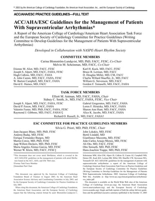
- 7. ACC - www.acc.org AHA - www.americanheart.org Blomström-Lundqvist and Scheinman et al. 2003 ESC - www.escardio.org ACC/AHA/ESC Practice Guidelines 7 Figure 1. Afterdepolarizations from myocytes. Action potentials recorded before (A) and after (B) application of a potassium chan- nel blocker. Potassium channel blockers lengthen action potential duration and encourage afterdepolarizations. The action potentials are prolonged in (B), and repolarization is interrupted by early afterdepolarizations (C). A series of DADs of decreasing amplitude occurs after completion of repolarization of the action potential, often in the setting of intracellular [Ca2+] overload. DADs indicates delayed afterdepolarizations; mV, millivolts; sec, seconds. IV. CLINICAL PRESENTATION, GENERAL Sinus tachycardia is, conversely, nonparoxysmal and accel- EVALUATION, AND MANAGEMENT OF erates and terminates gradually. Patients with sinus tachycar- PATIENTS WITH SUPRAVENTRICULAR dia may require evaluation for stressors such as infection or ARRHYTHMIA volume loss. Episodes of regular and paroxysmal palpita- tions with sudden onset and termination (also referred to as A. General Evaluation of Patients Without PSVT) most commonly result from AVRT or AVNRT. Documented Arrhythmia Termination by vagal maneuvers further suggests a re-entrant 1. Clinical History and Physical Examination tachycardia involving AV nodal tissue (eg, AVNRT, AVRT). Polyuria is caused by release of atrial natriuretic peptide in Patients with paroxysmal arrhythmias are most often asymp- response to increased atrial pressures from contraction of tomatic at the time of evaluation. Arrhythmia-related symp- atria against a closed AV valve, which is supportive of a sus- toms include palpitations; fatigue; lightheadedness; chest tained supraventricular arrhythmia. discomfort; dyspnea; presyncope; or, more rarely, syncope. With SVT, syncope is observed in approximately 15% of A history of arrhythmia-related symptoms may yield important clues to the type of arrhythmia. Premature beats patients, usually just after initiation of rapid SVT or with a are commonly described as pauses or nonconducted beats prolonged pause after abrupt termination of the tachycardia followed by a sensation of a strong heartbeat, or they are (55). Syncope may be associated with AF with rapid con- described as irregularities in heart rhythm. Supraventricular duction over an accessory AV pathway or may suggest con- tachycardias occur in all age groups and may be associated comitant structural abnormalities, such as valvular aortic with minimal symptoms, such as palpitations, or may present stenosis, hypertrophic cardiomyopathy, or cerebrovascular with syncope. The clinician should distinguish whether the disease. Symptoms vary with the ventricular rate, underlying palpitations are regular or irregular. Irregular palpitations heart disease, duration of SVT, and individual patient per- may be due to premature depolarizations, AF, or MAT. The ceptions. Supraventricular tachycardia that is persistent for latter are most commonly encountered in patients with pul- weeks to months and associated with a fast ventricular monary disease. If the arrhythmia is recurrent and has abrupt response may lead to a tachycardia-mediated cardiomyopa- onset and termination, then it is designated paroxysmal. thy (56-58).
- 8. ACC - www.acc.org Blomström-Lundqvist and Scheinman et al. 2003 AHA - www.americanheart.org 8 ACC/AHA/ESC Practice Guidelines ESC - www.escardio.org Of crucial importance in clinical decision making is a clin- otherwise made by careful analysis of the 12-lead ECG dur- ical history describing the pattern in terms of the number of ing tachycardia (see Section IV). Therefore, patients with a episodes, duration, frequency, mode of onset, and possible history of sustained arrhythmia should always be encouraged triggers. to have at least one 12-lead ECG taken during the arrhyth- Supraventricular tachycardia has a heterogeneous clinical mia. Automatic analysis systems of 12-lead ECGs are unre- presentation, most often occurring in the absence of liable and commonly suggest an incorrect arrhythmia diag- detectable heart disease in younger individuals. The presence nosis. of associated heart disease should, nevertheless, always be Indications for referral to a cardiac arrhythmia specialist sought and an echocardiogram may be helpful. While a phys- include presence of a wide complex tachycardia of unknown ical examination during tachycardia is standard, it usually origin. For those with narrow complex tachycardias, referral does not lead to a definitive diagnosis. If irregular cannon A is indicated for those with drug resistance or intolerance as waves and/or irregular variation in S1 intensity is present, well as for patients desiring to be free of drug therapy. then a ventricular origin of a regular tachycardia is strongly Because of the potential for lethal arrhythmias, all patients suggested. with Wolff-Parkinson-White (WPW) syndrome (ie, pre-exci- tation combined with arrhythmias) should be referred for fur- 2. Diagnostic Investigations ther evaluation. All patients with severe symptoms, such as A resting 12-lead ECG should be recorded and evaluated for syncope or dyspnea, during palpitations also should be the presence of abnormal rhythm, pre-excitation, prolonged referred for prompt evaluation by an arrhythmia specialist. QT interval, sinus tachycardia, segment abnormalities, or An echocardiographic examination should be considered in evidence of underlying heart disease. The presence of pre- patients with documented sustained SVT to exclude the pos- excitation on the resting ECG in a patient with a history of sibility of structural heart disease, which usually cannot be paroxysmal regular palpitations is sufficient for the pre- detected by physical examination or 12-lead ECG. sumptive diagnosis of AVRT, and attempts to record sponta- An ambulatory 24-hour Holter recording can be used in neous episodes are not required before referral to an arrhyth- patients with frequent (ie, several episodes per week) but mia specialist for therapy (Fig. 2). Specific therapy is dis- transient tachycardias (59-61). An event or wearable loop cussed in Section V–A clinical history of irregular and parox- recorder is often more useful than a 24-hour recording in ysmal palpitations in a patient with baseline pre-excitation patients with less frequent arrhythmias (62). Implantable strongly suggests episodes of AF, which requires immediate loop recorders may be helpful in selected cases with rare electrophysiological evaluation because these patients are at symptoms (ie, fewer than two episodes per month) associat- risk for sudden death (see Section V–D). The diagnosis is ed with severe symptoms of hemodynamic instability (63). Figure 2. Initial evaluation of patients with suspected tachycardia. AVRT indicates atrioventricular reciprocating tachycardia; ECG, electrocardiogram.
- 9. ACC - www.acc.org AHA - www.americanheart.org Blomström-Lundqvist and Scheinman et al. 2003 ESC - www.escardio.org ACC/AHA/ESC Practice Guidelines 9 Exercise testing is less often useful for diagnosis unless the empirically provided that significant bradycardia (less than arrhythmia is clearly triggered by exertion. 50 bpm) have been excluded. Due to the risk of proarrhyth- Transesophageal atrial recordings and stimulation may be mia, antiarrhythmic treatment with Class I or Class III drugs used in selected cases for diagnosis or to provoke paroxys- should not be initiated without a documented arrhythmia. mal tachyarrhythmias if the clinical history is insufficient or if other measures have failed to document an arrhythmia. B. General Evaluation of Patients With Documented Esophageal stimulation is not indicated if invasive electro- Arrhythmia physiological investigation is planned (64,65). Invasive elec- trophysiological investigation with subsequent catheter abla- 1. Diagnostic Evaluation tion may be used for diagnoses and therapy in cases with a Whenever possible, a 12-lead ECG should be taken during clear history of paroxysmal regular palpitations. It may also tachycardia but should not delay immediate therapy to termi- be used empirically in the presence of pre-excitation or dis- nate the arrhythmia if there is hemodynamic instability. At a abling symptoms (Fig. 2). minimum, a monitor strip should be obtained from the defib- rillator, even in cases with cardiogenic shock or cardiac 3. Management arrest, before direct current (DC) cardioversion is applied to The management of patients with symptoms suggestive of an terminate the arrhythmia. arrhythmia but without ECG documentation depends on the nature of the symptoms. If the surface ECG is normal and the a. Differential Diagnosis for Narrow QRS-Complex patient reports a history consistent with premature extra Tachycardia beats, then precipitating factors, such as excessive caffeine, alcohol, nicotine intake, recreational drugs, or hyperthy- If ventricular activation (QRS) is narrow (less than 120 mil- roidism, should be reviewed and eliminated (Table 3). liseconds [ms]), then the tachycardia is almost always Benign extrasystoles are often manifest at rest and tend to supraventricular and the differential diagnosis relates to its become less common with exercise. mechanism (Fig. 3) (66,67). If no P waves or evidence of If symptoms and the clinical history indicate that the atrial activity is apparent and the RR interval is regular, then arrhythmia is paroxysmal in nature and the resting 12-lead AVNRT is most commonly the mechanism (Fig. 4). P-wave ECG gives no clue for the arrhythmia mechanism, then fur- activity in AVNRT may be only partially hidden within the ther diagnostic tests for documentation may not be necessary QRS complex and may deform the QRS to give a pseudo-R before referral for an invasive electrophysiological study wave in lead V1 and/or a pseudo-S wave in inferior leads and/or catheter ablation. Patients should be taught to perform (Fig. 4). If a P wave is present in the ST segment and sepa- vagal maneuvers. A beta-blocking agent may be prescribed rated from the QRS by 70 ms, then AVRT is most likely. In tachycardias with RP longer than PR (Fig. 5), the most typi- Table 3. Predisposing or Precipitating Factors for Patients With cal diagnosis is atypical AVNRT, permanent form of junc- Palpitations tional reciprocating tachycardia (PJRT) (ie, AVRT via a Noncardiac Causes slowly conducting accessory pathway), or AT (see Sections Nicotine, alcohol, caffeine V–B, V–D, and V–E). Responses of narrow QRS-complex Physical or mental stress tachycardias to adenosine or carotid massage may aid in the Hyperthyroidism differential diagnosis (Fig. 6) (68-70). A 12-lead ECG Premenstrual or menstrual recording is desirable during use of adenosine or carotid Electrolyte disturbance massage. If P waves are not visible, then the use of Certain drugs (antiarrhythmic, antidepressant, antibiotic drugs; esophageal pill electrodes can also be helpful. stimulants; antihistamines; appetite suppressants) Anemia Anxiety or hypovolemia b. Differential Diagnosis for Wide QRS-Complex Fever, infection Tachycardia Lack of sleep If the QRS is wide (greater than 120 ms), then it is important Cardiac Causes to differentiate between SVT and ventricular tachycardia Coronary artery disease; old myocardial infarction, especially for (VT) (Fig. 7). Intravenous medications given for the treat- ventricular tachycardias ment of SVT, particularly verapamil or diltiazem, may be Congestive heart failure deleterious because they may precipitate hemodynamic col- Cardiomyopathy Valvular disease lapse for a patient with VT (71-73). Stable vital signs during Congenital heart disease tachycardias are not helpful for distinguishing SVT from VT. Other conditions that may cause myocardial scarring (ie, If the diagnosis of SVT cannot be proven or cannot be made sarcoidosis, tuberculosis) easily, then the patient should be treated as if VT were pres- Primary electrical disorders (ie, long QT syndrome, Brugada ent. Wide-QRS tachycardia can be divided into three groups: syndrome) SVT with bundle-branch block (BBB) or aberration, SVT Accessory pathways with AV conduction over an accessory pathway, and VT.
- 10. ACC - www.acc.org Blomström-Lundqvist and Scheinman et al. 2003 AHA - www.americanheart.org 10 ACC/AHA/ESC Practice Guidelines ESC - www.escardio.org Figure 3. Differential diagnosis for narrow QRS tachycardia. Patients with focal junctional tachycardia may mimic the pattern of slow- fast AVNRT and may show AV dissociation and/or marked irregularity in the junctional rate. AV indicates atrioventricular; AVNRT, atri- oventricular nodal reciprocating tachycardia; AVRT, atrioventricular reciprocating tachycardia; MAT, multifocal atrial tachycardia; ms, milliseconds; PJRT, permanent form of junctional reciprocating tachycardia; QRS, ventricular activation on electrocardiogram. SUPRAVENTRICULAR TACHYCARDIA WITH BUNDLE-BRANCH VENTRICULAR TACHYCARDIA. Several ECG criteria have been BLOCK. Bundle-branch block may be pre-existing or may described to differentiate the underlying mechanism of a occur only during tachycardia when one of the bundle wide-QRS tachycardia. branches is refractory due to the rapid rate. Most BBBs are VENTRICULAR ARRHYTHMIA DISSOCIATION. Ventricular arrhyth- not only rate-related, but are also due to a long-short mia dissociation with a ventricular rate faster than the atrial sequence of initiation. Bundle-branch block can occur with rate generally proves the diagnosis of VT (Fig. 8) but is clear- any supraventricular arrhythmia. If a rate-related BBB devel- ly discernible in only 30% of all VTs (74). Fusion complex- ops during orthodromic AVRT, then the tachycardia rate may es represent a merger between conducted sinus (or supraven- slow if the BBB is ipsilateral to the bypass tract location. tricular complexes) impulses and ventricular depolarization SUPRAVENTRICULAR TACHYCARDIA WITH ATRIOVENTRICULAR occurring during AV dissociation. These complexes are CONDUCTION OVER AN ACCESSORY PATHWAY. Supra- pathognomonic of VT. Retrograde VA block may be present ventricular tachycardia with AV conduction over an accesso- spontaneously or brought out by carotid massage. The ry pathway may occur during AT, atrial flutter, AF, AVNRT demonstration that P waves are not necessary for tachycardia or antidromic AVRT. The latter is defined as anterograde maintenance strongly suggests VT. P waves can be difficult conduction over the accessory pathway and retrograde con- to recognize during a wide-QRS tachycardia. Therefore, one duction over the AV node or a second accessory AV pathway. should also look for evidence of VA dissociation on exami- A wide-QRS complex with left bundle-branch block (LBBB) nation: irregular cannon A waves in the jugular venous pulse morphology may be seen with anterograde conduction over and variability in the loudness of the first heart sound and in other types of accessory pathways, such as atriofascicular, systolic blood pressure (75). If P waves are not visible, then nodofascicular, or nodoventricular tracts. the use of esophageal pill electrodes can also be useful.
- 11. ACC - www.acc.org AHA - www.americanheart.org Blomström-Lundqvist and Scheinman et al. 2003 ESC - www.escardio.org ACC/AHA/ESC Practice Guidelines 11 Figure 4. ECG pattern of typical AVNRT. Panel A: 12-Lead ECG shows a regular SVT recorded at an ECG paper speed of 25 mm/sec. Panel B: After conversion to sinus rhythm, the 12-lead ECG shows sinus rhythm with narrow QRS complexes. In compari- son with Panel A: Note the pseudo r´ in V1 (arrow) and accentuated S waves in 2, 3, aVF (arrow). These findings are pathognomonic for AVNRT. AVNRT indicates atrioventricular nodal reciprocating tachycardia; ECG, electrocardiogram; mm/sec, millimeters per sec- ond; QRS, ventricular activation on ECG; SVT, supraventricular tachycardia. WIDTH OF THE QRS COMPLEX. A QRS width of more than 0.14 • An RS (from the initial R to the nadir of S) interval seconds with right bundle-branch block (RBBB) or 0.16 sec- longer than 100 ms in any precordial lead is highly onds during LBBB pattern favors VT (74). The QRS width suggestive of VT (78). criteria are not helpful in differentiating VT from SVT with AV conduction over an accessory pathway. A patient with • A QRS pattern with negative concordance in the pre- SVT can have a QRS width of more than 0.14 (RBBB) or cordial leads is diagnostic for VT (“negative concor- 0.16 (LBBB) in the presence of either pre-existing BBB or dance” means that the QRS patterns in all of the pre AV conduction over an accessory pathway, or when class Ic cordial leads are similar, and with QS complexes). or class Ia antiarrhythmic drugs are used. Positive concordance does not exclude antidromic AVRT over a left posterior accessory pathway (79). CONFIGURATIONAL CHARACTERISTICS OF THE QRS COMPLEX DURING TACHYCARDIA. Leads V1 and V6 are helpful in differ- • The presence of ventricular fusion beats indicates a ven- entiating VT from SVT (74,76,77). tricular origin of the tachycardia. Figure 5. ECG tracing with limb leads I, II, and III, showing an RP (initial R to initial P) interval longer than the PR interval. The P wave differs from the sinus P wave. ECG indicates electrocardiogram.
- 12. ACC - www.acc.org Blomström-Lundqvist and Scheinman et al. 2003 AHA - www.americanheart.org 12 ACC/AHA/ESC Practice Guidelines ESC - www.escardio.org Figure 6. Responses of narrow complex tachycardias to adenosine. AT indicates atrial tachycardia; AV, atrioventricular; AVNRT, atri- oventricular nodal reciprocating tachycardia; AVRT, atrioventricular reciprocating tachycardia; IV, intravenous; QRS, ventricular acti- vation on electrocardiogram; VT, ventricular tachycardia. • QR complexes indicate a myocardial scar and are in cold water), should be initiated to terminate the arrhythmia present in approximately 40% of patients with VTs or to modify AV conduction. If this fails, IV antiarrhythmic after myocardial infarction (80). drugs should be administered for arrhythmia termination in hemodynamically stable patients. Adenosine or nondihy- The width and morphologic criteria are less specific for dropyridine calcium-channel antagonists are the drugs of patients taking certain antiarrhythmic agents and those with choice (Fig. 7). The advantage of adenosine relative to IV hyperkalemia or severe heart failure. Despite ECG criteria, calcium-channel or beta blockers relates to its rapid onset patients presenting with wide QRS-complex tachycardia are and short half-life. Intravenous adenosine is, therefore, the often misdiagnosed (71,72,81). A positive answer to two preferred agent except for patients with severe asthma. inquiries, namely the presence of a previous myocardial Patients treated with theophylline may require higher doses infarct and the first occurrence of a wide QRS-complex of adenosine for effect, and adenosine effects are potentiated tachycardia after an infarct, strongly indicates a diagnosis of by dipyridamole. In addition, higher rates of heart block may VT (82). be seen when adenosine is concomittantly administered with carbamazepine. Longer-acting agents (eg, IV calcium-chan- 2. Management nel blockers or beta blockers [ie, verapamil/diltiazem or When a definitive diagnosis can be made on the basis of ECG metoprolol]) are of value, particularly for patients with fre- and clinical criteria, acute and chronic treatment should be quent atrial premature beats or ventricular premature beats, initiated on the basis of the underlying mechanism (see the which may serve to trigger early recurrence of PSVT. sections on specific arrhythmias). Adenosine or DC cardioversion is preferred for those with If the specific diagnosis of a wide QRS-complex tachycar- PSVT in whom a rapid therapeutic effect is essential. dia cannot be made despite careful evaluation, then the Potential adverse effects of adenosine include initiation of patient should be treated for VT. Acute management of AF (1 to 15%), which is usually transient, and may be par- ticularly problematic for those with ventricular pre-excita- patients with hemodynamically stable and regular tachycar- tion. Adenosine should be avoided in patients with severe dia is outlined in Fig. 9. bronchial asthma. It is important to use extreme care with The most effective and rapid means of terminating any concomitant use of IV calcium-channel blockers and beta hemodynamically unstable narrow or wide QRS-complex blockers because of possible potentiation of hypotensive tachycardia is DC cardioversion. and/or bradycardic effects. An ECG should be recorded dur- ing vagal maneuvers or drug administration because the a. Acute Management of Narrow QRS-Complex response may aid in the diagnosis even if the arrhythmia does Tachycardia not terminate (Fig. 6). Termination of the tachycardia with a In regular narrow QRS-complex tachycardia, vagal maneu- P wave after the last QRS complex favors AVRT or AVNRT. vers (ie, Valsalva [83], carotid massage, and facial immersion Tachycardia termination with a QRS complex favors AT,
- 13. ACC - www.acc.org AHA - www.americanheart.org Blomström-Lundqvist and Scheinman et al. 2003 ESC - www.escardio.org ACC/AHA/ESC Practice Guidelines 13 Figure 7. Differential diagnosis for wide QRS-complex tachycardia (greater than 120 ms). A QRS conduction delay during sinus rhythm, when available for comparison, reduces the value of QRS morphology analysis. Adenosine should be used with caution when the diagnosis is unclear because it may produce VF in patients with coronary artery disease and AF with a rapid ventricular rate in pre-excited tachycardias. Various adenosine responses are shown in Fig. 6. *Concordant indicates that all precordial leads show either positive or negative deflections. Fusion complexes are diagnostic of VT. †In pre-excited tachycardias, the QRS is generally wider (ie, more pre-excited) compared with sinus rhythm. A indicates atrial; AF, atrial fibrillation; AP, accessory pathway; AT, atrial tachycardia; AV, atrioventricular; AVRT, atrioventricular reciprocating tachycardia; BBB, bundle-branch block; LBBB, left bundle- branch block; ms, milliseconds; QRS, ventricular activation on ECG; RBBB, right bundle-branch block; SR, sinus rhythm; SVT, supraventricular tachycardias; V, ventricular; VF, ventricular fibrillation; VT, ventricular tachycardia. which is often adenosine insensitive. Continuation of tachy- dations are in accord with the current Advanced cardia with AV block is virtually diagnostic of AT or atrial Cardiovascular Life Support guidelines (88). Special circum- flutter, excludes AVRT, and makes AVNRT very unlikely. stances may require alternative therapy (ie, pre-excited tachycardias and VT caused by digitalis toxicity). For termi- b. Acute Management of Wide QRS-Complex nation of an irregular wide QRS-complex tachycardia (ie, Tachycardia pre-excited AF), DC cardioversion is recommended. Or, if the patient is hemodynamically stable, pharmacologic con- Immediate DC cardioversion is the treatment for hemody- version using IV ibutilide, flecainide, or procainamide is namically unstable tachycardias. If the tachycardia is hemo- appropriate. dynamically stable and definitely supraventricular, then management is as described for narrow QRS-complex tachy- c. Further Management cardias (Fig. 6). For pharmacologic termination of a stable wide QRS-complex tachycardia, IV procainamide and/or After successful termination of a wide QRS-complex tachy- sotalol are recommended on the basis of randomized but cardia of unknown etiology, patients should be referred to an small studies (84,85). Amiodarone is also considered accept- arrhythmia specialist. Patients with stable narrow QRS-com- able. Amiodarone is preferred, compared to procainamide plex tachycardia, normal LV function, and a normal ECG and sotalol, in patients with impaired left ventricular (LV) during sinus rhythm (ie, no pre-excitation) may require no function (86,87) or signs of heart failure. These recommen- specific therapy. Referral is indicated for those with drug
- 14. ACC - www.acc.org Blomström-Lundqvist and Scheinman et al. 2003 AHA - www.americanheart.org 14 ACC/AHA/ESC Practice Guidelines ESC - www.escardio.org Figure 8. Electrocardiogram showing AV dissociation during VT in a patient with a wide QRS-complex tachycardia. The P waves are marked with arrows. resistance or intolerance as well as for patients desiring to be autonomic influences. Nevertheless, the sinus node is a ver- free of lifelong drug therapy. When treatment is indicated, satile structure and is influenced by many other factors, options include catheter ablation or drug therapy. Finally, including hypoxia, acidosis, stretch, temperature, and hor- because of the potential for lethal arrhythmias, all patients mones (eg, tri-iodothyronine, serotonin). with WPW syndrome (ie, pre-excitation and arrhythmias) should be referred for further evaluation (89). a. Definition Sinus tachycardia is defined as an increase in sinus rate to V. SPECIFIC ARRHYTHMIAS greater than 100 bpm in keeping with the level of physical, A. Sinus Tachyarrhythmias emotional, pathological, or pharmacologic stress. Pathological causes of sinus tachycardia include pyrexia, Sinus tachycardia usually occurs in response to an appropri- hypovolemia, or anemia, which may result from infections, ate physiological stimulus (eg, exercise) or to an excessive malignancies, myocardial ischemia, congestive cardiac fail- stimulus (eg, hyperthyroidism). Failure of the mechanisms ure, pulmonary emboli, shock, and thyrotoxicosis. Drugs that that control the sinus rate may lead to an inappropriate sinus induce sinus tachycardia include stimulants (eg, caffeine, tachycardia. Excessive sinus tachycardia may also occur in alcohol, nicotine); prescribed compounds (eg, salbutamol, response to upright posture (postural orthostatic tachycardia aminophylline, atropine, catecholamines); and certain recre- syndrome [POTS]). A re-entry mechanism may also occur ational/illicit drugs (eg, amphetamines, cocaine, “ecstasy,” within, or close to, the sinus node, resulting in so-called sinus cannabis) (100). Anticancer treatments, in particular anthra- node re-entrant tachycardia, which is also sometimes known cycline compounds such as doxorubicin (or Adriamycin) and as SA re-entry. daunorubicin, can also trigger sinus tachycardia as part of the acute cardiotoxic response that is predominantly cate- 1. Physiological Sinus Tachycardia cholamine/histamine induced (101) or part of a late car- The normally innervated sinus node generates an impulse diotoxic response (102,103). Sinus tachycardia may signal approximately 60 to 90 times per minute and responds to severe underlying pathologies and often requires comprehen-
- 15. ACC - www.acc.org AHA - www.americanheart.org Blomström-Lundqvist and Scheinman et al. 2003 ESC - www.escardio.org ACC/AHA/ESC Practice Guidelines 15 Figure 9. Acute management of patients with hemodynamically stable and regular tachycardia. *A 12-lead ECG during sinus rhythm must be available for diagnosis. †Adenosine should be used with caution in patients with severe coronary artery disease and may produce AF, which may result in rapid ventricular rates for patients with pre-excitation. **Ibutilide is especially effective for patients with atrial flutter but should not be used in patients with EF less than 30% due to increased risk of polymorphic VT. AF indicates atri- al fibrillation; AV, atrioventricular; BBB, bundle-branch block; DC, direct current; ECG, electrocardiogram; IV, intravenous; LV, left ven- tricle; QRS, ventricular activation on ECG; SVT, supraventricular tachycardia; VT, ventricular tachycardia. sive evaluation. Atrial and sinus tachycardias may be difficult plane, it is directed anteriorly and slightly leftward and can, to differentiate. therefore, be negative in leads V1 and V2 but positive in leads V3 to V6. The PR interval is normally between 120 ms b. Mechanism and 200 ms (220 ms in the elderly). The P waves have a nor- Sinus tachycardia results from physiological influences on mal contour, but a larger amplitude may develop and the individual pacemaker cells and from an anatomical shift in wave may become peaked (105). Sinus tachycardia is non- the site of origin of atrial depolarization superiorly within the paroxysmal, thus differentiating it from re-entry. sinus node (104). If current activity is one of several mecha- nisms by which phase 4 diastolic depolarization is hastened, d. Treatment therefore increasing the heart rate, then an increase in cyclic The mainstay in the management of sinus tachycardias pri- adenosine monophosphate triggers opening of ion channels marily involves identifying the cause and either eliminating responsible for the pacemaker, or funny current (if), resulting or treating it. However, beta blockade can be extremely use- in a faster heart rate due to more rapid attainment of thresh- ful and effective for physiological symptomatic sinus tachy- old potential. cardia triggered by emotional stress and other anxiety-relat- ed disorders (106-113); for prognostic benefit after myocar- c. Diagnosis dial infarction (114-117); for the symptomatic and prognos- In normal sinus rhythm, the P wave on a 12-lead ECG is pos- tic benefits in certain other irreversible causes of sinus tachy- itive in leads I, II, and aVF and negative in aVR. Its axis in cardias, such as congestive cardiac failure (118-120); and for the frontal plane lies between 0 and +90; in the horizontal symptomatic thyrotoxicosis in combination with carbima-
- 16. ACC - www.acc.org Blomström-Lundqvist and Scheinman et al. 2003 AHA - www.americanheart.org 16 ACC/AHA/ESC Practice Guidelines ESC - www.escardio.org Recommendations for Acute Management of Hemodynamically Stable and Regular Tachycardia ECG Recommendation* Classification Level of Evidence References Narrow QRS-complex Vagal maneuvers I B tachycardia (SVT) Adenosine I A (4,69,90) Verapamil, diltiazem I A (91) Beta blockers IIb C (92,93) Amiodarone IIb C (94) Digoxin IIb C Wide QRS-complex tachycardia •SVT and BBB See above •Pre-excited SVT/AF† Flecainide‡ I B (95) Ibutilide‡ I B (96) Procainamide‡ I B DC cardioversion I C •Wide QRS-complex Procainamide‡ I B (84,97) tachycardia of Sotalol‡ I B (85) unknown origin Amiodarone I B (25,86) DC cardioversion I B (98) Lidocaine IIb B (85,97) Adenosine§ IIb C (99) Beta blockers¶ III C (98) Verapamil** III B (73) Wide QRS-complex Amiodarone I B (25,86) tachycardia of unknown DC cardioversion, I B (98) origin in patients with lidocaine poor LV function The order in which treatment recommendations appear in this table within each class of recommendation does not necessarily reflect a preferred sequence of administration. Please refer to text for details. For pertinent drug dosing information, please refer to the ACC/AHA/ESC Guidelines on the Management of Patients With Atrial Fibrillation. *All listed drugs are administered intravenously. †See Section V, specific section. ‡Should not be taken by patients with reduced LV function. §Adenosine should be used with caution in patients with severe coronary artery disease because vasodilation of normal coronary vessels may produce ischemia in vulnerable territory. It should be used only with full resuscitative equipment available. ¶Beta blockers may be used as first-line therapy for those with catecholamine-sensitive tachycardias, such as right ventricular outflow tachycardia. **Verapamil may be used as first-line therapy for those with LV fascicular VT. AF indicates atrial fibrillation; BBB, bundle-branch block; DC, direct current; ECG, electrocardiogram; LV, left ventricular; QRS, ventricular activation on ECG; SVT, supraventricular zole or propylthiouracyl while these palliative agents take b. Mechanism effect (121,122). Nondihydropyridine calcium-channel The underlying pathological basis for inappropriate sinus blockers, such as diltiazem or verapamil, may be of benefit tachycardia is likely to be multifactorial, but two main mech- in patients with symptomatic thyrotoxicosis, if beta blockade anisms have been proposed: is contraindicated (123). 1. Enhanced automaticity of the sinus node (125) 2. Inappropriate Sinus Tachycardia 2. Abnormal autonomic regulation of the sinus node with excess sympathetic and reduced parasympathetic tone a. Definition (126,127) Inappropriate sinus tachycardia is a persistent increase in It is unclear whether these mechanisms are a direct result of resting heart rate or sinus rate unrelated to, or out of propor- impaired neural input into the sinus node or whether they tion with, the level of physical, emotional, pathological, or represent an inherent abnormality within the sinus node itself pharmacologic stress. (128).
- 17. ACC - www.acc.org AHA - www.americanheart.org Blomström-Lundqvist and Scheinman et al. 2003 ESC - www.escardio.org ACC/AHA/ESC Practice Guidelines 17 Recommendations for Treatment of Inappropriate Sinus Tachycardia Treatment Recommendation Classification Level of Evidence References Medical Beta blockers I C – Verapamil, diltiazem IIa C – Interventional Catheter ablation-sinus node IIb C (133-135,137-141) modification/elimination* The order in which treatment recommendations appear in this table within each class of recommendation does not necessarily reflect a preferred sequence of administration. Please refer to text for details. For pertinent drug dosing information, please refer to the ACC/AHA/ESC Guidelines on the Management of Patients With Atrial Fibrillation. *Used as a last resort. c. Presentation ry cases of inappropriate sinus tachycardia (133). Potential adverse effects include pericarditis, phrenic nerve injury, A high proportion of patients with inappropriate sinus tachy- superior vena cava (SVC) syndrome, or need for permanent cardia are healthcare professionals, and approximately 90% pacing. A number of case reports have recorded successful are female (129). The mean age of presentation is 38 plus or surgical excision (134,135) or radiofrequency (RF) ablation minus 12 years. Although the predominant symptom at pres- of the sinus node (136-138). There is also a case report of entation is palpitations, symptoms such as chest pain, short- successful obliteration of the sinus node artery for the man- ness of breath, dizziness, lightheadedness, and presyncope agement of this disorder (139). The diagnosis of POTS (see have also been reported. The degree of disability can vary Section V–A, 3) must be excluded before considering abla- tremendously, from totally asymptomatic patients identified tion. In a retrospective analysis of 29 cases undergoing sinus during routine medical examination to individuals who are fully incapacitated. Clinical examination and routine investi- node modification for inappropriate sinus tachycardia (140), gations allow elimination of a secondary cause for the tachy- a 76% acute success rate (22 out of 29 cases) was reported. cardia but are generally not helpful in establishing the diag- The long-term success rate has been reported to be 25% to nosis. 65%. d. Diagnosis 3. Postural Orthostatic Tachycardia Syndrome Inappropriate sinus tachycardia is diagnosed on the basis of Postural orthostatic tachycardia syndrome is part of a wide invasive and noninvasive criteria (128,129): spectrum of disorders that exhibit autonomic dysfunction (142). These include severe orthostatic hypotension in the 1. The presence of a persistent sinus tachycardia (heart rate presence of autonomic neuropathy and vasovagal syncope in greater than 100 bpm) during the day with excessive rate the absence of other evidence of autonomic dysfunction. increase in response to activity and nocturnal normaliza- Postural orthostatic tachycardia syndrome manifests as an tion of rate, as confirmed by a 24-hour Holter recording excessive orthostatic tachycardia without significant ortho- 2. The tachycardia (and symptoms) is nonparoxysmal static hypotension in those without overt autonomic neu- ropathy. It is associated with numerous other symptoms, 3. P-wave morphology and endocardial activation identical such as exercise intolerance, palpitations, weakness, and to sinus rhythm lightheadedness; most of these symptoms are also autonom- 4. Exclusion of a secondary systemic cause (eg, hyperthy- ically mediated (143-145). roidism, pheochromocytoma, physical deconditioning) a. Definition e. Treatment Postural orthostatic tachycardia syndrome is the diagnosis The treatment of inappropriate sinus tachycardia is predomi- applied to individuals who present with orthostatic intoler- nantly symptom driven. The risk of tachycardia-induced car- ance (ie, symptoms on standing that are relieved by recum- diomyopathy (130) in untreated patients is unknown but like- bency) in the presence of a demonstrable exaggerated, per- ly to be small. sistent postural sinus tachycardia (greater than 30 bpm from Although no randomized, double-blinded, placebo-con- baseline or greater than 120 bpm) within 10 minutes of an trolled clinical trials exist, beta blockers may be useful and upright tilt in the absence of postural hypotension and any should be prescribed as first-line therapy in the majority of demonstrable autonomic neuropathy. these patients. Anecdotal evidence suggests that nondihy- dropyridine calcium-channel blockers, such as verapamil and b. Mechanism diltiazem, are also effective (131). Specific bradycardic agents (eg, If inhibitor ivabradine) may be valuable, but these Many mechanisms have been proposed for POTS. These agents are still under investigation (132). range from idiopathic hypovolemia (146) and reduced circu- Sinus node modification by catheter ablation remains a lating blood volume (147) to splanchnic bed blood pooling potentially important therapeutic option in the most refracto- (148,149) and reduced red cell mass resulting from an