SlideShare uma empresa Scribd logo
1 de 21
Baixar para ler offline
Chapter 22
Intrapartum Fetal Surveillance
                                                                      Michael P. Nageotte, MD, and Larry C. Gilstrap III, MD


                                                                             ability. Hence, there are two possible vagal influences on the heart: a
                                                                             tonic influence tending to decrease FHR and an oscillatory influence
Factors Controlling Fetal                                                    that results in FHR variability.2 Further, vagal tone is not necessarily
Heart Rate                                                                   constant. Its influence increases with gestational age.3 In fetal sheep,
                                                                             vagal activity increases as much as fourfold during acute hypoxia4 or
Fetal heart rate (FHR) analysis is the most common means by which            experimentally produced fetal growth restriction.5
a fetus is evaluated for adequacy of oxygenation. Knowledge of the rate
and regulation of the fetal heart is therefore of great importance to the
obstetrician. At 20 weeks of gestation, the average FHR is 155 beats/        Sympathetic Nervous System
min; at 30 weeks, it is 144 beats/min; and at term, it is 140 beats/min.     Sympathetic nerves are widely distributed in the muscle of the heart
This progression is thought to be related to maturation of vagal tone,       at term. Stimulation of the sympathetic nerves releases norepinephrine
with consequent slowing of the baseline FHR. Variations of 20 beats/         and causes increases in both the rate and the strength of fetal cardiac
min faster or slower than these baseline values are present in normal        contractions, resulting in higher cardiac output. The sympathetic
fetuses.                                                                     nerves are a reserve mechanism to improve the pumping activity of
    The fetal heart is similar to the adult heart in that it has its own     the heart during intermittent stressful situations. There is normally a
intrinsic pacemaker activity which results in rhythmic myocardial con-       tonic sympathetic influence on the heart. Blocking the action of these
tractions. The sinoatrial node, found in one wall of the right atrium,       sympathetic nerves causes a decrease in FHR of approximately 10
has the fastest rate of conduction and sets the rate in the normal heart.    beats/min. As with the vagal tone, tonic sympathetic influence increases
The next fastest pacemaker rate is found in the atrium. Finally, the         as much as twofold during fetal hypoxia.
ventricle has a slower rate than either the sinoatrial node or the atrium.
In cases of complete or partial heart block in the fetus, variations in
rate markedly slower than normal can be seen. The rate in a fetus with       Chemoreceptors
a complete heart block is often 60 to 80 beats/min.                          Chemoreceptors are found in both the peripheral and the central
    Variability of the FHR is important clinically and is of prognostic      nervous system. They have their most dramatic effects on the regula-
value with respect to its specific amplitude as part of the FHR pattern.      tion of respiration but are also important in control of the circulation.
The heart rate is the result of many physiologic factors that modulate       The peripheral chemoreceptors are in the aortic and carotid bodies,
the intrinsic rate of the fetal heart, the most common being signals         which are located in the arch of the aorta and the area of the carotid
from the autonomic nervous system.                                           sinus, respectively. The central chemoreceptors in the medulla oblon-
                                                                             gata respond to changes in oxygen and carbon dioxide tension in the
                                                                             blood or in the cerebrospinal fluid perfusing this area.
Parasympathetic Nervous System                                                   In the adult, when oxygen is decreased or the carbon dioxide
The parasympathetic nervous system consists primarily of the vagus           content is increased in the arterial blood perfusing the central chemo-
nerve (10th cranial nerve), which originates in the medulla oblongata.       receptors, a reflex tachycardia occurs. There is also a substantial increase
The vagus nerve innervates both the sinoatrial and the atrioventricular      in arterial blood pressure, particularly when the carbon dioxide con-
nodes. Stimulation of the vagus nerve results in a decrease in FHR in        centration is increased. Both effects are thought to be protective, rep-
the normal fetus, because vagal influence on the sinoatrial node              resenting an attempt to circulate more blood through the affected areas
decreases its rate of firing. In a similar fashion, blockade of this nerve    and thereby bring about a decrease in carbon dioxide tension (PCO2)
in a normal fetus causes an increase in the FHR of approximately             or an increase in oxygen tension (PO2). Selective hypoxia or hypercap-
20 beats/min at term.1 This finding demonstrates a normally constant          nia of the peripheral chemoreceptors alone in the adult produces a
vagal influence on the FHR, which tends to decrease it from its intrinsic     bradycardia, in contrast to the tachycardia and hypertension that
rate.                                                                        results from central hypoxia or hypercapnia.
    The vagus nerve has another very important function: It is respon-           The interaction of the central and peripheral chemoreceptors in the
sible for transmission of impulses causing beat-to-beat variability of       fetus is poorly understood. It is known that the net result of hypoxia
FHR. Blockade of the vagus nerve results in disappearance of this vari-      or hypercapnia in the unanesthetized fetus is bradycardia with hyper-
398       CHAPTER 22             Intrapartum Fetal Surveillance

tension. During basal conditions, the chemoreceptors contribute to
stabilization of FHR and blood pressure.6                                     Blood Volume Control
                                                                              Capillary Fluid Shift
Baroreceptors                                                                 In the adult, when the blood pressure is elevated by excessive blood
In the arch of the aorta, and in the carotid sinus at the junction of the     volume, fluid moves out of the capillaries into interstitial spaces,
internal and external carotid arteries, are small stretch receptors in        thereby decreasing the blood volume toward normal. Conversely, if the
the vessel walls that are sensitive to increases in blood pressure. When      adult loses blood through hemorrhage, some fluid shifts out of the
pressure rises, impulses are sent from these receptors via the vagus or       interstitial spaces into the circulation, increasing the blood volume
glossopharyngeal nerve to the midbrain; this results in further impulses’     toward normal. There is normally a delicate balance between the pres-
being sent via the vagus nerve to the heart, tending to slow it. This is      sures inside and outside the capillaries. This mechanism of regulating
an extremely rapid response, occurring almost with the first systolic          blood pressure is slower than the almost instantaneous regulation
rise of blood pressure. It is a protective, stabilizing attempt by the body   observed with the reflex mechanisms discussed previously. Its role
to lower blood pressure by decreasing the heart rate and cardiac output       in the fetus is imperfectly understood, although imbalances may be
when blood pressure is increasing.                                            responsible for the hydrops seen in some cases of red cell alloimmu-
                                                                              nization and the high-output failure sometimes seen with supra-
                                                                              ventricular tachycardia.
Central Nervous System
In the adult, the higher centers of the brain influence the heart rate.        Intraplacental Pressures
Heart rate is increased by emotional stimuli such as fear and sexual          Fluid moves along hydrostatic pressure gradients and in response to
arousal. In fetal lambs and monkeys, the electroencephalogram or              osmotic pressure gradients. The specific role of these factors within the
electro-oculogram shows increased activity, at times in association           human placenta, where fetal and maternal blood closely approximate,
with increased variability of the FHR and body movements. At other            is unclear. It seems likely, however, that some delicate balancing mecha-
times, apparently when the fetus is sleeping, body movement slows and         nisms within the placenta prevent rapid fluid shifts between mother
FHR variability decreases, suggesting an association between these two        and fetus. The mean arterial blood pressure of the mother (approxi-
factors and central nervous system activity.7                                 mately 100 mm Hg) is much higher than that of the fetus (approxi-
    The medulla oblongata contains the vasomotor centers, integrative         mately 55 mm Hg), but the osmotic pressures are not substantially
centers in which the net result of all the inputs is either acceleration      different. Therefore, some compensatory mechanism must be present
or deceleration of the FHR. It is probably in these centers that the          to equalize the effective pressures at the exchange points so as to
net result of numerous central and peripheral inputs is processed             prevent dramatic fluid shifts.
to generate irregular oscillatory vagal impulses, giving rise to FHR
variability.
                                                                              Frank-Starling Mechanism
                                                                              The amount of blood pumped by the heart is determined by the
Hormonal Regulation                                                           amount of blood returning to the heart; that is, the heart normally
                                                                              pumps the blood that flows into it without excessive damming of blood
Adrenal Medulla                                                               in the venous circulation. When the cardiac muscle is stretched before
The fetal adrenal medulla produces epinephrine and norepinephrine             contraction by an increased inflow of blood, it contracts with a greater
in response to stress. Both substances act on the heart and cardiovas-        force and is able to pump out more blood. This mechanism of response
cular system in a way similar to sympathetic stimulation to produce a         to preload is apparently not the same in the fetal heart as in the adult
faster FHR, greater force of contraction of the heart, and higher arterial    heart. In the fetus, increases in preload produce minor, if any, changes
blood pressure.                                                               in combined ventricular output, suggesting that the fetal heart nor-
                                                                              mally operates near the peak of its function curve.
Renin-Angiotensin System                                                          The output of the fetal heart is essentially related to the FHR.
Angiotensin II may play a role in fetal circulatory regulation at rest,       Some researchers have shown that spontaneous variations of FHR
but its main activity is observed during hemorrhagic stress on a              relate directly to cardiac output (i.e., as the rate increases, output also
fetus.                                                                        increases). However, different responses have been observed during
                                                                              right or left atrial pacing studies.8 Clearly, additional factors are
Prostaglandins                                                                required to explain such differences. In addition to FHR and preload,
Various prostaglandins and arachidonic acid metabolites are found in          cardiac output depends on afterload and intrinsic contractility.8,9
the fetal circulation and in many fetal tissues. Their main roles with            The fetal heart appears to be highly sensitive to changes in afterload,
respect to cardiovascular function seem to be regulating umbilical            represented by the fetal arterial blood pressure. Increases in afterload
blood flow and maintaining the patency of the ductus arteriosus                dramatically reduce the stroke volume or cardiac output. As already
during fetal life.                                                            stated, the fetal heart is incompletely developed. Many ultrastructural
                                                                              differences between the fetal and adult heart account for a lower intrin-
Other Hormones                                                                sic capacity of the fetal heart to alter its contraction efficiency. The
Other hormones, such as nitric oxide, α-melanocyte–stimulating                determinants of cardiac output do not work separately; each interacts
hormone, atrial natriuretic hormone, neuropeptide Y, thyrotropin-             dynamically to modulate the fetal cardiac output during changing
releasing hormone, cortisol, and metabolites such as adenosine, are           physiologic conditions. In clinical practice, it is reasonable to assume
also present in the fetus and participate in the regulation of circulatory    that modest variations of FHR from the normal range produce rela-
function.                                                                     tively small effects on the cardiac output. However, at the extremes
CHAPTER 22             Intrapartum Fetal Surveillance         399
(e.g., tachycardia >240 beats/min, bradycardia <60 beats/min), cardiac             Although this device is simple to apply, it is often inconsistent in
output and umbilical blood flow are likely to be substantially decreased.        obtaining a signal because of interference caused by maternal and fetal
                                                                                movements. Improvements in the logic and technology of the moni-
                                                                                tors have made the external devices more accurate and easier to use.
Umbilical Blood Flow                                                            In particular, the technique of “autocorrelation” is now used to define
                                                                                the timing of the cardiac contraction more accurately. A very large
The umbilical blood flow is approximately 40% of the combined fetal              number of points on the “curve” depicting the Doppler frequency shift
ventricular output, and not all of this blood flow to the placenta               are analyzed, and this produces a signal that much more accurately
exchanges with maternal blood. Umbilical blood flow is unaffected by             represents the FHR variability.
acute moderate hypoxia but is decreased by severe hypoxia affecting
the myocardial function. There is no innervation of the umbilical cord,
and there are no known means of increasing umbilical flow. How-                  Uterine Activity Detection
ever, variable decelerations in the FHR commonly occur with
transient umbilical cord compression, and flow is certainly diminished           Intra-amniotic Catheter
or stopped for a time, depending on the degree and duration of cord             The internal means of detecting uterine activity typically uses a soft
compression or occlusion.                                                       plastic, transducer-tipped catheter placed transcervically into the
                                                                                amniotic cavity. The pressure of the baseline uterine tone and that of
                                                                                any uterine contraction is translated into an electrical signal, which
Fetal Heart Rate Monitor                                                        is calibrated and displayed directly (in millimeters of mercury of
                                                                                pressure).
The electronic FHR monitor is a device with two components: One
establishes the FHR, and the other measures uterine contractions.10 To          Tocodynamometer
recognize FHR, the device uses either the R wave of the fetal electro-          The tocodynamometer is an external device that is placed on the
cardiogram (ECG) complex (fetal scalp electrode) or the modulation              maternal abdominal wall, over the uterine fundus. The tightening of
of an ultrasound signal generated by movement of a cardiovascular               the fundus with each contraction is detected by pressure on a small
structure (Doppler ultrasound transducer or cardiotachometer).                  button in the center of the transducer, and uterine activity is displayed
Uterine contractions are detected either directly, by a pressure trans-         on the recorder. It acts just like a hand placed on the uterine fundus
ducer attached to a catheter within the amniotic cavity (intrauterine           through the abdominal wall to detect uterine activity. This device
pressure catheter), or by an belt-like external device (tocodynamome-           detects the frequency and duration of uterine contractions but not true
ter) that recognizes the tightening of the uterus during a contraction.         contraction intensity. One disadvantage of the tocodynamometer is
Monitoring with devices attached directly to the fetus or placed within         that it works best with the mother in the supine position. This limita-
the uterine cavity is called “internal,” and monitoring with devices that       tion may not always be compatible with maternal comfort, fetal well-
are on the maternal abdomen is called “external.”                               being, or progression of labor. With repositioning of the patient, it is
                                                                                important to reestablish accurate monitoring of both the fetal heart
                                                                                and uterine activity.
Fetal Heart Rate Detection
Fetal Electrode
The fetal electrode consists of a small, stainless steel, spiral wire that is   Fetal Responses to
typically attached to the fetal scalp. A second contact is bathed by the
vaginal fluids. The wires traverse the vaginal canal and are connected
                                                                                Hypoxia/Acidemia
to a maternal leg plate, which in turn is attached to the fetal monitor.        Studies of chronically prepared animals have shown that a number of
The internal mode gives the most accurate FHR tracing, because this             responses occur during acute hypoxia or acidemia in the previously
technique directly measures the fetal cardiac electrical signal and true        normoxemic fetus. Little or no change in combined cardiac output and
beat-to-beat variability.                                                       umbilical (placental) blood flow occurs, but there is a redistribution
                                                                                of blood flow favoring certain vital organs—namely, heart, brain, and
Doppler Ultrasound Transducer                                                   adrenal gland—and a decrease in blood flow to the gut, spleen, kidneys,
The FHR monitoring device most commonly employed is the cardio-                 and carcass.11 This initial response is presumed to be advantageous to
tachometer or Doppler ultrasound transducer. This device emits a                a fetus in the same way as the diving reflex is advantageous to an adult
high-frequency ultrasound signal (approximately 2.5 MHz) that is                seal, in that the blood containing the available oxygen and other nutri-
reflected from any moving structure (e.g., ventricle wall, valvular leaf-        ents is supplied preferentially to vital organs. This series of responses
lets), with the reflected signal being altered in frequency. The change          may be thought of as temporary compensatory mechanisms that
in frequency with each systole is recognized as a cardiac contraction           enable a fetus to survive for moderately long periods (e.g., up to 30
and is processed by the transducer. The interval between cardiac events         minutes) of limited oxygen supply without decompensation of vital
is measured (in seconds) and then divided into 60 to yield a rate for           organs, particularly the brain and heart.
each interval between beats. These calculated rates are transcribed onto            The close matching of blood flow to oxygen availability to achieve
a paper strip that is moving at a specific speed (usually 3 cm/min). The         a constancy of oxygen consumption has been demonstrated in the fetal
resulting tracing appears as a jiggly line and is a very close representa-      cerebral circulation12 and in the fetal myocardium.13 In studies on
tion of true FHR variability. If the intervals between heartbeats are           hypoxic lamb fetuses, cerebral oxygen consumption was constant over
persistently identical, the resultant FHR line is straight, suggesting          a wide range of arterial oxygen concentrations, because the decrease
minimal or absent variability.                                                  in arteriovenous oxygen content accompanying hypoxia was offset by
400      CHAPTER 22             Intrapartum Fetal Surveillance

an increase in cerebral blood flow. However, during more severe aci-         placenta, where they diffuse slowly (in contradistinction to carbon
demia or sustained hypoxemia, these responses were no longer main-          dioxide) into the maternal circulation. The maternal kidney excretes
tained, and decreases in cardiac output, arterial blood pressure, and       noncarbonic acids produced by both maternal and fetal metabolism
blood flow to the brain and heart resulted.14 These changes may be           and thus helps regenerate bicarbonate. Because the maternal glomeru-
considered to be a stage of decompensation after which tissue damage        lar filtration rate increases significantly during normal pregnancy, the
and even fetal death may follow.15 The implications of such a scenario      maternal kidney filters and reabsorbs large quantities of bicarbonate
are of obvious importance to the fetus in utero.                            daily.
                                                                                The fetus does have the ability to metabolize accumulated lactate
                                                                            in the presence of sufficient oxygen; however, this is a slow process, and
                                                                            for practical purposes it is not thought to account for a large propor-
Fetal Acid-Base Balance                                                     tion of lactic acid elimination from the fetal compartment.

Physiology                                                                  Buffers
Normal metabolism in the fetus results in the production of both car-       Dramatic changes in pH are minimized by the action of buffers. The
bonic and organic acids. These acids are buffered by various mecha-         two major buffers are plasma bicarbonate and hemoglobin. Other,
nisms which regulate the fetal pH within a very narrow range. Although      quantitatively less important buffers include erythrocyte bicarbonate
the concentration of hydrogen ions is extremely low, changes in fetal       and inorganic phosphates.19
pH as small as 0.1 unit can have profound effects on metabolic activity        Terms that are used for the expression of buffering capacity include
and on the cardiovascular and central nervous systems. Extreme              the following:
changes in pH can be fatal.
    The maternal acid-base status can adversely affect fetal acid-base      1. Delta base—a measure of “change” in the buffering capacity of
status. In normal pregnancies, the difference between maternal and             bicarbonate
fetal pH is usually 0.05 to 0.10 units.16                                   2. Base deficit—bicarbonate values lower than normal
                                                                            3. Base excess—bicarbonate values higher than normal
Carbonic Acid
Carbonic acid (H2CO3) is a volatile acid that is produced from the              Although the fetus has a limited ability to buffer an increase in acid
metabolism of glucose and fatty acids. During fetal oxidative metabo-       production with bicarbonate and hemoglobin, the placental bicarbon-
lism (i.e., aerobic glycolysis or cellular respiration), the oxidation of   ate pool could also play a role in buffering the fetus against changes in
glucose uses oxygen (O2) and produces carbon dioxide (CO2).                 maternal pH or blood gas status. Aarnoudse and colleagues20 studied
    From a practical standpoint, carbonic acid formation is equivalent      bicarbonate permeability in the perfused human placental cotyledon
to carbon dioxide generation, and most of the free hydrogen ion             model and found that acidification of the maternal circulation to pH
formed is buffered intracellularly. As blood passes through the placenta    7.06 for 30 minutes did not significantly alter fetal pH. Instead, there
(or through the lung in the adult), bicarbonate ion (HCO3−) reenters        was an efflux of total carbon dioxide from the placenta into the mater-
erythrocytes and combines with hydrogen ions to form carbonic acid,         nal circulation in the form of bicarbonate, which was not matched by
which then dissociates to carbon dioxide and water. The carbon dioxide      an influx of total carbon dioxide from the fetal circulation. By this
thus formed in the fetus diffuses across the placenta and is excreted by    mechanism, bicarbonate transfer could take place between the placen-
the maternal lung. Carbon dioxide diffuses rapidly across the human         tal tissue pool and the maternal circulation, while the transmission of
placenta, so that even large quantities produced by the fetus can be        maternal pH and blood gas changes to the fetal circulation would be
eliminated rapidly if maternal respiration, uteroplacental blood flow,       minimized.
and umbilical blood flow are normal.
    The rate of fetal carbon dioxide production is roughly equivalent       pH Determination
to the fetal oxygen consumption rate.17 In order for carbon dioxide to      The pH is a measure of the acid-base status of various body fluids.
diffuse from fetus to mother, a gradient must be maintained between         Specifically, pH is the negative logarithm of the hydrogen ion con-
PCO2 in fetal umbilical blood and that in maternal uteroplacental           centration. It is directly related to the concentration of bicarbonate
blood; in addition, adequate perfusion of both sides of the placenta        (base) and inversely related to the concentration of carbonic acid
must be preserved. Secondary to progesterone-stimulated maternal            (acid). The H2CO3 equals 0.03 × PCO2 and the pK equals 6.11
hyperventilation, the maternal arterial PCO2 is reduced from a mean         for normal plasma at 37°C. This relation is best illustrated by the
of 39 mm Hg during nonpregnancy to a mean of 31 mm Hg during                Henderson-Hasselbalch equation for determining the pH of a buffered
pregnancy. Renal compensation, in turn, results in an increase in bicar-    system, in which pK is the negative logarithm of the acid dissociation
bonate excretion, resulting in plasma levels of 18 to 22 mEq/L during       constant:
pregnancy.18
                                                                                                                     [ base]
                                                                                                     pH = pK + log
Noncarbonic Acids                                                                                                    [acid ]
Anaerobic metabolism in the fetus results in the production of non-
volatile or noncarbonic acids via two mechanisms: (1) use of non–              In the case of fetal acid-base balance determinations,
sulfur-containing amino acids, which results in uric acid formation,
and (2) incomplete combustion of carbohydrates and fatty acids,
                                                                                                    pH = pK + log
                                                                                                                    [HCO3− ]
which results in the production of lactic acid and the ketoacids (e.g.,                                             [H2CO3 ]
β-hydroxybutyric acid).
    Because of relatively immature renal function, the fetus is unable                        pH = pK + log
                                                                                                               [HCO3− ](mEq/L )
to effectively excrete these acids; instead, they are transported to the                                      0.03[PCO2 ](mm Hg )
CHAPTER 22             Intrapartum Fetal Surveillance        401
   In simplest terms, the HCO3− represents the “metabolic” com-                Removal of carbon dioxide depends on fetal blood flow to the
ponent, whereas the H2CO3 (or PCO2) represents the “respiratory”            placenta and transport across the placenta. Fixed-acid equilibrium
component.21                                                                depends on a continued state of balance between production and
                                                                            removal.

Terminology                                                                 Respiratory Factors
Acidemia refers to an increase in hydrogen ions in the blood; acidosis      Respiratory acidosis results from increased PCO2 and subsequently
refers to an increase in hydrogen ions in tissue. Similarly, hypoxemia is   decreased pH. In the fetus, this picture is usually associated with a
a decrease in oxygen content in blood, whereas hypoxia is a decrease        decrease in PO2 as well. The most common cause of acute respiratory
in oxygen content in tissue (Table 22-1).                                   acidosis in the fetus is a sudden decrease in placental or umbilical
    Although an umbilical artery pH of less than 7.20 has traditionally     perfusion. Umbilical cord compression, uterine hyperstimulation, and
been used to define newborn acidemia, most clinicians define acidemia         abruptio placentae are good examples, with transient cord compres-
as two standard deviations below the mean umbilical artery pH (7.10         sion being the most common factor.
to 7.18). The concept of clinically significant or “pathologic” acidemia         Conditions associated with maternal hypoventilation or hypoxia
is discussed later in this chapter.                                         can also result in respiratory acidosis in the fetus (and in metabolic
    Acidemia in the newborn can be classified into three basic types:        acidosis, if severe enough). Coleman and Rund23 reviewed the
metabolic, respiratory, and mixed. The type is based primarily on the       association of maternal hypoxia with nonobstetric conditions (e.g.,
levels of HCO3− and PCO2 (Table 22-2). With marked elevations of the        asthma, epilepsy) during pregnancy, noting that the normal physio-
PCO2, there is a compensatory increase in HCO3− of 1 mEq/L for each         logic changes that occur during pregnancy may make early recognition
10 mm Hg increase in PCO2.22                                                of maternal hypoxia difficult. For example, in a mother with asthma,
                                                                            a pH of less than 7.35 and a PCO2 higher than 38 mm Hg could indicate
                                                                            respiratory compromise.24 To minimize the risk of concurrent hypox-
Factors Affecting Acid-Base Balance                                         emia in the fetus, early intubation in mothers who have borderline
With regard to acid-base balance in the fetus, the placenta acts as both    or poor blood gas values or evidence of respiratory compromise is
“lungs” and “kidneys” by supplying oxygen and removing carbon               recommended.
dioxide and various metabolites. The pH in the fetus is thus controlled         Other conditions can result in maternal hypoventilation (acute or
within a very tight range. Umbilical blood oxygen content and satura-       chronic) during pregnancy. Induction of anesthesia or narcotic over-
tion and fetal arterial delta base depend primarily on uterine blood        dose can depress the medullary respiratory center. Hypokalemia, neu-
flow. Oxygen supply, in turn, depends on the following:                      romuscular disorders (e.g., myasthenia gravis), and drugs that impair
                                                                            neuromuscular transmission (e.g., magnesium sulfate) can, in toxic
      Adequate maternal oxygenation                                         doses, result in hypoventilation or even paralysis of the respiratory
      Blood flow to the placenta                                             muscles. Finally, airway obstruction from foreign bodies can also result
      Transfer across the placenta                                          in maternal respiratory acidosis. Restoration of normal fetal acid-base
      Fetal oxygenation                                                     balance depends on the reversibility of maternal etiologic factors.
      Delivery to fetal tissues                                                 Maternal respiratory alkalosis may occur when hyperventilation
                                                                            reduces the PCO2 and subsequently increases pH. Severe anxiety, acute
                                                                            salicylate toxicity, fever, sepsis, pneumonia, pulmonary emboli, and
                                                                            acclimation to high altitudes are etiologic factors. As in respiratory
  TABLE 22-1          TERMINOLOGY
                                                                            acidosis, restoration of the maternal acid-base balance by appropriate
 Acidemia         Increased concentration of hydrogen ions in blood         treatment of causative factors results in normalization of fetal blood
 Acidosis         Increased concentration of hydrogen ions in tissue        gases.
 Asphyxia         Hypoxia with metabolic acidosis
 Base deficit      HCO3− concentration lower than normal                     Metabolic Factors
 Base excess      HCO3− concentration higher than normal                    Fetal metabolic acidosis is characterized by loss of bicarbonate, high
 Delta base       Measure of change in buffering capacity of
                                                                            base deficit, and a subsequent fall in pH. This type of acidosis results
                    bicarbonate
                                                                            from protracted periods of oxygen deficiency to a degree that results
 Hypoxemia        Decreased oxygen content in blood
 Hypoxia          Decreased level of oxygen in tissue                       in anaerobic metabolism. The etiology can be fetal or maternal and
 pH               Negative logarithm of hydrogen ion concentration          usually implies the existence of a chronic metabolic derangement.
                                                                            Conditions such as growth restriction resulting from chronic uteropla-
 Adapted from American College of Obstetricians and Gynecologists:          cental hypoperfusion can be associated with fetal metabolic acidosis
 Umbilical Artery Blood Acid-Base Analysis. Technical Bulletin No. 216.     secondary to decreased oxygen delivery.
 Washington, DC, ACOG, November 1995.
                                                                                Maternal metabolic acidosis can also cause fetal metabolic acidosis
                                                                            and is classified according to the status of the anion gap. In addition
                                                                            to bicarbonate and chloride, the remaining anions required to balance
  TABLE 22-2          TYPES OF ACIDEMIA*                                    the plasma sodium concentration are referred to as “unmeasured
                                                                            anions” or the anion gap. Reduced excretion of inorganic acids (as
 Metabolic        Normal PCO2 and decreased HCO3−                           in renal failure) or accumulation of organic acids (as in alcoholic,
 Respiratory      Increased PCO2 and normal HCO3− (after correction
                                                                            diabetic, or starvation ketoacidosis and lactic acidosis) results in an
                    of PCO2)
 Mixed            Increased PCO2 and decreased HCO3−
                                                                            increased anion gap metabolic acidosis. Bicarbonate loss (as in renal
                                                                            tubular acidosis, hyperparathyroidism, and diarrheal states) or failure
 *Umbilical artery pH <7.10.                                                of bicarbonate regeneration results in metabolic acidosis characterized
402      CHAPTER 22              Intrapartum Fetal Surveillance

                           TABLE 22-3          FETAL SCALP BLOOD VALUES IN LABOR*

                           Measurement                    Early First Stage          Late First Stage          Second Stage
                           pH                                 7.33   ±   0.03           7.32 ± 0.02              7.29 ± 0.04
                           PCO2 (mm Hg)                      44.00   ±   4.05          42.00 ± 5.1              46.30 ± 4.2
                           PO2 (mm Hg)                        21.8   ±   2.6            21.3 ± 2.1               16.5 ± 1.4
                           Bicarbonate (mMol/L)               20.1   ±   1.2            19.1 ± 2.1                 17 ± 2
                           Base excess (mMol/L)                3.9   ±   1.9             4.1 ± 2.5                6.4 ± 1.8

                           *Mean ± standard deviation.
                           From Huch R, Huch A: Maternal-fetal acid-base balance and blood gas measurement. In Beard RW,
                           Nathanielsz PW (eds): Fetal Physiology and Medicine. New York, Marcel Dekker, 1984, p 713.


by a normal anion gap. Fetal responses to these maternal conditions             10,000 U/mL of heparin to 0.2 mL of blood significantly decreased the
are manifested by a pure metabolic acidosis with normal respiratory             pH, PCO2, and bicarbonate. Therefore, any residual heparin (as well as
gas exchange as long as placental perfusion remains normal.                     air) should be ejected, and the needle should be capped.
   Prolonged fetal respiratory acidosis (as in cord compression and                 A few practical points merit mention. First, it is not necessary to
abruptio placentae) can also result in accumulation of noncarbonic              draw the sample from the umbilical artery immediately, provided that
acids produced by anaerobic metabolism; this condition is character-            the cord is clamped. Adequate specimens have been obtained from a
ized by blood gas measurements that reflect a mixed respiratory and              clamped segment of cord as long as 60 minutes after delivery without
metabolic acidosis.                                                             significant changes in pH or PCO2.30 Moreover, once the specimens
                                                                                have been drawn into the syringe, they are relatively stable at room
Effects of Labor                                                                temperature for up to 60 minutes31 and do not need to be transported
Each uterine contraction transiently diminishes uterine blood flow,              to the laboratory on ice.32 The same may not be true for specimens
reduces placental perfusion, and impairs transplacental gaseous                 obtained from placental vessels.33
exchange. A sample of blood may be obtained from the fetal presenting               Chauhan and colleagues34 prepared a mathematical model that
part to help evaluate fetal status during labor. Typical fetal scalp blood      allows for the calculation of umbilical artery pH for up to 60 hours
values during labor are shown in Table 22-3.                                    after delivery. This model permits the estimation of fetal pH at birth.


Umbilical Cord Blood                                                            Normal Values
                                                                                Although there is no consensus as to what the most appropriate umbil-
Acid-Base Analysis                                                              ical artery pH cutoff should be to define acidemia, the mean pH values
Umbilical cord blood acid-base analysis provides an objective method            from four studies are shown in Table 22-4. The mean value for umbili-
of evaluating a newborn’s condition, especially with regard to hypoxia          cal artery pH appears to be very close to 7.28. The subjects in the study
and acidemia.16 Assessing umbilical cord blood pH has become an                 reported by Riley and Johnson27 were from 3522 unselected women
important adjunct in defining perinatal hypoxia that is severe enough            undergoing vaginal delivery.
to result in acute neurologic injury.25 Moreover, the technique is simple           The mean pH for umbilical venous blood has been reported to be
and relatively inexpensive.                                                     7.32 to 7.35. In a study of umbilical venous blood pH, D’Souza and
                                                                                associates28 reported a mean venous pH of 7.34 (± 0.07). Huisjes and
Technique                                                                       Aarnoudse35 also reported good correlation between umbilical venous
A segment of cord (approximately 10 to 20 cm) should be doubly                  and arterial pH.
clamped immediately after delivery in all women, in the event that cord             Although the Apgar scores of premature infants may be low because
blood analysis is desired or deemed necessary. The cord is clamped              of immaturity, mean values for both arterial and venous pH and blood
immediately because a delay as short as 20 seconds can significantly             gas values are similar to those of the term infant. Mean values for
alter the arterial pH and PCO2.26 Specimens should be obtained ideally          almost 2000 premature infants are summarized in Table 22-5.
from the umbilical artery and the umbilical vein, but the umbilical
artery sample provides a more direct assessment of fetal condition,             Pathologic Fetal Acidemia
whereas the umbilical vein reflects placental acid-base status. In cases         What level of umbilical artery pH should be considered abnormal,
such as cord prolapse, the umbilical artery pH may be extremely low,            “pathologic,” or clinically significant? It is now well established that the
even in the presence of a normal umbilical vein pH.27 Nevertheless,             former pH cutoff of 7.20 is not appropriate.16,36 Most newborns with
some clinicians still prefer to use the umbilical vein, which is easier to      an umbilical artery pH lower than 7.20 are vigorous and without sys-
access for drawing blood, especially in the very premature infant. In           temic evidence of hypoxia. Recent evidence suggests that significant
one study of 453 term infants, D’Souza and colleagues28 determined              neonatal morbidity is more likely to occur in neonates with umbilical
that umbilical venous and arterial blood pH are significantly related to         artery pH values lower than 7.00, especially if associated with a low
each other and that umbilical venous pH measurements do provide                 Apgar score (i.e., ≤3). For example, in a study of 2738 term newborns,
useful information regarding newborn acidemia at birth.                         hypotonia, seizures, and need for intubation were significantly corre-
    Samples should be drawn in plastic or glass syringes that have been         lated with an umbilical artery pH of less than 7.00 and an Apgar score
flushed with heparin (1000 U/mL). Commercial syringes (1 to 2 mL)                of 3 or less at 1 minute.37 The authors concluded that a newborn must
containing lyophilized heparin are also available for obtaining speci-          be severely depressed at birth for birth hypoxia to be implicated as the
mens. Kirshon and Moise29 reported that the addition of 0.2 mL of               cause of seizures.
CHAPTER 22             Intrapartum Fetal Surveillance            403

  TABLE 22-4         NORMAL UMBILICAL CORD BLOOD pH AND BLOOD GAS VALUES IN TERM NEWBORNS
                     (MEAN ± SD)

                                    Yeomans et al., 1985            Ramin et al., 1989          Riley and Johnson, 1993            Thorp et al., 1989
 Measurement                            (n = 146)                      (n = 1292)                      (n = 3520)                     (n = 1924)
 Arterial Blood (n = 1694)
 pH                                       7.28 ± 0.05                   7.28 ± 0.07                     7.27 ± 0.07                    7.24 ± 0.07
 PCO2 (mm Hg)                            49.20 ± 8.4                   49.90 ± 14.2                    50.30 ± 11.1                   56.30 ± 8.6
 HCO3− (mEq/L)                           22.30 ± 2.5                   23.10 ± 2.8                     22.00 ± 3.6                    24.10 ± 2.2
 Base excess (mEq/L)                          —                        −3.60 ± 2.8                     −2.70 ± 2.8                    −3.60 ± 2.7

 Venous Blood (n = 1820)
 pH                                       7.35 ± 0.05                       —                           7.34 ± 0.06                    7.32 ± 0.06
 PCO2 (mm Hg)                            38.20 ± 5.6                        —                          40.70 ± 7.9                    43.80 ± 6.7
 HCO3− (mEq/L)                           20.40 ± 4.1                        —                          21.40 ± 2.5                    22.60 ± 2.1
 Base excess (mEq/L)                          —                             —                          −2.40 ± 2.0                     2.90 ± 2.4

 Data from Yeomans ER, Hauth JC, Gilstrap LC, et al: Umbilical cord pH, PCO2 and bicarbonate following uncomplicated term vaginal deliveries. Am J
 Obstet Gynecol 151:798, 1985; Ramin SM, Gilstrap LC, Leveno KJ, et al: Umbilical artery acid-base status in the preterm infant. Obstet Gynecol 74:256,
 1989; Riley RJ, Johnson JWC: Collecting and analyzing cord blood gases. Clin Obstet Gynecol 36:13, 1993; Thorp JA, Boylan PC, Parisi VM, et al:
 Effects of high-dose oxytocin augmentation on umbilical cord blood gas values in primigravid women. Am J Obstet Gynecol 159:670, 1988.


  TABLE 22-5         NORMAL ARTERY BLOOD GAS                                     TABLE 22-6          NEONATAL MORBIDITY AND
                     VALUES FOR PREMATURE INFANTS                                                    MORTALITY ACCORDING TO
                     (MEAN ± SD)                                                                     pH CUTOFF

                           Dickenson et al.,      Riley and Johnson,
 Measurement                1992 (n = 949)          1993 (n = 1015)
 pH                            7.27 ± 0.07             7.28 ± 0.089
 PCO2 (mm Hg)                 51.60 ± 9.4             50.20 ± 12.3
 HCO3− (mEq/L)                23.90 ± 2.1             22.40 ± 3.5
 Base excess (mEq/L)          −3.00 ± 2.5             −2.50 ± 3.0

 From Dickenson JE, Eriksen NL, Meyer BA, et al: The effect of preterm
 birth on umbilical cord blood gases. Obstet Gynecol 79:575, 1992; Riley        From Goldaber KG, Gilstrap LC, Leveno KJ, et al: Pathologic fetal
 RJ, Johnson JWC: Collecting and analyzing cord blood gases. Clin               acidemia. Obstet Gynecol 78:1103, 1991.
 Obstet Gynecol 36:13, 1993.


   Goldaber and coworkers,22 in an attempt to better define the critical       those without this diagnosis (6.93; 25th to 75th percentile, 6.85 to 6.97;
cutoff for pathologic fetal acidemia, studied the neonatal outcome of         P = .03). The median pH was also less than 6.90 in newborns who
3506 term newborns. They determined the critical pH cutoff point to           required intubation (6.83) or cardiopulmonary resuscitation (6.83)
be 7.00 (Table 22-6). Many of these babies, however, had no complica-         and was significantly lower (P < .05) than in newborns without these
tions and went to the newborn nursery. In a follow-up study from the          complications It should also be noted that the median PCO2 and base
same institution, King and associates38 reported on 35 term newborns          deficit were significantly higher in neonates with these morbidities.40
with an umbilical artery pH greater than 7.00 who were triaged to the
newborn nursery. These authors concluded that term newborns who               Acute Neurologic Injury
had this degree of acidemia at birth but had a stable appearance in the       There is poor correlation between neurologic outcome and the
delivery room (and were without other complications) did not have             1-minute and 5-minute Apgar scores. The correlation does improve if
evidence of hypoxia or ischemia during the 48 hours after birth. It has       the scores remain between 0 and 3 at 10, 15, and 20 minutes; however,
been reported that fewer than half of neonates with an umbilical artery       many such babies will still be “normal” if they survive. Similarly, a low
pH lower than 7.00 actually have neonatal complications.39                    umbilical artery pH in and of itself also has poor correlation with
   The critical pH cutoff for neonatal morbidity may actually be even         adverse outcome. The American College of Obstetricians and Gyne-
lower than 7.00. Data presented by Andres and colleagues40 suggest a          cologists (ACOG)41 has established the following essential criteria (all
value closer to 6.90. In a review of 93 neonates (>34 weeks’ gestational      four must be met) to indicate hypoxia proximate to delivery severe
age) with an umbilical artery pH greater than 7.00, the median pH for         enough to be associated with acute neurologic injury:
the group was 6.92 (range, 6.62 to 6.99); however, the median pH was
6.75 for neonates with seizures (25th to 75th percentile, 6.72 to 6.88),      1. Evidence of a metabolic acidosis in fetal umbilical cord arterial
compared with 6.93 for those without seizures (P = .02). The median              blood obtained at delivery (pH <7 and base deficit ≥12 mmol/L)
pH for newborns with hypoxic-ischemic encephalopathy was also sig-            2. Early onset of severe or moderate neonatal encephalopathy in
nificantly lower (6.69; 25th to 75th percentile, 6.62 to 6.75) than for           infants born at 34 or more weeks of gestation
404      CHAPTER 22             Intrapartum Fetal Surveillance

3. Cerebral palsy of the spastic quadriplegic or dyskinetic type           versus 0%; P < .05). These authors, however, used an umbilical artery
4. Exclusion of other identifiable etiologies, such as trauma, coagula-     pH cutoff of 7.25 to define acidemia.
   tion disorders, infectious conditions, or genetic disorders                 In another report of 323 newborns with meconium by Yeomans
                                                                           and associates,49 there was a significantly increased frequency of meco-
   In two publications, Low and associates42,43 reported on the associa-   nium below the cords in acidemic fetuses than in nonacidemic fetuses
tion of severe or significant metabolic acidosis (as determined by the      (31% versus 18%; P < .05). Meconium aspiration syndrome, however,
umbilical artery blood gas profile) and newborn complications. Low43        was an uncommon event, occurring in only 3% of newborns. Ramin
proposed a classification of intrapartum fetal asphyxia, the severity of    and colleagues,50 in a study of meconium, reported that 55% of meco-
which is based on newborn encephalopathy and other organ system            nium aspiration syndrome cases occurred in newborns with an umbili-
dysfunction.                                                               cal artery pH greater than 7.20.
                                                                               In a review of 4985 term neonates born to mothers with meco-
Other Clinical Events and Umbilical Blood                                  nium-stained amniotic fluid, Blackwell and colleagues51 identified 48
Acid-Base Analysis                                                         cases of severe meconium aspiration syndrome in which umbilical
                                                                           artery pH measurements were obtained; the pH was 7.20 or higher in
Beyond its use in assessing prematurity and neurologic injury, um-         29 of these patients and less than 7.20 in 19. There was no difference
bilical blood gas analysis has been reported in a variety of clinical      in frequency of seizures between the two pH groups. The authors
situations, such as acute chorioamnionitis, nuchal cords, meconium,        concluded that severe meconium aspiration syndrome occurred in the
prolonged pregnancy, FHR anomalies, operative vaginal delivery,            presence of normal acid-base status at delivery in many of the cases,
breech delivery, and use of oxytocin.36 Such analysis may also prove       suggesting that a “preexisting injury or a nonhypoxic mechanism is
useful in assessing the interval to delivery in shoulder dystocia cases.   often involved.”51

   ACUTE CHORIOAMNIONITIS                                                     PROLONGED PREGNANCY
   In one study of 123 women with acute chorioamnionitis, compared             In a study of 108 women with a prolonged pregnancy, Silver and
with more than 6000 noninfected women, Maberry and coauthors44             colleagues52 reported a mean umbilical artery pH of 7.25. Moreover,
found no significant association between infection and fetal acidemia       significantly more newborns who were delivered for FHR indications
(Table 22-7). Hankins and colleagues45 found no association between        had acidemia than newborns who were not (45% versus 13%;
acute chorioamnionitis and newborn acidemia. Meyer and colleagues,46       P < .05).
however, reported an association of fetal sepsis with a decrease in
umbilical artery pH compared with controls (7.21 versus 7.26).                FETAL HEART RATE ABNORMALITIES
                                                                               Gilstrap and colleagues,53 in a study of 403 term newborns with
   NUCHAL CORDS                                                            FHR abnormalities in the second stage of labor compared with 430
   In a study of 110 newborns with nuchal cords, Hankins and col-          control newborns, reported an association of abnormalities and acide-
leagues47 reported that significantly more newborns with nuchal cords       mia (Table 22-8). This was confirmed in a follow-up study.54 Honjo
were acidemic (umbilical artery pH <7.20), compared with controls          and Yamaguchi55 also reported a correlation between second-stage
(20% versus 12%; P < .05); however, there were no significant differ-       baseline FHR abnormalities and fetal acidemia at birth. Although there
ences in mean pH (7.25 versus 7.27); PCO2 (49 versus 48 mm Hg), or         may be an association between FHR abnormalities and acidemia, the
HCO3− (20.5 versus 21.0 mEq/L).                                            association with adverse long-term neurologic outcome is uncommon.
                                                                           For example, Nelson and colleagues,56 in a population-based study of
   MECONIUM                                                                children with cerebral palsy and a birth weight of 2500 g or more,
   In one study of 53 term pregnancies with moderate to thick meco-        reported that specific FHR abnormalities (i.e., late deceleration and
nium, Mitchell and colleagues48 reported that approximately half of the    decreased beat-to-beat variability) were associated with an increased
newborns were acidemic, and that significantly more acidemic new-           risk of cerebral palsy. However, of all the children with these abnormal
borns had meconium below the cords, compared with controls (32%            FHR findings only 0.19% had cerebral palsy. Therefore, the false-posi-
                                                                           tive rate was 99.8%.

  TABLE 22-7          COMPARISON OF UMBILICAL
                      ARTERY pH IN PATIENTS WITH                             TABLE 22-8         ASSOCIATION OF UMBILICAL
                      AND WITHOUT ACUTE                                                         ARTERY ACIDEMIA AND SECOND-
                      CHORIOAMNIONITIS                                                          STAGE FETAL HEART RATE (FHR)
                                                                                                ABNORMALITIES IN NEWBORNS
                                Patients with
                               Chorioamnionitis            Controls         FHR Pattern                         N    Umbilical Artery pH <7.20
 Umbilical Artery pH              (n = 123)               (n = 6769)
                                                                            Tachycardia*                 117                     15%
 <7.20                             18 (15.0%)            701 (10.0%)        Mild bradycardia*            165                     18%
 <7.15                              4 (3.0%)             242 (4.0%)         Moderate/marked bradycardia* 121                     27%
 <7.00                              0                      6 (0.1%)         Normal                       430                      4%
 Metabolic acidemia                 1 (0.8%)               9 (0.1%)
                                                                            *P < .0001 compared with normals.
 From Maberry MC, Ramin SM, Gilstrap LC, et al: Intrapartum asphyxia        Modified from Gilstrap LC, Hauth JC, Toussaint S: Second stage fetal
 in pregnancies complicated by intraamniotic infection. Obstet Gynecol      heart rate abnormalities and neonatal acidosis. Obstet Gynecol 63:209,
 76:351, 1990. Reprinted with permission from the American College of       1984. Reprinted with permission from the American College of
 Obstetricians and Gynecologists.                                           Obstetricians and Gynecologists.
CHAPTER 22             Intrapartum Fetal Surveillance         405

  TABLE 22-9          METHOD OF DELIVERY AND                                 cord blood measurements for all cesarean deliveries and instrumental
                      FETAL ACIDEMIA                                         deliveries for fetal distress. The ACOG41 recommends umbilical cord
                                                                             blood acid-base analysis in the following situations:
 Method of Delivery                     N          % with Acidemia*
                                                                                   Cesarean delivery for fetal compromise
 Spontaneous                           303                   7
                                                                                   Low 5-minute Apgar score
 Elective outlet/low forceps           177                   9
 Indicated outlet/low forceps          293                  18                     Severe growth restriction
 Indicated midforceps                  234                  21                     Abnormal FHR tracing
 Cesarean delivery                     111                  18                     Maternal thyroid disease
                                                                                   Intrapartum fever
 *Umbilical artery pH <7.20.                                                       Multifetal gestations
 From Gilstrap LC, Hauth JC, Shiano S, et al: Neonatal acidosis and
 method of delivery. Obstet Gynecol 63:681, 1984. Reprinted with
 permission from the American College of Obstetricians and
 Gynecologists.
                                                                             Characteristics of Fetal
   OPERATIVE VAGINAL DELIVERY                                                Heart Rate Patterns
    Gilstrap and coworkers57 found no significant difference in the fre-
quency of newborn acidemia according to method of delivery (Table            Basic Patterns
22-9). This was true even when the indication for delivery was fetal         The characteristics of the FHR pattern are classified as baseline or
distress.                                                                    periodic/episodic.65,66 The baseline features—rate and variability—are
                                                                             those recorded between uterine contractions. Periodic changes occur
   BREECH DELIVERY                                                           in association with uterine contractions, and episodic changes are those
    Although the mean umbilical artery pH was lower for infants deliv-       not obviously associated with uterine contractions.
ered vaginally in breech presentations compared with cephalic presen-
tations in two studies,58,59 pH levels were not significantly low from a      Baseline Features
clinical standpoint (7.23 and 7.16, respectively). It seems unlikely that    The baseline features of the FHR are those predominant characteristics
delivery of breech presentations by the vaginal route is related to sig-     that can be recognized between uterine contractions. They are the
nificant newborn acidemia.                                                    baseline rate and variability of the FHR.

   SHOULDER DYSTOCIA                                                            BASELINE RATE
    Most adverse outcomes associated with shoulder dystocia are due              The definition of baseline rate is the FHR recorded between con-
to actual physical injury to the brachial plexus60 and not to acidemia       tractions. More rigidly described, it is the approximate mean FHR
or asphyxia (unless an extremely protracted period is needed to extract      rounded to 5 beats/min during a 10-minute segment, excluding the
the fetus). In a review of 134 infants born after shoulder dystocia,         following: (1) periodic or episodic changes, (2) periods of marked FHR
Stallings and colleagues61 reported that this complication was associ-       variability, and (3) segments of the baseline that differ by at least 25
ated with a “statistically significant but clinically insignificant” reduc-    beats/min. In any 10-minute window, the minimum baseline duration
tion in mean umbilical artery pH levels, compared to their obstetric         must be at least 2 minutes; otherwise, the baseline for that period is
population (7.23 versus 7.27).                                               indeterminate.
                                                                                 The normal baseline FHR is between 110 and 160 beats/min. Rates
   OXYTOCIN                                                                  slower than 110 beats/min are termed bradycardia, and rates faster than
    In a study of 556 women who received oxytocin compared with              160 beats/min are termed tachycardia. Baseline bradycardia and tachy-
704 who did not, Thorp and colleagues62 found no significant dif-             cardia are quantified by the actual rate observed in keeping with the
ference in mean umbilical artery pH measurements (7.23 versus 7.24,          definition of baseline rate.
respectively).
                                                                                FETAL HEART RATE VARIABILITY
                                                                                 Electronic fetal heart rate monitoring (EFM) in most cases pro-
Summary                                                                      duces a tracing with an irregular line, demonstrating the FHR
The fetus maintains its pH within a very limited range and is depen-         variability. The irregularities represent the slight differences in time
dent on the placenta and the maternal circulation to maintain acid-          interval, and therefore in calculated FHR, that occur from beat to beat.
base balance. Several methods for assessing fetal-newborn acid-base          If all intervals between heartbeats were identical, the line would be
status have been described. Of these methods, umbilical blood gas            straight. Fluctuations in the baseline FHR are irregular in amplitude
analysis is probably the most useful and the easiest (and relatively the     and frequency. (A pattern called “sinusoidal,” discussed later, differs
least expensive) to perform.                                                 from variability in that it has a smooth sine wave of regular frequency
    There are few data to justify a policy of umbilical blood gas analysis   and amplitude and therefore is excluded from the definition of FHR
for all newborns. In a survey of 133 universities in the United States,      variability.)
Johnson and Riley63 reported that approximately 27% of centers used              Baseline variability is defined as fluctuations in the FHR of 2 or
cord blood for assessing newborn acid-base status in all deliveries.         more cycles per minute and is quantitated as the peak-to-trough
Two thirds of the programs used them for tracing abnormal FHRs               amplitude of the FHR in beats per minute. Variability is absent when
or for low Apgar scores. The Royal College of Obstetricians and              the amplitude range is undetectable. It is minimal when there is an
Gynecologists and the Royal College of Midwives64 recommend routine          amplitude range but it is less than 5 beats/min. Variability is normal or
406      CHAPTER 22              Intrapartum Fetal Surveillance

moderate when the amplitude is between 6 and 25 beats/min. Vari-             and seconds from the beginning to the end of the deceleration. Accel-
ability is marked when the amplitude ranges greater than 25 beats/           eration is quantified similarly. Decelerations are defined as recurrent
min67 (see Classification and Significance of Baseline Variability).           or persistent if they occur with more than 50% of uterine contractions
                                                                             in any 20-minute period. Bradycardia and tachycardia are quantified
Periodic Heart Rate Patterns                                                 by the actual FHR in beats per minute.
Periodic patterns are the alterations in FHR that are associated with
uterine contractions or changes in blood flow within the umbilical
cord vessels. These patterns are termed late decelerations, early decel-
                                                                             Normal and Abnormal Heart
erations, variable decelerations, and accelerations. In each case, the       Rate Patterns
decrease or increase in FHR is calculated from the most recently deter-      The normal or reassuring FHR pattern (Fig. 22-1) is accepted as
mined portion of the baseline.                                               that with a baseline FHR of between 110 and 160 beats/min; an FHR
                                                                             variability amplitude between 6 and 25 beats/min; and no decelerative
   LATE DECELERATIONS                                                        periodic changes (although there may be periodic or episodic accelera-
    In late deceleration of the FHR, there is a visually apparent decrease   tions). It is widely accepted in clinical practice that a fetus born with
and subsequent return to baseline FHR that is associated with a uterine      this FHR pattern is normally oxygenated if delivery occurs when the
contraction. The decrease is gradual, with the time from onset of            normal FHR pattern is traced.4,68,69
deceleration to nadir being at least 30 seconds, and is delayed in timing,       In contrast to the high predictability of fetal normoxia and vigor
with the nadir of the deceleration occurring late in relation to the peak    in the presence of the normal pattern, variant patterns are not as
of the uterine contraction. In most cases, the onset, nadir, and recovery    accurately predictive of fetal compromise. However, when these non-
are all late in relation to the beginning, peak, and ending of the con-      reassuring patterns are placed in the context of the clinical case (e.g.,
traction, respectively.                                                      the progressive change in the patterns, the duration of the variant pat-
                                                                             terns), one can make reasonable judgments about the likelihood of
   EARLY DECELERATIONS                                                       fetal decompensation. With this screening approach, impending intol-
   Early deceleration of the FHR is similar to late deceleration             erable fetal acidosis can be presumed or, in certain cases, ruled out by
except that the decrease is coincident in timing, with the nadir of the      the use of ancillary techniques (e.g., fetal scalp stimulation, vibro-
deceleration occurring at the same time as the peak of the uterine           acoustic stimulation, fetal scalp blood sampling).
contraction. In most cases, the onset, nadir, and recovery are all coin-         As a predictor for significant neurologic morbidity such as cerebral
cident with the beginning, peak, and ending of the contraction,              palsy, EFM has a very poor specificity and positive predictive value.
respectively.                                                                The positive predictive value of a nonreassuring FHR is stated to be
                                                                             0.14%. This means that for every 1000 fetuses born with a nonreassur-
   VARIABLE DECELERATIONS                                                    ing FHR tracing, 1 or 2 of them develop cerebral palsy.70 Thus, the
   Variable deceleration is defined as a visually apparent abrupt             false-positive rate is greater than 99%.
decrease (less than 30 seconds from onset of deceleration to beginning
of nadir) in FHR from the baseline. The decrease in FHR is at least 15       Baseline Rate
beats/min, and its duration (from baseline to baseline) is at least 15          BRADYCARDIA
seconds but not more than 2 minutes. When variable decelerations are             Bradycardia is defined as a baseline FHR slower than 110 beats/min.
associated with uterine contractions, their onset, depth, and duration       Certain fetuses have a baseline FHR of less than 110 beats/min and are
commonly vary with successive contractions.                                  cardiovascularly normal. Their baseline FHR simply represents a varia-
   Prolonged deceleration is a visually apparent abrupt decrease in FHR      tion outside the limits of normal. Others with an FHR slower than 110
below the baseline of at least 15 beats/min that has a duration between      beats/min may have congenital heart block and a well-compensated
2 and 10 minutes from onset to return to baseline.                           status despite a low FHR.
                                                                                 Bradycardia is a term that relates to baseline FHR and is distin-
   ACCELERATIONS                                                             guished from a deceleration. However, a prolonged deceleration result-
   Acceleration is defined as a visually apparent abrupt increase (<30        ing in a new baseline bradycardia may result from vagal activity in
seconds from onset of acceleration to peak) in FHR above the baseline.       response to fetal hypoxia (Fig. 22-2). Reasons for such decreases in
The acme is at least 15 beats/min above the baseline, and the accelera-      FHR include the following:
tion lasts between 15 seconds and 2 minutes from onset to return to
baseline. Before 32 weeks of gestation, accelerations are defined as          1. A sudden drop in oxygenation, such as occurs with placental abrup-
having an acme of at least 10 beats/min above the baseline and a dura-          tion, maternal apnea, or amniotic fluid embolus
tion of at least 10 seconds. Prolonged acceleration is an acceleration       2. A decrease or cessation in umbilical blood flow, such as occurs with
that lasts at least 2 minutes but less than 10 minutes.                         a prolapsed cord or uterine rupture
   There is a close association between the presence of accelerations        3. A decrease in uterine blood flow, such as occurs with severe mater-
and normal FHR variability. At times it may be difficult to decide               nal hypotension
whether a recorded pattern represents “acceleration” or a normal long-
term variability complex. The final decision is not important, because           TACHYCARDIA
both accelerations and normal variability have the same positive prog-           Tachycardia is defined as a baseline FHR of more than 160 beats/
nostic significance, indicating normal fetal oxygenation.                     min; it is distinguished from an acceleration in that its duration is at
                                                                             least 10 minutes. With tachycardia, there is commonly a loss of vari-
   QUANTIFICATION                                                            ability of the FHR. Although fetal tachycardia is potentially associated
   Any deceleration is quantified by the depth of the nadir in beats          with fetal hypoxia, particularly when it is accompanied by decelera-
per minute below the baseline. The duration is quantified in minutes          tions of the FHR, the more common association is with maternal or
CHAPTER 22             Intrapartum Fetal Surveillance        407


                  FIGURE 22-1 Normal fetal heart rate pattern. The tracing exhibits normal rate (approximately 130 beats/
                  min), normal variability (amplitude range about 15 beats/min), and absence of periodic changes. This pattern
                  represents a nonacidemic fetus without evidence of hypoxic stress. Uterine contractions are 2 to 3 minutes
                  apart and about 60 to 70 mm Hg in intensity.


                  FIGURE 22-2 Prolonged fetal bradycardia resulting from excessive oxytocin-induced hyperstimulation
                  of the uterus after intravenous infusion of meperidine (Demerol) and promethazine (Phenergan)
                  into the same tubing. The heart rate is returning to normal at the end of the tracing, after appropriate
                  treatment (signified by the notes “Pit off,” “O2 6 L/min,” and “side”). Note that fetal heart rate variability
                  was maintained throughout this asphyxial stress, signifying adequate central oxygenation.


fetal infection (e.g., chorioamnionitis). In most of these instances, the   Classification and Significance of
fetus is not hypoxic but has an elevated baseline FHR.                      Baseline Variability
    It is not uncommon for the FHR baseline to rise in the second stage     As described earlier, FHR variability may be absent, minimal, moder-
of labor. Certain drugs also cause tachycardia, such as β-mimetic           ate, or marked, based on the amplitude range. The moderate (normal)
agents used for attempted tocolysis or illicit drugs such as metham-        amplitude range is between 6 and 25 beats/min. If the FHR variability
phetamine and cocaine.                                                      is normal, regardless of what other FHR patterns may be present, the
    Tachycardia should not be confused with the rarer finding of a           fetus is not experiencing cerebral tissue acidemia. This is because the
fetal cardiac tachyarrhythmia, in which the FHR is faster than 240          fetus is able to centralize the available oxygen and is thus physiologi-
beats/min. These arrhythmias may be intermittent or persistent and          cally compensated. If excessive hypoxic stress persists, however, this
are the result of abnormalities of the intrinsic determinants of cardiac    compensation may break down, and the fetus may have progressive
rhythm. Such findings of supraventricular tachyarrhythmias need to           hypoxia in cerebral and myocardial tissues. In such cases, the FHR
be monitored closely and possibly treated with medical therapy or           variability decreases and eventually is lost.
delivery, because they may be associated with deterioration of the fetal        There are several possible nonhypoxic causes of decreased or absent
status.                                                                     FHR variability:
408     CHAPTER 22            Intrapartum Fetal Surveillance


                 FIGURE 22-3 No variability of fetal heart rate. The mother had severe preeclampsia and was receiving
                 magnesium sulfate and narcotics. The normal scalp blood pH (7.28) ensures that the absence of variability
                 is nonasphyxic in origin and that the fetus is not chronically asphyxiated and decompensating. The uterine
                 activity channel has an inaccurate trace in the first half.


                 FIGURE 22-4 Unremitting fetal bradycardia. This tracing does not signify asphyxia, because this fetus
                 had complete heart block, with a ventricular rate of about 55 beats/min. Note the absence of fetal heart rate
                 variability. There were serious cardiac structural defects, and the fetus died shortly after birth.


1. Absence of the cortex (anencephaly)                                        Reflex late deceleration is sometimes seen when an acute insult (e.g.,
2. Narcotized or drugged higher centers (e.g., by morphine, meperi-       reduced uterine blood flow resulting from maternal hypotension) is
   dine, diazepam) (Fig. 22-3)                                            superimposed on a previously normally oxygenated fetus in the pres-
3. Vagal blockade (e.g., by atropine or scopolamine)                      ence of contractions. These late decelerations are caused by a decrease
4. Defective cardiac conduction system (e.g., complete heart block)       in uterine blood flow (with the uterine contraction) beyond the capac-
   (Fig. 22-4)                                                            ity of the fetus to extract sufficient oxygen. The relatively deoxygenated
                                                                          blood is carried from the fetal placenta through the umbilical vein to
Periodic Changes in Fetal Heart Rate                                      the heart and is distributed to the aorta, neck vessels, and head. Here,
   LATE DECELERATIONS                                                     the low PO2 is sensed by chemoreceptors, and neuronal activity results
  Late decelerations (Fig. 22-5) are of two varieties: reflex and          in a vagal discharge, which causes the transient deceleration. The decel-
nonreflex.4,71-73                                                          eration is presumed to be “late” because of the circulation time from
CHAPTER 22            Intrapartum Fetal Surveillance        409


                  FIGURE 22-5 Late decelerations. These were recorded via Doppler ultrasound in the antepartum period in
                  a severely growth-restricted (1700-g) term infant born to a 32-year-old preeclamptic primipara. Delivery was
                  by cesarean section because neither a direct fetal electrocardiogram nor a fetal blood sample could be
                  obtained due to a firm closed posterior cervix. The infant subsequently did well.


                  FIGURE 22-6 Reflex late decelerations. The fetal heart rate pattern was previously normal, but late
                  decelerations appeared after severe maternal hypotension (70/30 mm Hg), which was a result of
                  sympathetic blockade caused by a caudal anesthetic agent.


the fetal placental site to the chemoreceptors and because the progres-    death.74 The animals were initially observed with normal blood gases,
sively decreasing PO2 must reach a certain threshold before vagal activ-   normal FHR variability, presence of accelerations, and no persistent
ity occurs. There may also be baroreceptor activity causing the vagal      periodic changes. After a variable period of time, they first demon-
discharge.71 Between contractions, oxygen delivery is adequate and         strated late decelerations and retained accelerations. This period was
there is no additional vagal activity, so the baseline FHR is normal.      associated with a small decline in ascending aortic PO2, from 28 to
These reflex late decelerations are accompanied by normal FHR               24 mm Hg, and a normal acid-base state. These late decelerations were
variability and thus signify normal central nervous system integrity       probably of the vagal reflex type caused by chemoreceptor activity. At
(i.e., the fetus is physiologically “compensated” in the vital organs)     an average of more than 3 days after the onset of these reflex decelera-
(Fig. 22-6).                                                               tions, accelerations were lost in the presence of worsening hypoxia
    The second type of late deceleration results from the same initial     (PO2, 19 mm Hg) and acidemia (pH, 7.22). Fetal death followed an
mechanism, except that the deoxygenated bolus of blood from the            average of 36 hours of persistent late decelerations, and these latter
placenta is presumed to be insufficient to support myocardial action.       decelerations without accelerations were presumed to be of the non-
Thus, for the period of the contraction, there is direct myocardial        reflex myocardial depression type.
hypoxic depression (or failure) and vagal activity.71,73 These nonreflex        When late decelerations are present, one should make efforts to
late decelerations are seen without variability (Fig. 22-7), signifying    optimize placental blood flow and maternal oxygenation and ensure
fetal “decompensation” (i.e., inadequate fetal cerebral and myocardial     that maternal blood pressure is normal.
oxygenation). They are seen most commonly in states of decreased
placental reserve (e.g., preeclampsia, intrauterine growth restriction)       VARIABLE DECELERATIONS
or after prolonged hypoxic stress (e.g., a long period of severe reflex        Variable decelerations      (Fig.   22-8),   have   the   following
late decelerations).                                                       characteristics:
    Further support for the two etiologic mechanisms of late decelera-
tions comes from observations on chronically catheterized fetal            1. They are variable in duration, depth, and shape.
monkeys in spontaneous labor during the course of intrauterine             2. They are usually abrupt in onset and cessation.
410      CHAPTER 22             Intrapartum Fetal Surveillance


                  FIGURE 22-7 Nonreflex late decelerations with virtual absence of fetal heart rate (FHR) variability. The
                  decelerations represent transient asphyxic myocardial failure as well as intermittent vagal decreases in heart
                  rate. The lack of FHR variability also signifies decreased cerebral oxygenation. Note the acidemia in fetal
                  scalp blood (7.07). The infant, a 3340-g girl with Apgar scores of 3 (1 minute) and 4 (5 minutes) was
                  delivered soon after this tracing. Cesarean section was considered to be contraindicated because of a
                  severe preeclamptic coagulopathy.


                  FIGURE 22-8 Variable decelerations. Intrapartum recording using fetal scalp electrode and
                  tocodynamometer. The spikes in the uterine activity channel represent maternal pushing efforts in the
                  second stage of labor. The normal baseline variability between contractions signifies normal central
                  oxygenation despite the intermittent hypoxic stress represented by the moderate variable decelerations.


3. They are not reassuring when there is either a slow return to base-      significant period), further diagnostic steps or delivery may be indi-
   line, a rise in baseline, or an absence of variability in the baseline   cated. Certain patterns are of such a severe character that immediate
   between decelerations.                                                   delivery is warranted if they cannot rapidly be relieved (Figs. 22-9 and
                                                                            22-10).
   EFFECT OF IN UTERO TREATMENT
   Fetal oxygenation can be improved, acidemia relieved, and variant        Other Patterns
FHR patterns abolished by certain modes of treatment. The events that          SINUSOIDAL PATTERN
result in fetal stress (recognized by FHR patterns) are presented in            The sinusoidal pattern is a regular, smooth, sine wave–like baseline,
Table 22-10 together with the recommended treatment maneuvers and           with a frequency of approximately 3 to 6 cycles per minute and an
presumed mechanisms for improving fetal oxygenation. These should           amplitude range of up to 30 beats/min. The regularity of the waves
be the primary maneuvers carried out; if the hypoxic event is acute         distinguishes this pattern from long-term variability complexes, which
and the fetus was previously normoxic, there is an excellent chance that    are crudely shaped and irregular. Another distinguishing feature is the
the undesired FHR pattern will be abolished.                                absence of beat-to-beat or short-term variability (Fig. 22-11).
   If the FHR pattern cannot be improved (i.e., if the stress patterns          The sinusoidal pattern was first described in a group of severely
indicative of peripheral tissue or central tissue hypoxia persist for a     affected Rh-alloimmunized fetuses but was subsequently noted in
CHAPTER 22              Intrapartum Fetal Surveillance              411

TABLE 22-10          INTRAUTERINE TREATMENT FOR VARIANT FETAL HEART RATE (FHR) PATTERNS

Causes                              Possible Resulting FHR Patterns                   Corrective Maneuver                            Mechanism
Hypotension (e.g., supine           Bradycardia, late decelerations           Intravenous fluids, position change,          Return of uterine blood flow
  hypotension, conduction                                                       ephedrine                                    toward normal
  anesthesia)
Excessive uterine activity          Bradycardia, late decelerations           Decrease in oxytocin, lateral position       Same as above
Transient umbilical cord            Variable decelerations                    Change in maternal position (e.g.,           Presumably removes fetal part
  compression                                                                   left or right lateral, Trendelenburg)        from cord
                                                                              Amnioinfusion                                Relieves compression of cord
Head compression                    Early or variable decelerations           Push only with alternate                     Allows fetal recovery
                                                                                contractions
Decreased uterine blood flow         Late decelerations                        Change in maternal position (e.g.,           Enhancement of uterine blood
 associated with uterine                                                        left lateral or Trendelenburg)               flow toward optimum
 contraction                                                                  Tocolytic agents (e.g., terbutaline)         Decrease in contractions or
                                                                                                                             tone
Prolonged asphyxia                  Decreasing FHR variability*               Change in maternal position (e.g.,           Enhancement of uterine blood
                                                                                left lateral or Trendelenburg),              flow to optimum, increase
                                                                                establishment of maternal                    in maternal-fetal oxygen
                                                                                hyperoxia                                    gradient

*During labor, this is virtually always preceded by a heart rate pattern signifying asphyxial stress (e.g., late decelerations, usually severe), severe
variable decelerations, or a prolonged bradycardia. This is not necessarily so in the antepartum period, before the onset of uterine contractions.


                  FIGURE 22-9 Sinister heart rate pattern in a 28-week fetus (gestational age determined after delivery)
                  with baseline tachycardia, absence of heart rate variability, and severe periodic changes. The scalp
                  blood pH was 7.0, and the fetus died shortly after this tracing was made. Cesarean section was not
                  performed because the fetus was believed to be previable, although in fact it weighed 1100 g. There is
                  much artifact in the uterine activity channel.


                  FIGURE 22-10 Bradycardia resulting from cord prolapse. The infant was delivered by cesarean section
                  and did well.
412      CHAPTER 22              Intrapartum Fetal Surveillance


                    A


                    B
                   FIGURE 22-11 Sinusoidal pattern. A and B, Sinusoidal pattern in a term fetus with severe hemolysis
                   caused by Rh disease. Cord hematocrit was 20%, and the infant, delivered by cesarean section, was
                   subsequently normal. Recording by direct fetal electrode.


association with fetuses that were anemic for other reasons and in              or lower. Many severely anemic Rh-affected fetuses do not have a
severely depressed infants. It was also described in cases of normal            sinusoidal pattern but rather have a rounded, blunted pattern, and
infants born without depression or acid-base abnormalities, but in              accelerations are usually absent.
these cases there is dispute about whether the patterns were truly                  If a sinusoidal pattern is seen in an Rh-sensitized patient and severe
sinusoidal or whether, because of the moderately irregular pattern,             hemolysis is confirmed (by peak systolic velocity measurement of flow
they were variants of long-term variability. Such patterns, often called        in the middle cerebral artery of the fetus, by cordocentesis, or by the
pseudosinusoidal, may also be seen after administration of narcotics            deviation in optical density at 450 nm determination by spectropho-
to the mother.                                                                  tometry of amniotic fluid), rapid intervention is needed. This step may
    It is believed that an essential characteristic of the sinusoidal pattern   take the form of delivery or intrauterine transfusion, depending on
is extreme regularity and smoothness. Murata and colleagues75 impli-            gestational age and the fetal status (see Chapter 26).
cated arginine vasopressin in the sinusoidal pattern. The presence of a             Management of a sinusoidal pattern in the absence of alloimmu-
sinusoidal pattern or variant of this in an Rh-sensitized patient usually       nization is somewhat more difficult to recommend. If the pattern is
suggests anemia with a fetal hematocrit value of less than 30%. The             persistent, monotonously regular, and unaccompanied by short-term
presence of hydrops in such a fetus suggests a fetal hematocrit of 15%          variability and cannot be abolished by the maneuvers just described,
CHAPTER 22             Intrapartum Fetal Surveillance         413


                    FIGURE 22-12 Saltatory pattern. Saltatory pattern showing excessive fetal heart rate variability of up to
                    60 beats/min in brief intervals, probably representing mild hypoxic stress.


further workup and evaluation of the adequacy of fetal oxygenation
(e.g., contraction stress test, fetal stimulation test, biophysical profile,
fetal blood sampling) are indicated. Nonalloimmune sinusoidal pat-            Efficacy, Risks, and
terns have been associated with severe fetal acidemia and with fetal
anemia resulting from fetal-maternal bleeding. The latter diagnosis is
                                                                              Recommendations for Usage
supported by the identification of fetal red blood cells in maternal
                                                                              Electronic Monitoring
blood, detected by the Kleihauer-Betke test. If the pattern is irregularly
sinusoidal or pseudosinusoidal, intermittently present, and not associ-       versus Auscultation
ated with intervening periodic decelerations, fetal compromise is             Because there are no prospective randomized clinical trials comparing
unlikely, and immediate delivery is not warranted.                            EFM with no fetal heart monitoring during labor, most efforts to
                                                                              suggest its efficacy have relied on research reports comparing EFM
   SALTATORY PATTERN                                                          with intermittent auscultation. Efficacy is generally held to be an
    The saltatory pattern consists of rapid variations in FHR with a          expected decrease in complications, which, for FHR monitoring, could
frequency of 3 to 6 cycles per minute and an amplitude range greater          include fetal death in labor or severe neonatal and pediatric morbidity
than 25 beats/min (Fig. 22-12). It is qualitatively described as a marked     (e.g., neonatal seizures, cerebral palsy). Ideally, the improved outcomes
variability, and the variations have a strikingly bizarre appearance. The     would be accompanied by appropriate interventions and appropriate
saltatory pattern is seen during labor rather than in the antepartum          noninterventions.
period. The etiology is uncertain, but it may be similar to that of the           In a meta-analysis of the nine published clinical studies comparing
increased FHR variability seen in animal experiments with brief and           EFM with intermittent auscultation of the FHR, several conclusions
acute hypoxia in a previously normoxic fetus. Therefore, efforts should       were reached.77 (It should be noted that in several of these trials,
be made to optimize placental blood flow and fetal oxygenation if such         patients with conditions considered to be high risk were not random-
a pattern appears during labor.                                               ized for study inclusion.) The use of EFM was associated with signifi-
                                                                              cant increases in the rate of cesarean delivery for fetal intolerance
                                                                              to labor, the overall cesarean delivery rate, and the use of instrumenta-
Congenital Anomalies                                                          tion for vaginal delivery (both vacuum and forceps). However,
Except as described for the dysrhythmias, most fetuses with congenital        there was no reduced overall perinatal mortality in these patients.
anomalies have essentially normal FHR patterns and respond to                 As a result of such reports, either option of monitoring the fetus
hypoxia in a manner similar to the normal fetus. There are several            during labor is acceptable for patients not considered to be at high
exceptions, including complete heart block and anencephaly. Aneu-             risk.67 However, it should be noted that the optimal frequency for
ploid fetuses and fetuses with aplastic lungs, meningomyelocele, and/         intermittent auscultation in such low-risk patients has not been estab-
or hydrocephalus may give no FHR warning of such underlying defects,          lished. At a minimum, the FHR should be assessed at least every 30
because they are not necessarily experiencing hypoxia or acidosis.            minutes in the first stage of labor and every 15 minutes in the second
However, even though there was no pathognomonic pattern in such               stage.67 Another method is to auscultate and record FHR every
fetuses, the rate of cesarean section for fetal intolerance to labor was      15 minutes in the active first stage of labor and every 5 minutes
reported to be significantly increased, presumably because of nonreas-         in the second stage, without limiting such a modality to low-risk
suring patterns during or preceding labor.76                                  patients.78
4 u1.0-b978-1-4160-4224-2..50024-7..docpdf

Mais conteúdo relacionado

Mais procurados

Autoregulation of cerebral blood flow part 1/2
Autoregulation of cerebral blood flow part 1/2Autoregulation of cerebral blood flow part 1/2
Autoregulation of cerebral blood flow part 1/2Sameep Koshti
 
Intra cranial pressure
Intra cranial pressureIntra cranial pressure
Intra cranial pressurepochamkeshav
 
Effects of anesthetics on cerebral blood flow
Effects of anesthetics on cerebral blood flowEffects of anesthetics on cerebral blood flow
Effects of anesthetics on cerebral blood flowZIKRULLAH MALLICK
 
Dr Anzil A R( CEREBRAL PHYSIOLOGY AND ANAESTHESIA DRUGS)
Dr Anzil A R( CEREBRAL PHYSIOLOGY AND ANAESTHESIA DRUGS)Dr Anzil A R( CEREBRAL PHYSIOLOGY AND ANAESTHESIA DRUGS)
Dr Anzil A R( CEREBRAL PHYSIOLOGY AND ANAESTHESIA DRUGS)AnzilAR
 
Cerebral blood flow & stroke
Cerebral blood flow & strokeCerebral blood flow & stroke
Cerebral blood flow & strokeBuddika Sadaruwan
 
Icp smith
Icp smithIcp smith
Icp smithccy888
 
Autoregulation of cerebral blood flow
Autoregulation of cerebral blood flowAutoregulation of cerebral blood flow
Autoregulation of cerebral blood flowAhmed Mahmood
 
Acute pressure syndromes
Acute pressure syndromesAcute pressure syndromes
Acute pressure syndromesRISHIKESAN K V
 
new approach to Hypotension in neonates
new approach to Hypotension in neonatesnew approach to Hypotension in neonates
new approach to Hypotension in neonatesTarek Kotb
 
Congenital heart block
Congenital heart blockCongenital heart block
Congenital heart blockChandan Gowda
 
Autoregulation : Role and mechanism
Autoregulation : Role and mechanismAutoregulation : Role and mechanism
Autoregulation : Role and mechanismParul Gupta
 
Regional circulation
Regional circulationRegional circulation
Regional circulationDr Sara Sadiq
 
Management of Increased intracranial pressure in cerebellar stroke
Management of Increased intracranial pressure in cerebellar strokeManagement of Increased intracranial pressure in cerebellar stroke
Management of Increased intracranial pressure in cerebellar strokeNeurology Residency
 
2. cardiac output
2. cardiac output2. cardiac output
2. cardiac outputAmyEmtage
 
Cardiac arrest management
Cardiac arrest managementCardiac arrest management
Cardiac arrest managementMEEQAT HOSPITAL
 
Shock & Inotropes in Neonates - Dr Padmesh - Neonatology
Shock & Inotropes in Neonates  - Dr Padmesh - NeonatologyShock & Inotropes in Neonates  - Dr Padmesh - Neonatology
Shock & Inotropes in Neonates - Dr Padmesh - NeonatologyDr Padmesh Vadakepat
 

Mais procurados (20)

Cns blood flow
Cns blood flowCns blood flow
Cns blood flow
 
Autoregulation of cerebral blood flow part 1/2
Autoregulation of cerebral blood flow part 1/2Autoregulation of cerebral blood flow part 1/2
Autoregulation of cerebral blood flow part 1/2
 
Intra cranial pressure
Intra cranial pressureIntra cranial pressure
Intra cranial pressure
 
Effects of anesthetics on cerebral blood flow
Effects of anesthetics on cerebral blood flowEffects of anesthetics on cerebral blood flow
Effects of anesthetics on cerebral blood flow
 
Dr Anzil A R( CEREBRAL PHYSIOLOGY AND ANAESTHESIA DRUGS)
Dr Anzil A R( CEREBRAL PHYSIOLOGY AND ANAESTHESIA DRUGS)Dr Anzil A R( CEREBRAL PHYSIOLOGY AND ANAESTHESIA DRUGS)
Dr Anzil A R( CEREBRAL PHYSIOLOGY AND ANAESTHESIA DRUGS)
 
Cerebral blood flow & stroke
Cerebral blood flow & strokeCerebral blood flow & stroke
Cerebral blood flow & stroke
 
Cardio Vascular Reflex
Cardio Vascular ReflexCardio Vascular Reflex
Cardio Vascular Reflex
 
Icp smith
Icp smithIcp smith
Icp smith
 
Autoregulation of cerebral blood flow
Autoregulation of cerebral blood flowAutoregulation of cerebral blood flow
Autoregulation of cerebral blood flow
 
Acute pressure syndromes
Acute pressure syndromesAcute pressure syndromes
Acute pressure syndromes
 
new approach to Hypotension in neonates
new approach to Hypotension in neonatesnew approach to Hypotension in neonates
new approach to Hypotension in neonates
 
Congenital heart block
Congenital heart blockCongenital heart block
Congenital heart block
 
Autoregulation : Role and mechanism
Autoregulation : Role and mechanismAutoregulation : Role and mechanism
Autoregulation : Role and mechanism
 
Regional circulation
Regional circulationRegional circulation
Regional circulation
 
Management of Increased intracranial pressure in cerebellar stroke
Management of Increased intracranial pressure in cerebellar strokeManagement of Increased intracranial pressure in cerebellar stroke
Management of Increased intracranial pressure in cerebellar stroke
 
arrhythmias
arrhythmias arrhythmias
arrhythmias
 
2. cardiac output
2. cardiac output2. cardiac output
2. cardiac output
 
Cardiac arrest management
Cardiac arrest managementCardiac arrest management
Cardiac arrest management
 
Cerebral Edema
Cerebral EdemaCerebral Edema
Cerebral Edema
 
Shock & Inotropes in Neonates - Dr Padmesh - Neonatology
Shock & Inotropes in Neonates  - Dr Padmesh - NeonatologyShock & Inotropes in Neonates  - Dr Padmesh - Neonatology
Shock & Inotropes in Neonates - Dr Padmesh - Neonatology
 

Semelhante a 4 u1.0-b978-1-4160-4224-2..50024-7..docpdf

Cardiotocograph
Cardiotocograph Cardiotocograph
Cardiotocograph hkdt
 
Shock in Neonates
Shock in NeonatesShock in Neonates
Shock in NeonatesKing_maged
 
neonatal shock and hypitension in neonatal units
neonatal shock and hypitension in neonatal unitsneonatal shock and hypitension in neonatal units
neonatal shock and hypitension in neonatal unitsFayrouz Essawi
 
1 2009 Fetal Surveillance During Labor
1 2009   Fetal  Surveillance  During  Labor1 2009   Fetal  Surveillance  During  Labor
1 2009 Fetal Surveillance During LaborDeep Deep
 
8.Fetal Surveillance During Labor
8.Fetal Surveillance During Labor8.Fetal Surveillance During Labor
8.Fetal Surveillance During LaborDeep Deep
 
What you should know about Intracranial pressure
 What you should know about Intracranial pressure What you should know about Intracranial pressure
What you should know about Intracranial pressureNapoleon Abonales
 
Intrapartum assessment 2021
Intrapartum assessment 2021Intrapartum assessment 2021
Intrapartum assessment 2021OBGYN Notes
 
NEUROCRITICAL CARE BASICS ANATOMY25M.pptx
NEUROCRITICAL CARE BASICS  ANATOMY25M.pptxNEUROCRITICAL CARE BASICS  ANATOMY25M.pptx
NEUROCRITICAL CARE BASICS ANATOMY25M.pptxdrpratikcriticalcare
 
Functional echocardiography in the fetus with non cardiac disease
Functional echocardiography in the fetus with non cardiac diseaseFunctional echocardiography in the fetus with non cardiac disease
Functional echocardiography in the fetus with non cardiac diseaseLuis Carlos Murillo Valencia
 
Functional echocardiography in the fetus with non cardiac disease
Functional echocardiography in the fetus with non cardiac diseaseFunctional echocardiography in the fetus with non cardiac disease
Functional echocardiography in the fetus with non cardiac diseasegisa_legal
 
Functional echocardiography in the fetus with non cardiac disease
Functional echocardiography in the  fetus with non cardiac diseaseFunctional echocardiography in the  fetus with non cardiac disease
Functional echocardiography in the fetus with non cardiac diseasegisa_legal
 
Pathophysiology of hypertension
Pathophysiology of hypertensionPathophysiology of hypertension
Pathophysiology of hypertensionAnusha Rameshwaram
 
Persistent pulmonary hypertension of newborn
Persistent pulmonary hypertension of newbornPersistent pulmonary hypertension of newborn
Persistent pulmonary hypertension of newbornNavdeep Sidhu
 
BibliographyHall, J. E. (2015). Guyton and Hall textbook of medic.pdf
BibliographyHall, J. E. (2015). Guyton and Hall textbook of medic.pdfBibliographyHall, J. E. (2015). Guyton and Hall textbook of medic.pdf
BibliographyHall, J. E. (2015). Guyton and Hall textbook of medic.pdfakkhan101
 

Semelhante a 4 u1.0-b978-1-4160-4224-2..50024-7..docpdf (20)

Cardiotocograph
Cardiotocograph Cardiotocograph
Cardiotocograph
 
Shock in Neonates
Shock in NeonatesShock in Neonates
Shock in Neonates
 
neonatal shock and hypitension in neonatal units
neonatal shock and hypitension in neonatal unitsneonatal shock and hypitension in neonatal units
neonatal shock and hypitension in neonatal units
 
1 2009 Fetal Surveillance During Labor
1 2009   Fetal  Surveillance  During  Labor1 2009   Fetal  Surveillance  During  Labor
1 2009 Fetal Surveillance During Labor
 
8.Fetal Surveillance During Labor
8.Fetal Surveillance During Labor8.Fetal Surveillance During Labor
8.Fetal Surveillance During Labor
 
What you should know about Intracranial pressure
 What you should know about Intracranial pressure What you should know about Intracranial pressure
What you should know about Intracranial pressure
 
Intrapartum assessment 2021
Intrapartum assessment 2021Intrapartum assessment 2021
Intrapartum assessment 2021
 
NEUROCRITICAL CARE BASICS ANATOMY25M.pptx
NEUROCRITICAL CARE BASICS  ANATOMY25M.pptxNEUROCRITICAL CARE BASICS  ANATOMY25M.pptx
NEUROCRITICAL CARE BASICS ANATOMY25M.pptx
 
Hie ppt
Hie pptHie ppt
Hie ppt
 
Hie ppt
Hie pptHie ppt
Hie ppt
 
Functional echocardiography in the fetus with non cardiac disease
Functional echocardiography in the fetus with non cardiac diseaseFunctional echocardiography in the fetus with non cardiac disease
Functional echocardiography in the fetus with non cardiac disease
 
Functional echocardiography in the fetus with non cardiac disease
Functional echocardiography in the fetus with non cardiac diseaseFunctional echocardiography in the fetus with non cardiac disease
Functional echocardiography in the fetus with non cardiac disease
 
Functional echocardiography in the fetus with non cardiac disease
Functional echocardiography in the  fetus with non cardiac diseaseFunctional echocardiography in the  fetus with non cardiac disease
Functional echocardiography in the fetus with non cardiac disease
 
ICP management.pptx
ICP management.pptxICP management.pptx
ICP management.pptx
 
Cardiotocography
CardiotocographyCardiotocography
Cardiotocography
 
birth asphyxia
 birth asphyxia  birth asphyxia
birth asphyxia
 
Art. hemodinamia
Art. hemodinamiaArt. hemodinamia
Art. hemodinamia
 
Pathophysiology of hypertension
Pathophysiology of hypertensionPathophysiology of hypertension
Pathophysiology of hypertension
 
Persistent pulmonary hypertension of newborn
Persistent pulmonary hypertension of newbornPersistent pulmonary hypertension of newborn
Persistent pulmonary hypertension of newborn
 
BibliographyHall, J. E. (2015). Guyton and Hall textbook of medic.pdf
BibliographyHall, J. E. (2015). Guyton and Hall textbook of medic.pdfBibliographyHall, J. E. (2015). Guyton and Hall textbook of medic.pdf
BibliographyHall, J. E. (2015). Guyton and Hall textbook of medic.pdf
 

Mais de Loveis1able Khumpuangdee (20)

Rollup01
Rollup01Rollup01
Rollup01
 
Protec
ProtecProtec
Protec
 
Factsheet hfm
Factsheet hfmFactsheet hfm
Factsheet hfm
 
Factsheet
FactsheetFactsheet
Factsheet
 
Eidnotebook54
Eidnotebook54Eidnotebook54
Eidnotebook54
 
Data l3 148
Data l3 148Data l3 148
Data l3 148
 
Data l3 147
Data l3 147Data l3 147
Data l3 147
 
Data l3 127
Data l3 127Data l3 127
Data l3 127
 
Data l3 126
Data l3 126Data l3 126
Data l3 126
 
Data l3 113
Data l3 113Data l3 113
Data l3 113
 
Data l3 112
Data l3 112Data l3 112
Data l3 112
 
Data l3 92
Data l3 92Data l3 92
Data l3 92
 
Data l3 89
Data l3 89Data l3 89
Data l3 89
 
Data l2 80
Data l2 80Data l2 80
Data l2 80
 
Hfm reccomment10072555
Hfm reccomment10072555Hfm reccomment10072555
Hfm reccomment10072555
 
Hfm work2550
Hfm work2550Hfm work2550
Hfm work2550
 
Factsheet hfm
Factsheet hfmFactsheet hfm
Factsheet hfm
 
Publichealth
PublichealthPublichealth
Publichealth
 
แนวทางการดาเน ํ นงานป ิ องก ้ นควบค ั มการระบาดของโรคม ุ ือ เท้า ปาก สําหรบแพ...
แนวทางการดาเน ํ นงานป ิ องก ้ นควบค ั มการระบาดของโรคม ุ ือ เท้า ปาก สําหรบแพ...แนวทางการดาเน ํ นงานป ิ องก ้ นควบค ั มการระบาดของโรคม ุ ือ เท้า ปาก สําหรบแพ...
แนวทางการดาเน ํ นงานป ิ องก ้ นควบค ั มการระบาดของโรคม ุ ือ เท้า ปาก สําหรบแพ...
 
hand foot mouth
hand foot mouthhand foot mouth
hand foot mouth
 

Último

Presentation on General Anesthetics pdf.
Presentation on General Anesthetics pdf.Presentation on General Anesthetics pdf.
Presentation on General Anesthetics pdf.Prerana Jadhav
 
Study on the Impact of FOCUS-PDCA Management Model on the Disinfection Qualit...
Study on the Impact of FOCUS-PDCA Management Model on the Disinfection Qualit...Study on the Impact of FOCUS-PDCA Management Model on the Disinfection Qualit...
Study on the Impact of FOCUS-PDCA Management Model on the Disinfection Qualit...MehranMouzam
 
Biomechanics- Shoulder Joint!!!!!!!!!!!!
Biomechanics- Shoulder Joint!!!!!!!!!!!!Biomechanics- Shoulder Joint!!!!!!!!!!!!
Biomechanics- Shoulder Joint!!!!!!!!!!!!ibtesaam huma
 
Radiation Dosimetry Parameters and Isodose Curves.pptx
Radiation Dosimetry Parameters and Isodose Curves.pptxRadiation Dosimetry Parameters and Isodose Curves.pptx
Radiation Dosimetry Parameters and Isodose Curves.pptxDr. Dheeraj Kumar
 
Nutrition of OCD for my Nutritional Neuroscience Class
Nutrition of OCD for my Nutritional Neuroscience ClassNutrition of OCD for my Nutritional Neuroscience Class
Nutrition of OCD for my Nutritional Neuroscience Classmanuelazg2001
 
Music Therapy's Impact in Palliative Care| IAPCON2024| Dr. Tara Rajendran
Music Therapy's Impact in Palliative Care| IAPCON2024| Dr. Tara RajendranMusic Therapy's Impact in Palliative Care| IAPCON2024| Dr. Tara Rajendran
Music Therapy's Impact in Palliative Care| IAPCON2024| Dr. Tara RajendranTara Rajendran
 
PULMONARY EDEMA AND ITS MANAGEMENT.pdf
PULMONARY EDEMA AND  ITS  MANAGEMENT.pdfPULMONARY EDEMA AND  ITS  MANAGEMENT.pdf
PULMONARY EDEMA AND ITS MANAGEMENT.pdfDolisha Warbi
 
Monoclonal antibody production by hybridoma technology
Monoclonal antibody production by hybridoma technologyMonoclonal antibody production by hybridoma technology
Monoclonal antibody production by hybridoma technologyHasnat Tariq
 
L1.INTRODUCTION to ENDOCRINOLOGY MEDICINE.pptx
L1.INTRODUCTION to ENDOCRINOLOGY MEDICINE.pptxL1.INTRODUCTION to ENDOCRINOLOGY MEDICINE.pptx
L1.INTRODUCTION to ENDOCRINOLOGY MEDICINE.pptxDr Bilal Natiq
 
PERFECT BUT PAINFUL TKR -ROLE OF SYNOVECTOMY.pptx
PERFECT BUT PAINFUL TKR -ROLE OF SYNOVECTOMY.pptxPERFECT BUT PAINFUL TKR -ROLE OF SYNOVECTOMY.pptx
PERFECT BUT PAINFUL TKR -ROLE OF SYNOVECTOMY.pptxdrashraf369
 
VarSeq 2.6.0: Advancing Pharmacogenomics and Genomic Analysis
VarSeq 2.6.0: Advancing Pharmacogenomics and Genomic AnalysisVarSeq 2.6.0: Advancing Pharmacogenomics and Genomic Analysis
VarSeq 2.6.0: Advancing Pharmacogenomics and Genomic AnalysisGolden Helix
 
SYNDESMOTIC INJURY- ANATOMICAL REPAIR.pptx
SYNDESMOTIC INJURY- ANATOMICAL REPAIR.pptxSYNDESMOTIC INJURY- ANATOMICAL REPAIR.pptx
SYNDESMOTIC INJURY- ANATOMICAL REPAIR.pptxdrashraf369
 
MedDRA-A-Comprehensive-Guide-to-Standardized-Medical-Terminology.pdf
MedDRA-A-Comprehensive-Guide-to-Standardized-Medical-Terminology.pdfMedDRA-A-Comprehensive-Guide-to-Standardized-Medical-Terminology.pdf
MedDRA-A-Comprehensive-Guide-to-Standardized-Medical-Terminology.pdfSasikiranMarri
 
Plant Fibres used as Surgical Dressings PDF.pdf
Plant Fibres used as Surgical Dressings PDF.pdfPlant Fibres used as Surgical Dressings PDF.pdf
Plant Fibres used as Surgical Dressings PDF.pdfDivya Kanojiya
 
Informed Consent Empowering Healthcare Decision-Making.pptx
Informed Consent Empowering Healthcare Decision-Making.pptxInformed Consent Empowering Healthcare Decision-Making.pptx
Informed Consent Empowering Healthcare Decision-Making.pptxSasikiranMarri
 
Apiculture Chapter 1. Introduction 2.ppt
Apiculture Chapter 1. Introduction 2.pptApiculture Chapter 1. Introduction 2.ppt
Apiculture Chapter 1. Introduction 2.pptkedirjemalharun
 
SGK HÓA SINH NĂNG LƯỢNG SINH HỌC 2006.pdf
SGK HÓA SINH NĂNG LƯỢNG SINH HỌC 2006.pdfSGK HÓA SINH NĂNG LƯỢNG SINH HỌC 2006.pdf
SGK HÓA SINH NĂNG LƯỢNG SINH HỌC 2006.pdfHongBiThi1
 
Measurement of Radiation and Dosimetric Procedure.pptx
Measurement of Radiation and Dosimetric Procedure.pptxMeasurement of Radiation and Dosimetric Procedure.pptx
Measurement of Radiation and Dosimetric Procedure.pptxDr. Dheeraj Kumar
 
History and Development of Pharmacovigilence.pdf
History and Development of Pharmacovigilence.pdfHistory and Development of Pharmacovigilence.pdf
History and Development of Pharmacovigilence.pdfSasikiranMarri
 
systemic bacteriology (7)............pptx
systemic bacteriology (7)............pptxsystemic bacteriology (7)............pptx
systemic bacteriology (7)............pptxEyobAlemu11
 

Último (20)

Presentation on General Anesthetics pdf.
Presentation on General Anesthetics pdf.Presentation on General Anesthetics pdf.
Presentation on General Anesthetics pdf.
 
Study on the Impact of FOCUS-PDCA Management Model on the Disinfection Qualit...
Study on the Impact of FOCUS-PDCA Management Model on the Disinfection Qualit...Study on the Impact of FOCUS-PDCA Management Model on the Disinfection Qualit...
Study on the Impact of FOCUS-PDCA Management Model on the Disinfection Qualit...
 
Biomechanics- Shoulder Joint!!!!!!!!!!!!
Biomechanics- Shoulder Joint!!!!!!!!!!!!Biomechanics- Shoulder Joint!!!!!!!!!!!!
Biomechanics- Shoulder Joint!!!!!!!!!!!!
 
Radiation Dosimetry Parameters and Isodose Curves.pptx
Radiation Dosimetry Parameters and Isodose Curves.pptxRadiation Dosimetry Parameters and Isodose Curves.pptx
Radiation Dosimetry Parameters and Isodose Curves.pptx
 
Nutrition of OCD for my Nutritional Neuroscience Class
Nutrition of OCD for my Nutritional Neuroscience ClassNutrition of OCD for my Nutritional Neuroscience Class
Nutrition of OCD for my Nutritional Neuroscience Class
 
Music Therapy's Impact in Palliative Care| IAPCON2024| Dr. Tara Rajendran
Music Therapy's Impact in Palliative Care| IAPCON2024| Dr. Tara RajendranMusic Therapy's Impact in Palliative Care| IAPCON2024| Dr. Tara Rajendran
Music Therapy's Impact in Palliative Care| IAPCON2024| Dr. Tara Rajendran
 
PULMONARY EDEMA AND ITS MANAGEMENT.pdf
PULMONARY EDEMA AND  ITS  MANAGEMENT.pdfPULMONARY EDEMA AND  ITS  MANAGEMENT.pdf
PULMONARY EDEMA AND ITS MANAGEMENT.pdf
 
Monoclonal antibody production by hybridoma technology
Monoclonal antibody production by hybridoma technologyMonoclonal antibody production by hybridoma technology
Monoclonal antibody production by hybridoma technology
 
L1.INTRODUCTION to ENDOCRINOLOGY MEDICINE.pptx
L1.INTRODUCTION to ENDOCRINOLOGY MEDICINE.pptxL1.INTRODUCTION to ENDOCRINOLOGY MEDICINE.pptx
L1.INTRODUCTION to ENDOCRINOLOGY MEDICINE.pptx
 
PERFECT BUT PAINFUL TKR -ROLE OF SYNOVECTOMY.pptx
PERFECT BUT PAINFUL TKR -ROLE OF SYNOVECTOMY.pptxPERFECT BUT PAINFUL TKR -ROLE OF SYNOVECTOMY.pptx
PERFECT BUT PAINFUL TKR -ROLE OF SYNOVECTOMY.pptx
 
VarSeq 2.6.0: Advancing Pharmacogenomics and Genomic Analysis
VarSeq 2.6.0: Advancing Pharmacogenomics and Genomic AnalysisVarSeq 2.6.0: Advancing Pharmacogenomics and Genomic Analysis
VarSeq 2.6.0: Advancing Pharmacogenomics and Genomic Analysis
 
SYNDESMOTIC INJURY- ANATOMICAL REPAIR.pptx
SYNDESMOTIC INJURY- ANATOMICAL REPAIR.pptxSYNDESMOTIC INJURY- ANATOMICAL REPAIR.pptx
SYNDESMOTIC INJURY- ANATOMICAL REPAIR.pptx
 
MedDRA-A-Comprehensive-Guide-to-Standardized-Medical-Terminology.pdf
MedDRA-A-Comprehensive-Guide-to-Standardized-Medical-Terminology.pdfMedDRA-A-Comprehensive-Guide-to-Standardized-Medical-Terminology.pdf
MedDRA-A-Comprehensive-Guide-to-Standardized-Medical-Terminology.pdf
 
Plant Fibres used as Surgical Dressings PDF.pdf
Plant Fibres used as Surgical Dressings PDF.pdfPlant Fibres used as Surgical Dressings PDF.pdf
Plant Fibres used as Surgical Dressings PDF.pdf
 
Informed Consent Empowering Healthcare Decision-Making.pptx
Informed Consent Empowering Healthcare Decision-Making.pptxInformed Consent Empowering Healthcare Decision-Making.pptx
Informed Consent Empowering Healthcare Decision-Making.pptx
 
Apiculture Chapter 1. Introduction 2.ppt
Apiculture Chapter 1. Introduction 2.pptApiculture Chapter 1. Introduction 2.ppt
Apiculture Chapter 1. Introduction 2.ppt
 
SGK HÓA SINH NĂNG LƯỢNG SINH HỌC 2006.pdf
SGK HÓA SINH NĂNG LƯỢNG SINH HỌC 2006.pdfSGK HÓA SINH NĂNG LƯỢNG SINH HỌC 2006.pdf
SGK HÓA SINH NĂNG LƯỢNG SINH HỌC 2006.pdf
 
Measurement of Radiation and Dosimetric Procedure.pptx
Measurement of Radiation and Dosimetric Procedure.pptxMeasurement of Radiation and Dosimetric Procedure.pptx
Measurement of Radiation and Dosimetric Procedure.pptx
 
History and Development of Pharmacovigilence.pdf
History and Development of Pharmacovigilence.pdfHistory and Development of Pharmacovigilence.pdf
History and Development of Pharmacovigilence.pdf
 
systemic bacteriology (7)............pptx
systemic bacteriology (7)............pptxsystemic bacteriology (7)............pptx
systemic bacteriology (7)............pptx
 

4 u1.0-b978-1-4160-4224-2..50024-7..docpdf

  • 1. Chapter 22 Intrapartum Fetal Surveillance Michael P. Nageotte, MD, and Larry C. Gilstrap III, MD ability. Hence, there are two possible vagal influences on the heart: a tonic influence tending to decrease FHR and an oscillatory influence Factors Controlling Fetal that results in FHR variability.2 Further, vagal tone is not necessarily Heart Rate constant. Its influence increases with gestational age.3 In fetal sheep, vagal activity increases as much as fourfold during acute hypoxia4 or Fetal heart rate (FHR) analysis is the most common means by which experimentally produced fetal growth restriction.5 a fetus is evaluated for adequacy of oxygenation. Knowledge of the rate and regulation of the fetal heart is therefore of great importance to the obstetrician. At 20 weeks of gestation, the average FHR is 155 beats/ Sympathetic Nervous System min; at 30 weeks, it is 144 beats/min; and at term, it is 140 beats/min. Sympathetic nerves are widely distributed in the muscle of the heart This progression is thought to be related to maturation of vagal tone, at term. Stimulation of the sympathetic nerves releases norepinephrine with consequent slowing of the baseline FHR. Variations of 20 beats/ and causes increases in both the rate and the strength of fetal cardiac min faster or slower than these baseline values are present in normal contractions, resulting in higher cardiac output. The sympathetic fetuses. nerves are a reserve mechanism to improve the pumping activity of The fetal heart is similar to the adult heart in that it has its own the heart during intermittent stressful situations. There is normally a intrinsic pacemaker activity which results in rhythmic myocardial con- tonic sympathetic influence on the heart. Blocking the action of these tractions. The sinoatrial node, found in one wall of the right atrium, sympathetic nerves causes a decrease in FHR of approximately 10 has the fastest rate of conduction and sets the rate in the normal heart. beats/min. As with the vagal tone, tonic sympathetic influence increases The next fastest pacemaker rate is found in the atrium. Finally, the as much as twofold during fetal hypoxia. ventricle has a slower rate than either the sinoatrial node or the atrium. In cases of complete or partial heart block in the fetus, variations in rate markedly slower than normal can be seen. The rate in a fetus with Chemoreceptors a complete heart block is often 60 to 80 beats/min. Chemoreceptors are found in both the peripheral and the central Variability of the FHR is important clinically and is of prognostic nervous system. They have their most dramatic effects on the regula- value with respect to its specific amplitude as part of the FHR pattern. tion of respiration but are also important in control of the circulation. The heart rate is the result of many physiologic factors that modulate The peripheral chemoreceptors are in the aortic and carotid bodies, the intrinsic rate of the fetal heart, the most common being signals which are located in the arch of the aorta and the area of the carotid from the autonomic nervous system. sinus, respectively. The central chemoreceptors in the medulla oblon- gata respond to changes in oxygen and carbon dioxide tension in the blood or in the cerebrospinal fluid perfusing this area. Parasympathetic Nervous System In the adult, when oxygen is decreased or the carbon dioxide The parasympathetic nervous system consists primarily of the vagus content is increased in the arterial blood perfusing the central chemo- nerve (10th cranial nerve), which originates in the medulla oblongata. receptors, a reflex tachycardia occurs. There is also a substantial increase The vagus nerve innervates both the sinoatrial and the atrioventricular in arterial blood pressure, particularly when the carbon dioxide con- nodes. Stimulation of the vagus nerve results in a decrease in FHR in centration is increased. Both effects are thought to be protective, rep- the normal fetus, because vagal influence on the sinoatrial node resenting an attempt to circulate more blood through the affected areas decreases its rate of firing. In a similar fashion, blockade of this nerve and thereby bring about a decrease in carbon dioxide tension (PCO2) in a normal fetus causes an increase in the FHR of approximately or an increase in oxygen tension (PO2). Selective hypoxia or hypercap- 20 beats/min at term.1 This finding demonstrates a normally constant nia of the peripheral chemoreceptors alone in the adult produces a vagal influence on the FHR, which tends to decrease it from its intrinsic bradycardia, in contrast to the tachycardia and hypertension that rate. results from central hypoxia or hypercapnia. The vagus nerve has another very important function: It is respon- The interaction of the central and peripheral chemoreceptors in the sible for transmission of impulses causing beat-to-beat variability of fetus is poorly understood. It is known that the net result of hypoxia FHR. Blockade of the vagus nerve results in disappearance of this vari- or hypercapnia in the unanesthetized fetus is bradycardia with hyper-
  • 2. 398 CHAPTER 22 Intrapartum Fetal Surveillance tension. During basal conditions, the chemoreceptors contribute to stabilization of FHR and blood pressure.6 Blood Volume Control Capillary Fluid Shift Baroreceptors In the adult, when the blood pressure is elevated by excessive blood In the arch of the aorta, and in the carotid sinus at the junction of the volume, fluid moves out of the capillaries into interstitial spaces, internal and external carotid arteries, are small stretch receptors in thereby decreasing the blood volume toward normal. Conversely, if the the vessel walls that are sensitive to increases in blood pressure. When adult loses blood through hemorrhage, some fluid shifts out of the pressure rises, impulses are sent from these receptors via the vagus or interstitial spaces into the circulation, increasing the blood volume glossopharyngeal nerve to the midbrain; this results in further impulses’ toward normal. There is normally a delicate balance between the pres- being sent via the vagus nerve to the heart, tending to slow it. This is sures inside and outside the capillaries. This mechanism of regulating an extremely rapid response, occurring almost with the first systolic blood pressure is slower than the almost instantaneous regulation rise of blood pressure. It is a protective, stabilizing attempt by the body observed with the reflex mechanisms discussed previously. Its role to lower blood pressure by decreasing the heart rate and cardiac output in the fetus is imperfectly understood, although imbalances may be when blood pressure is increasing. responsible for the hydrops seen in some cases of red cell alloimmu- nization and the high-output failure sometimes seen with supra- ventricular tachycardia. Central Nervous System In the adult, the higher centers of the brain influence the heart rate. Intraplacental Pressures Heart rate is increased by emotional stimuli such as fear and sexual Fluid moves along hydrostatic pressure gradients and in response to arousal. In fetal lambs and monkeys, the electroencephalogram or osmotic pressure gradients. The specific role of these factors within the electro-oculogram shows increased activity, at times in association human placenta, where fetal and maternal blood closely approximate, with increased variability of the FHR and body movements. At other is unclear. It seems likely, however, that some delicate balancing mecha- times, apparently when the fetus is sleeping, body movement slows and nisms within the placenta prevent rapid fluid shifts between mother FHR variability decreases, suggesting an association between these two and fetus. The mean arterial blood pressure of the mother (approxi- factors and central nervous system activity.7 mately 100 mm Hg) is much higher than that of the fetus (approxi- The medulla oblongata contains the vasomotor centers, integrative mately 55 mm Hg), but the osmotic pressures are not substantially centers in which the net result of all the inputs is either acceleration different. Therefore, some compensatory mechanism must be present or deceleration of the FHR. It is probably in these centers that the to equalize the effective pressures at the exchange points so as to net result of numerous central and peripheral inputs is processed prevent dramatic fluid shifts. to generate irregular oscillatory vagal impulses, giving rise to FHR variability. Frank-Starling Mechanism The amount of blood pumped by the heart is determined by the Hormonal Regulation amount of blood returning to the heart; that is, the heart normally pumps the blood that flows into it without excessive damming of blood Adrenal Medulla in the venous circulation. When the cardiac muscle is stretched before The fetal adrenal medulla produces epinephrine and norepinephrine contraction by an increased inflow of blood, it contracts with a greater in response to stress. Both substances act on the heart and cardiovas- force and is able to pump out more blood. This mechanism of response cular system in a way similar to sympathetic stimulation to produce a to preload is apparently not the same in the fetal heart as in the adult faster FHR, greater force of contraction of the heart, and higher arterial heart. In the fetus, increases in preload produce minor, if any, changes blood pressure. in combined ventricular output, suggesting that the fetal heart nor- mally operates near the peak of its function curve. Renin-Angiotensin System The output of the fetal heart is essentially related to the FHR. Angiotensin II may play a role in fetal circulatory regulation at rest, Some researchers have shown that spontaneous variations of FHR but its main activity is observed during hemorrhagic stress on a relate directly to cardiac output (i.e., as the rate increases, output also fetus. increases). However, different responses have been observed during right or left atrial pacing studies.8 Clearly, additional factors are Prostaglandins required to explain such differences. In addition to FHR and preload, Various prostaglandins and arachidonic acid metabolites are found in cardiac output depends on afterload and intrinsic contractility.8,9 the fetal circulation and in many fetal tissues. Their main roles with The fetal heart appears to be highly sensitive to changes in afterload, respect to cardiovascular function seem to be regulating umbilical represented by the fetal arterial blood pressure. Increases in afterload blood flow and maintaining the patency of the ductus arteriosus dramatically reduce the stroke volume or cardiac output. As already during fetal life. stated, the fetal heart is incompletely developed. Many ultrastructural differences between the fetal and adult heart account for a lower intrin- Other Hormones sic capacity of the fetal heart to alter its contraction efficiency. The Other hormones, such as nitric oxide, α-melanocyte–stimulating determinants of cardiac output do not work separately; each interacts hormone, atrial natriuretic hormone, neuropeptide Y, thyrotropin- dynamically to modulate the fetal cardiac output during changing releasing hormone, cortisol, and metabolites such as adenosine, are physiologic conditions. In clinical practice, it is reasonable to assume also present in the fetus and participate in the regulation of circulatory that modest variations of FHR from the normal range produce rela- function. tively small effects on the cardiac output. However, at the extremes
  • 3. CHAPTER 22 Intrapartum Fetal Surveillance 399 (e.g., tachycardia >240 beats/min, bradycardia <60 beats/min), cardiac Although this device is simple to apply, it is often inconsistent in output and umbilical blood flow are likely to be substantially decreased. obtaining a signal because of interference caused by maternal and fetal movements. Improvements in the logic and technology of the moni- tors have made the external devices more accurate and easier to use. Umbilical Blood Flow In particular, the technique of “autocorrelation” is now used to define the timing of the cardiac contraction more accurately. A very large The umbilical blood flow is approximately 40% of the combined fetal number of points on the “curve” depicting the Doppler frequency shift ventricular output, and not all of this blood flow to the placenta are analyzed, and this produces a signal that much more accurately exchanges with maternal blood. Umbilical blood flow is unaffected by represents the FHR variability. acute moderate hypoxia but is decreased by severe hypoxia affecting the myocardial function. There is no innervation of the umbilical cord, and there are no known means of increasing umbilical flow. How- Uterine Activity Detection ever, variable decelerations in the FHR commonly occur with transient umbilical cord compression, and flow is certainly diminished Intra-amniotic Catheter or stopped for a time, depending on the degree and duration of cord The internal means of detecting uterine activity typically uses a soft compression or occlusion. plastic, transducer-tipped catheter placed transcervically into the amniotic cavity. The pressure of the baseline uterine tone and that of any uterine contraction is translated into an electrical signal, which Fetal Heart Rate Monitor is calibrated and displayed directly (in millimeters of mercury of pressure). The electronic FHR monitor is a device with two components: One establishes the FHR, and the other measures uterine contractions.10 To Tocodynamometer recognize FHR, the device uses either the R wave of the fetal electro- The tocodynamometer is an external device that is placed on the cardiogram (ECG) complex (fetal scalp electrode) or the modulation maternal abdominal wall, over the uterine fundus. The tightening of of an ultrasound signal generated by movement of a cardiovascular the fundus with each contraction is detected by pressure on a small structure (Doppler ultrasound transducer or cardiotachometer). button in the center of the transducer, and uterine activity is displayed Uterine contractions are detected either directly, by a pressure trans- on the recorder. It acts just like a hand placed on the uterine fundus ducer attached to a catheter within the amniotic cavity (intrauterine through the abdominal wall to detect uterine activity. This device pressure catheter), or by an belt-like external device (tocodynamome- detects the frequency and duration of uterine contractions but not true ter) that recognizes the tightening of the uterus during a contraction. contraction intensity. One disadvantage of the tocodynamometer is Monitoring with devices attached directly to the fetus or placed within that it works best with the mother in the supine position. This limita- the uterine cavity is called “internal,” and monitoring with devices that tion may not always be compatible with maternal comfort, fetal well- are on the maternal abdomen is called “external.” being, or progression of labor. With repositioning of the patient, it is important to reestablish accurate monitoring of both the fetal heart and uterine activity. Fetal Heart Rate Detection Fetal Electrode The fetal electrode consists of a small, stainless steel, spiral wire that is Fetal Responses to typically attached to the fetal scalp. A second contact is bathed by the vaginal fluids. The wires traverse the vaginal canal and are connected Hypoxia/Acidemia to a maternal leg plate, which in turn is attached to the fetal monitor. Studies of chronically prepared animals have shown that a number of The internal mode gives the most accurate FHR tracing, because this responses occur during acute hypoxia or acidemia in the previously technique directly measures the fetal cardiac electrical signal and true normoxemic fetus. Little or no change in combined cardiac output and beat-to-beat variability. umbilical (placental) blood flow occurs, but there is a redistribution of blood flow favoring certain vital organs—namely, heart, brain, and Doppler Ultrasound Transducer adrenal gland—and a decrease in blood flow to the gut, spleen, kidneys, The FHR monitoring device most commonly employed is the cardio- and carcass.11 This initial response is presumed to be advantageous to tachometer or Doppler ultrasound transducer. This device emits a a fetus in the same way as the diving reflex is advantageous to an adult high-frequency ultrasound signal (approximately 2.5 MHz) that is seal, in that the blood containing the available oxygen and other nutri- reflected from any moving structure (e.g., ventricle wall, valvular leaf- ents is supplied preferentially to vital organs. This series of responses lets), with the reflected signal being altered in frequency. The change may be thought of as temporary compensatory mechanisms that in frequency with each systole is recognized as a cardiac contraction enable a fetus to survive for moderately long periods (e.g., up to 30 and is processed by the transducer. The interval between cardiac events minutes) of limited oxygen supply without decompensation of vital is measured (in seconds) and then divided into 60 to yield a rate for organs, particularly the brain and heart. each interval between beats. These calculated rates are transcribed onto The close matching of blood flow to oxygen availability to achieve a paper strip that is moving at a specific speed (usually 3 cm/min). The a constancy of oxygen consumption has been demonstrated in the fetal resulting tracing appears as a jiggly line and is a very close representa- cerebral circulation12 and in the fetal myocardium.13 In studies on tion of true FHR variability. If the intervals between heartbeats are hypoxic lamb fetuses, cerebral oxygen consumption was constant over persistently identical, the resultant FHR line is straight, suggesting a wide range of arterial oxygen concentrations, because the decrease minimal or absent variability. in arteriovenous oxygen content accompanying hypoxia was offset by
  • 4. 400 CHAPTER 22 Intrapartum Fetal Surveillance an increase in cerebral blood flow. However, during more severe aci- placenta, where they diffuse slowly (in contradistinction to carbon demia or sustained hypoxemia, these responses were no longer main- dioxide) into the maternal circulation. The maternal kidney excretes tained, and decreases in cardiac output, arterial blood pressure, and noncarbonic acids produced by both maternal and fetal metabolism blood flow to the brain and heart resulted.14 These changes may be and thus helps regenerate bicarbonate. Because the maternal glomeru- considered to be a stage of decompensation after which tissue damage lar filtration rate increases significantly during normal pregnancy, the and even fetal death may follow.15 The implications of such a scenario maternal kidney filters and reabsorbs large quantities of bicarbonate are of obvious importance to the fetus in utero. daily. The fetus does have the ability to metabolize accumulated lactate in the presence of sufficient oxygen; however, this is a slow process, and for practical purposes it is not thought to account for a large propor- Fetal Acid-Base Balance tion of lactic acid elimination from the fetal compartment. Physiology Buffers Normal metabolism in the fetus results in the production of both car- Dramatic changes in pH are minimized by the action of buffers. The bonic and organic acids. These acids are buffered by various mecha- two major buffers are plasma bicarbonate and hemoglobin. Other, nisms which regulate the fetal pH within a very narrow range. Although quantitatively less important buffers include erythrocyte bicarbonate the concentration of hydrogen ions is extremely low, changes in fetal and inorganic phosphates.19 pH as small as 0.1 unit can have profound effects on metabolic activity Terms that are used for the expression of buffering capacity include and on the cardiovascular and central nervous systems. Extreme the following: changes in pH can be fatal. The maternal acid-base status can adversely affect fetal acid-base 1. Delta base—a measure of “change” in the buffering capacity of status. In normal pregnancies, the difference between maternal and bicarbonate fetal pH is usually 0.05 to 0.10 units.16 2. Base deficit—bicarbonate values lower than normal 3. Base excess—bicarbonate values higher than normal Carbonic Acid Carbonic acid (H2CO3) is a volatile acid that is produced from the Although the fetus has a limited ability to buffer an increase in acid metabolism of glucose and fatty acids. During fetal oxidative metabo- production with bicarbonate and hemoglobin, the placental bicarbon- lism (i.e., aerobic glycolysis or cellular respiration), the oxidation of ate pool could also play a role in buffering the fetus against changes in glucose uses oxygen (O2) and produces carbon dioxide (CO2). maternal pH or blood gas status. Aarnoudse and colleagues20 studied From a practical standpoint, carbonic acid formation is equivalent bicarbonate permeability in the perfused human placental cotyledon to carbon dioxide generation, and most of the free hydrogen ion model and found that acidification of the maternal circulation to pH formed is buffered intracellularly. As blood passes through the placenta 7.06 for 30 minutes did not significantly alter fetal pH. Instead, there (or through the lung in the adult), bicarbonate ion (HCO3−) reenters was an efflux of total carbon dioxide from the placenta into the mater- erythrocytes and combines with hydrogen ions to form carbonic acid, nal circulation in the form of bicarbonate, which was not matched by which then dissociates to carbon dioxide and water. The carbon dioxide an influx of total carbon dioxide from the fetal circulation. By this thus formed in the fetus diffuses across the placenta and is excreted by mechanism, bicarbonate transfer could take place between the placen- the maternal lung. Carbon dioxide diffuses rapidly across the human tal tissue pool and the maternal circulation, while the transmission of placenta, so that even large quantities produced by the fetus can be maternal pH and blood gas changes to the fetal circulation would be eliminated rapidly if maternal respiration, uteroplacental blood flow, minimized. and umbilical blood flow are normal. The rate of fetal carbon dioxide production is roughly equivalent pH Determination to the fetal oxygen consumption rate.17 In order for carbon dioxide to The pH is a measure of the acid-base status of various body fluids. diffuse from fetus to mother, a gradient must be maintained between Specifically, pH is the negative logarithm of the hydrogen ion con- PCO2 in fetal umbilical blood and that in maternal uteroplacental centration. It is directly related to the concentration of bicarbonate blood; in addition, adequate perfusion of both sides of the placenta (base) and inversely related to the concentration of carbonic acid must be preserved. Secondary to progesterone-stimulated maternal (acid). The H2CO3 equals 0.03 × PCO2 and the pK equals 6.11 hyperventilation, the maternal arterial PCO2 is reduced from a mean for normal plasma at 37°C. This relation is best illustrated by the of 39 mm Hg during nonpregnancy to a mean of 31 mm Hg during Henderson-Hasselbalch equation for determining the pH of a buffered pregnancy. Renal compensation, in turn, results in an increase in bicar- system, in which pK is the negative logarithm of the acid dissociation bonate excretion, resulting in plasma levels of 18 to 22 mEq/L during constant: pregnancy.18 [ base] pH = pK + log Noncarbonic Acids [acid ] Anaerobic metabolism in the fetus results in the production of non- volatile or noncarbonic acids via two mechanisms: (1) use of non– In the case of fetal acid-base balance determinations, sulfur-containing amino acids, which results in uric acid formation, and (2) incomplete combustion of carbohydrates and fatty acids, pH = pK + log [HCO3− ] which results in the production of lactic acid and the ketoacids (e.g., [H2CO3 ] β-hydroxybutyric acid). Because of relatively immature renal function, the fetus is unable pH = pK + log [HCO3− ](mEq/L ) to effectively excrete these acids; instead, they are transported to the 0.03[PCO2 ](mm Hg )
  • 5. CHAPTER 22 Intrapartum Fetal Surveillance 401 In simplest terms, the HCO3− represents the “metabolic” com- Removal of carbon dioxide depends on fetal blood flow to the ponent, whereas the H2CO3 (or PCO2) represents the “respiratory” placenta and transport across the placenta. Fixed-acid equilibrium component.21 depends on a continued state of balance between production and removal. Terminology Respiratory Factors Acidemia refers to an increase in hydrogen ions in the blood; acidosis Respiratory acidosis results from increased PCO2 and subsequently refers to an increase in hydrogen ions in tissue. Similarly, hypoxemia is decreased pH. In the fetus, this picture is usually associated with a a decrease in oxygen content in blood, whereas hypoxia is a decrease decrease in PO2 as well. The most common cause of acute respiratory in oxygen content in tissue (Table 22-1). acidosis in the fetus is a sudden decrease in placental or umbilical Although an umbilical artery pH of less than 7.20 has traditionally perfusion. Umbilical cord compression, uterine hyperstimulation, and been used to define newborn acidemia, most clinicians define acidemia abruptio placentae are good examples, with transient cord compres- as two standard deviations below the mean umbilical artery pH (7.10 sion being the most common factor. to 7.18). The concept of clinically significant or “pathologic” acidemia Conditions associated with maternal hypoventilation or hypoxia is discussed later in this chapter. can also result in respiratory acidosis in the fetus (and in metabolic Acidemia in the newborn can be classified into three basic types: acidosis, if severe enough). Coleman and Rund23 reviewed the metabolic, respiratory, and mixed. The type is based primarily on the association of maternal hypoxia with nonobstetric conditions (e.g., levels of HCO3− and PCO2 (Table 22-2). With marked elevations of the asthma, epilepsy) during pregnancy, noting that the normal physio- PCO2, there is a compensatory increase in HCO3− of 1 mEq/L for each logic changes that occur during pregnancy may make early recognition 10 mm Hg increase in PCO2.22 of maternal hypoxia difficult. For example, in a mother with asthma, a pH of less than 7.35 and a PCO2 higher than 38 mm Hg could indicate respiratory compromise.24 To minimize the risk of concurrent hypox- Factors Affecting Acid-Base Balance emia in the fetus, early intubation in mothers who have borderline With regard to acid-base balance in the fetus, the placenta acts as both or poor blood gas values or evidence of respiratory compromise is “lungs” and “kidneys” by supplying oxygen and removing carbon recommended. dioxide and various metabolites. The pH in the fetus is thus controlled Other conditions can result in maternal hypoventilation (acute or within a very tight range. Umbilical blood oxygen content and satura- chronic) during pregnancy. Induction of anesthesia or narcotic over- tion and fetal arterial delta base depend primarily on uterine blood dose can depress the medullary respiratory center. Hypokalemia, neu- flow. Oxygen supply, in turn, depends on the following: romuscular disorders (e.g., myasthenia gravis), and drugs that impair neuromuscular transmission (e.g., magnesium sulfate) can, in toxic Adequate maternal oxygenation doses, result in hypoventilation or even paralysis of the respiratory Blood flow to the placenta muscles. Finally, airway obstruction from foreign bodies can also result Transfer across the placenta in maternal respiratory acidosis. Restoration of normal fetal acid-base Fetal oxygenation balance depends on the reversibility of maternal etiologic factors. Delivery to fetal tissues Maternal respiratory alkalosis may occur when hyperventilation reduces the PCO2 and subsequently increases pH. Severe anxiety, acute salicylate toxicity, fever, sepsis, pneumonia, pulmonary emboli, and acclimation to high altitudes are etiologic factors. As in respiratory TABLE 22-1 TERMINOLOGY acidosis, restoration of the maternal acid-base balance by appropriate Acidemia Increased concentration of hydrogen ions in blood treatment of causative factors results in normalization of fetal blood Acidosis Increased concentration of hydrogen ions in tissue gases. Asphyxia Hypoxia with metabolic acidosis Base deficit HCO3− concentration lower than normal Metabolic Factors Base excess HCO3− concentration higher than normal Fetal metabolic acidosis is characterized by loss of bicarbonate, high Delta base Measure of change in buffering capacity of base deficit, and a subsequent fall in pH. This type of acidosis results bicarbonate from protracted periods of oxygen deficiency to a degree that results Hypoxemia Decreased oxygen content in blood Hypoxia Decreased level of oxygen in tissue in anaerobic metabolism. The etiology can be fetal or maternal and pH Negative logarithm of hydrogen ion concentration usually implies the existence of a chronic metabolic derangement. Conditions such as growth restriction resulting from chronic uteropla- Adapted from American College of Obstetricians and Gynecologists: cental hypoperfusion can be associated with fetal metabolic acidosis Umbilical Artery Blood Acid-Base Analysis. Technical Bulletin No. 216. secondary to decreased oxygen delivery. Washington, DC, ACOG, November 1995. Maternal metabolic acidosis can also cause fetal metabolic acidosis and is classified according to the status of the anion gap. In addition to bicarbonate and chloride, the remaining anions required to balance TABLE 22-2 TYPES OF ACIDEMIA* the plasma sodium concentration are referred to as “unmeasured anions” or the anion gap. Reduced excretion of inorganic acids (as Metabolic Normal PCO2 and decreased HCO3− in renal failure) or accumulation of organic acids (as in alcoholic, Respiratory Increased PCO2 and normal HCO3− (after correction diabetic, or starvation ketoacidosis and lactic acidosis) results in an of PCO2) Mixed Increased PCO2 and decreased HCO3− increased anion gap metabolic acidosis. Bicarbonate loss (as in renal tubular acidosis, hyperparathyroidism, and diarrheal states) or failure *Umbilical artery pH <7.10. of bicarbonate regeneration results in metabolic acidosis characterized
  • 6. 402 CHAPTER 22 Intrapartum Fetal Surveillance TABLE 22-3 FETAL SCALP BLOOD VALUES IN LABOR* Measurement Early First Stage Late First Stage Second Stage pH 7.33 ± 0.03 7.32 ± 0.02 7.29 ± 0.04 PCO2 (mm Hg) 44.00 ± 4.05 42.00 ± 5.1 46.30 ± 4.2 PO2 (mm Hg) 21.8 ± 2.6 21.3 ± 2.1 16.5 ± 1.4 Bicarbonate (mMol/L) 20.1 ± 1.2 19.1 ± 2.1 17 ± 2 Base excess (mMol/L) 3.9 ± 1.9 4.1 ± 2.5 6.4 ± 1.8 *Mean ± standard deviation. From Huch R, Huch A: Maternal-fetal acid-base balance and blood gas measurement. In Beard RW, Nathanielsz PW (eds): Fetal Physiology and Medicine. New York, Marcel Dekker, 1984, p 713. by a normal anion gap. Fetal responses to these maternal conditions 10,000 U/mL of heparin to 0.2 mL of blood significantly decreased the are manifested by a pure metabolic acidosis with normal respiratory pH, PCO2, and bicarbonate. Therefore, any residual heparin (as well as gas exchange as long as placental perfusion remains normal. air) should be ejected, and the needle should be capped. Prolonged fetal respiratory acidosis (as in cord compression and A few practical points merit mention. First, it is not necessary to abruptio placentae) can also result in accumulation of noncarbonic draw the sample from the umbilical artery immediately, provided that acids produced by anaerobic metabolism; this condition is character- the cord is clamped. Adequate specimens have been obtained from a ized by blood gas measurements that reflect a mixed respiratory and clamped segment of cord as long as 60 minutes after delivery without metabolic acidosis. significant changes in pH or PCO2.30 Moreover, once the specimens have been drawn into the syringe, they are relatively stable at room Effects of Labor temperature for up to 60 minutes31 and do not need to be transported Each uterine contraction transiently diminishes uterine blood flow, to the laboratory on ice.32 The same may not be true for specimens reduces placental perfusion, and impairs transplacental gaseous obtained from placental vessels.33 exchange. A sample of blood may be obtained from the fetal presenting Chauhan and colleagues34 prepared a mathematical model that part to help evaluate fetal status during labor. Typical fetal scalp blood allows for the calculation of umbilical artery pH for up to 60 hours values during labor are shown in Table 22-3. after delivery. This model permits the estimation of fetal pH at birth. Umbilical Cord Blood Normal Values Although there is no consensus as to what the most appropriate umbil- Acid-Base Analysis ical artery pH cutoff should be to define acidemia, the mean pH values Umbilical cord blood acid-base analysis provides an objective method from four studies are shown in Table 22-4. The mean value for umbili- of evaluating a newborn’s condition, especially with regard to hypoxia cal artery pH appears to be very close to 7.28. The subjects in the study and acidemia.16 Assessing umbilical cord blood pH has become an reported by Riley and Johnson27 were from 3522 unselected women important adjunct in defining perinatal hypoxia that is severe enough undergoing vaginal delivery. to result in acute neurologic injury.25 Moreover, the technique is simple The mean pH for umbilical venous blood has been reported to be and relatively inexpensive. 7.32 to 7.35. In a study of umbilical venous blood pH, D’Souza and associates28 reported a mean venous pH of 7.34 (± 0.07). Huisjes and Technique Aarnoudse35 also reported good correlation between umbilical venous A segment of cord (approximately 10 to 20 cm) should be doubly and arterial pH. clamped immediately after delivery in all women, in the event that cord Although the Apgar scores of premature infants may be low because blood analysis is desired or deemed necessary. The cord is clamped of immaturity, mean values for both arterial and venous pH and blood immediately because a delay as short as 20 seconds can significantly gas values are similar to those of the term infant. Mean values for alter the arterial pH and PCO2.26 Specimens should be obtained ideally almost 2000 premature infants are summarized in Table 22-5. from the umbilical artery and the umbilical vein, but the umbilical artery sample provides a more direct assessment of fetal condition, Pathologic Fetal Acidemia whereas the umbilical vein reflects placental acid-base status. In cases What level of umbilical artery pH should be considered abnormal, such as cord prolapse, the umbilical artery pH may be extremely low, “pathologic,” or clinically significant? It is now well established that the even in the presence of a normal umbilical vein pH.27 Nevertheless, former pH cutoff of 7.20 is not appropriate.16,36 Most newborns with some clinicians still prefer to use the umbilical vein, which is easier to an umbilical artery pH lower than 7.20 are vigorous and without sys- access for drawing blood, especially in the very premature infant. In temic evidence of hypoxia. Recent evidence suggests that significant one study of 453 term infants, D’Souza and colleagues28 determined neonatal morbidity is more likely to occur in neonates with umbilical that umbilical venous and arterial blood pH are significantly related to artery pH values lower than 7.00, especially if associated with a low each other and that umbilical venous pH measurements do provide Apgar score (i.e., ≤3). For example, in a study of 2738 term newborns, useful information regarding newborn acidemia at birth. hypotonia, seizures, and need for intubation were significantly corre- Samples should be drawn in plastic or glass syringes that have been lated with an umbilical artery pH of less than 7.00 and an Apgar score flushed with heparin (1000 U/mL). Commercial syringes (1 to 2 mL) of 3 or less at 1 minute.37 The authors concluded that a newborn must containing lyophilized heparin are also available for obtaining speci- be severely depressed at birth for birth hypoxia to be implicated as the mens. Kirshon and Moise29 reported that the addition of 0.2 mL of cause of seizures.
  • 7. CHAPTER 22 Intrapartum Fetal Surveillance 403 TABLE 22-4 NORMAL UMBILICAL CORD BLOOD pH AND BLOOD GAS VALUES IN TERM NEWBORNS (MEAN ± SD) Yeomans et al., 1985 Ramin et al., 1989 Riley and Johnson, 1993 Thorp et al., 1989 Measurement (n = 146) (n = 1292) (n = 3520) (n = 1924) Arterial Blood (n = 1694) pH 7.28 ± 0.05 7.28 ± 0.07 7.27 ± 0.07 7.24 ± 0.07 PCO2 (mm Hg) 49.20 ± 8.4 49.90 ± 14.2 50.30 ± 11.1 56.30 ± 8.6 HCO3− (mEq/L) 22.30 ± 2.5 23.10 ± 2.8 22.00 ± 3.6 24.10 ± 2.2 Base excess (mEq/L) — −3.60 ± 2.8 −2.70 ± 2.8 −3.60 ± 2.7 Venous Blood (n = 1820) pH 7.35 ± 0.05 — 7.34 ± 0.06 7.32 ± 0.06 PCO2 (mm Hg) 38.20 ± 5.6 — 40.70 ± 7.9 43.80 ± 6.7 HCO3− (mEq/L) 20.40 ± 4.1 — 21.40 ± 2.5 22.60 ± 2.1 Base excess (mEq/L) — — −2.40 ± 2.0 2.90 ± 2.4 Data from Yeomans ER, Hauth JC, Gilstrap LC, et al: Umbilical cord pH, PCO2 and bicarbonate following uncomplicated term vaginal deliveries. Am J Obstet Gynecol 151:798, 1985; Ramin SM, Gilstrap LC, Leveno KJ, et al: Umbilical artery acid-base status in the preterm infant. Obstet Gynecol 74:256, 1989; Riley RJ, Johnson JWC: Collecting and analyzing cord blood gases. Clin Obstet Gynecol 36:13, 1993; Thorp JA, Boylan PC, Parisi VM, et al: Effects of high-dose oxytocin augmentation on umbilical cord blood gas values in primigravid women. Am J Obstet Gynecol 159:670, 1988. TABLE 22-5 NORMAL ARTERY BLOOD GAS TABLE 22-6 NEONATAL MORBIDITY AND VALUES FOR PREMATURE INFANTS MORTALITY ACCORDING TO (MEAN ± SD) pH CUTOFF Dickenson et al., Riley and Johnson, Measurement 1992 (n = 949) 1993 (n = 1015) pH 7.27 ± 0.07 7.28 ± 0.089 PCO2 (mm Hg) 51.60 ± 9.4 50.20 ± 12.3 HCO3− (mEq/L) 23.90 ± 2.1 22.40 ± 3.5 Base excess (mEq/L) −3.00 ± 2.5 −2.50 ± 3.0 From Dickenson JE, Eriksen NL, Meyer BA, et al: The effect of preterm birth on umbilical cord blood gases. Obstet Gynecol 79:575, 1992; Riley From Goldaber KG, Gilstrap LC, Leveno KJ, et al: Pathologic fetal RJ, Johnson JWC: Collecting and analyzing cord blood gases. Clin acidemia. Obstet Gynecol 78:1103, 1991. Obstet Gynecol 36:13, 1993. Goldaber and coworkers,22 in an attempt to better define the critical those without this diagnosis (6.93; 25th to 75th percentile, 6.85 to 6.97; cutoff for pathologic fetal acidemia, studied the neonatal outcome of P = .03). The median pH was also less than 6.90 in newborns who 3506 term newborns. They determined the critical pH cutoff point to required intubation (6.83) or cardiopulmonary resuscitation (6.83) be 7.00 (Table 22-6). Many of these babies, however, had no complica- and was significantly lower (P < .05) than in newborns without these tions and went to the newborn nursery. In a follow-up study from the complications It should also be noted that the median PCO2 and base same institution, King and associates38 reported on 35 term newborns deficit were significantly higher in neonates with these morbidities.40 with an umbilical artery pH greater than 7.00 who were triaged to the newborn nursery. These authors concluded that term newborns who Acute Neurologic Injury had this degree of acidemia at birth but had a stable appearance in the There is poor correlation between neurologic outcome and the delivery room (and were without other complications) did not have 1-minute and 5-minute Apgar scores. The correlation does improve if evidence of hypoxia or ischemia during the 48 hours after birth. It has the scores remain between 0 and 3 at 10, 15, and 20 minutes; however, been reported that fewer than half of neonates with an umbilical artery many such babies will still be “normal” if they survive. Similarly, a low pH lower than 7.00 actually have neonatal complications.39 umbilical artery pH in and of itself also has poor correlation with The critical pH cutoff for neonatal morbidity may actually be even adverse outcome. The American College of Obstetricians and Gyne- lower than 7.00. Data presented by Andres and colleagues40 suggest a cologists (ACOG)41 has established the following essential criteria (all value closer to 6.90. In a review of 93 neonates (>34 weeks’ gestational four must be met) to indicate hypoxia proximate to delivery severe age) with an umbilical artery pH greater than 7.00, the median pH for enough to be associated with acute neurologic injury: the group was 6.92 (range, 6.62 to 6.99); however, the median pH was 6.75 for neonates with seizures (25th to 75th percentile, 6.72 to 6.88), 1. Evidence of a metabolic acidosis in fetal umbilical cord arterial compared with 6.93 for those without seizures (P = .02). The median blood obtained at delivery (pH <7 and base deficit ≥12 mmol/L) pH for newborns with hypoxic-ischemic encephalopathy was also sig- 2. Early onset of severe or moderate neonatal encephalopathy in nificantly lower (6.69; 25th to 75th percentile, 6.62 to 6.75) than for infants born at 34 or more weeks of gestation
  • 8. 404 CHAPTER 22 Intrapartum Fetal Surveillance 3. Cerebral palsy of the spastic quadriplegic or dyskinetic type versus 0%; P < .05). These authors, however, used an umbilical artery 4. Exclusion of other identifiable etiologies, such as trauma, coagula- pH cutoff of 7.25 to define acidemia. tion disorders, infectious conditions, or genetic disorders In another report of 323 newborns with meconium by Yeomans and associates,49 there was a significantly increased frequency of meco- In two publications, Low and associates42,43 reported on the associa- nium below the cords in acidemic fetuses than in nonacidemic fetuses tion of severe or significant metabolic acidosis (as determined by the (31% versus 18%; P < .05). Meconium aspiration syndrome, however, umbilical artery blood gas profile) and newborn complications. Low43 was an uncommon event, occurring in only 3% of newborns. Ramin proposed a classification of intrapartum fetal asphyxia, the severity of and colleagues,50 in a study of meconium, reported that 55% of meco- which is based on newborn encephalopathy and other organ system nium aspiration syndrome cases occurred in newborns with an umbili- dysfunction. cal artery pH greater than 7.20. In a review of 4985 term neonates born to mothers with meco- Other Clinical Events and Umbilical Blood nium-stained amniotic fluid, Blackwell and colleagues51 identified 48 Acid-Base Analysis cases of severe meconium aspiration syndrome in which umbilical artery pH measurements were obtained; the pH was 7.20 or higher in Beyond its use in assessing prematurity and neurologic injury, um- 29 of these patients and less than 7.20 in 19. There was no difference bilical blood gas analysis has been reported in a variety of clinical in frequency of seizures between the two pH groups. The authors situations, such as acute chorioamnionitis, nuchal cords, meconium, concluded that severe meconium aspiration syndrome occurred in the prolonged pregnancy, FHR anomalies, operative vaginal delivery, presence of normal acid-base status at delivery in many of the cases, breech delivery, and use of oxytocin.36 Such analysis may also prove suggesting that a “preexisting injury or a nonhypoxic mechanism is useful in assessing the interval to delivery in shoulder dystocia cases. often involved.”51 ACUTE CHORIOAMNIONITIS PROLONGED PREGNANCY In one study of 123 women with acute chorioamnionitis, compared In a study of 108 women with a prolonged pregnancy, Silver and with more than 6000 noninfected women, Maberry and coauthors44 colleagues52 reported a mean umbilical artery pH of 7.25. Moreover, found no significant association between infection and fetal acidemia significantly more newborns who were delivered for FHR indications (Table 22-7). Hankins and colleagues45 found no association between had acidemia than newborns who were not (45% versus 13%; acute chorioamnionitis and newborn acidemia. Meyer and colleagues,46 P < .05). however, reported an association of fetal sepsis with a decrease in umbilical artery pH compared with controls (7.21 versus 7.26). FETAL HEART RATE ABNORMALITIES Gilstrap and colleagues,53 in a study of 403 term newborns with NUCHAL CORDS FHR abnormalities in the second stage of labor compared with 430 In a study of 110 newborns with nuchal cords, Hankins and col- control newborns, reported an association of abnormalities and acide- leagues47 reported that significantly more newborns with nuchal cords mia (Table 22-8). This was confirmed in a follow-up study.54 Honjo were acidemic (umbilical artery pH <7.20), compared with controls and Yamaguchi55 also reported a correlation between second-stage (20% versus 12%; P < .05); however, there were no significant differ- baseline FHR abnormalities and fetal acidemia at birth. Although there ences in mean pH (7.25 versus 7.27); PCO2 (49 versus 48 mm Hg), or may be an association between FHR abnormalities and acidemia, the HCO3− (20.5 versus 21.0 mEq/L). association with adverse long-term neurologic outcome is uncommon. For example, Nelson and colleagues,56 in a population-based study of MECONIUM children with cerebral palsy and a birth weight of 2500 g or more, In one study of 53 term pregnancies with moderate to thick meco- reported that specific FHR abnormalities (i.e., late deceleration and nium, Mitchell and colleagues48 reported that approximately half of the decreased beat-to-beat variability) were associated with an increased newborns were acidemic, and that significantly more acidemic new- risk of cerebral palsy. However, of all the children with these abnormal borns had meconium below the cords, compared with controls (32% FHR findings only 0.19% had cerebral palsy. Therefore, the false-posi- tive rate was 99.8%. TABLE 22-7 COMPARISON OF UMBILICAL ARTERY pH IN PATIENTS WITH TABLE 22-8 ASSOCIATION OF UMBILICAL AND WITHOUT ACUTE ARTERY ACIDEMIA AND SECOND- CHORIOAMNIONITIS STAGE FETAL HEART RATE (FHR) ABNORMALITIES IN NEWBORNS Patients with Chorioamnionitis Controls FHR Pattern N Umbilical Artery pH <7.20 Umbilical Artery pH (n = 123) (n = 6769) Tachycardia* 117 15% <7.20 18 (15.0%) 701 (10.0%) Mild bradycardia* 165 18% <7.15 4 (3.0%) 242 (4.0%) Moderate/marked bradycardia* 121 27% <7.00 0 6 (0.1%) Normal 430 4% Metabolic acidemia 1 (0.8%) 9 (0.1%) *P < .0001 compared with normals. From Maberry MC, Ramin SM, Gilstrap LC, et al: Intrapartum asphyxia Modified from Gilstrap LC, Hauth JC, Toussaint S: Second stage fetal in pregnancies complicated by intraamniotic infection. Obstet Gynecol heart rate abnormalities and neonatal acidosis. Obstet Gynecol 63:209, 76:351, 1990. Reprinted with permission from the American College of 1984. Reprinted with permission from the American College of Obstetricians and Gynecologists. Obstetricians and Gynecologists.
  • 9. CHAPTER 22 Intrapartum Fetal Surveillance 405 TABLE 22-9 METHOD OF DELIVERY AND cord blood measurements for all cesarean deliveries and instrumental FETAL ACIDEMIA deliveries for fetal distress. The ACOG41 recommends umbilical cord blood acid-base analysis in the following situations: Method of Delivery N % with Acidemia* Cesarean delivery for fetal compromise Spontaneous 303 7 Low 5-minute Apgar score Elective outlet/low forceps 177 9 Indicated outlet/low forceps 293 18 Severe growth restriction Indicated midforceps 234 21 Abnormal FHR tracing Cesarean delivery 111 18 Maternal thyroid disease Intrapartum fever *Umbilical artery pH <7.20. Multifetal gestations From Gilstrap LC, Hauth JC, Shiano S, et al: Neonatal acidosis and method of delivery. Obstet Gynecol 63:681, 1984. Reprinted with permission from the American College of Obstetricians and Gynecologists. Characteristics of Fetal OPERATIVE VAGINAL DELIVERY Heart Rate Patterns Gilstrap and coworkers57 found no significant difference in the fre- quency of newborn acidemia according to method of delivery (Table Basic Patterns 22-9). This was true even when the indication for delivery was fetal The characteristics of the FHR pattern are classified as baseline or distress. periodic/episodic.65,66 The baseline features—rate and variability—are those recorded between uterine contractions. Periodic changes occur BREECH DELIVERY in association with uterine contractions, and episodic changes are those Although the mean umbilical artery pH was lower for infants deliv- not obviously associated with uterine contractions. ered vaginally in breech presentations compared with cephalic presen- tations in two studies,58,59 pH levels were not significantly low from a Baseline Features clinical standpoint (7.23 and 7.16, respectively). It seems unlikely that The baseline features of the FHR are those predominant characteristics delivery of breech presentations by the vaginal route is related to sig- that can be recognized between uterine contractions. They are the nificant newborn acidemia. baseline rate and variability of the FHR. SHOULDER DYSTOCIA BASELINE RATE Most adverse outcomes associated with shoulder dystocia are due The definition of baseline rate is the FHR recorded between con- to actual physical injury to the brachial plexus60 and not to acidemia tractions. More rigidly described, it is the approximate mean FHR or asphyxia (unless an extremely protracted period is needed to extract rounded to 5 beats/min during a 10-minute segment, excluding the the fetus). In a review of 134 infants born after shoulder dystocia, following: (1) periodic or episodic changes, (2) periods of marked FHR Stallings and colleagues61 reported that this complication was associ- variability, and (3) segments of the baseline that differ by at least 25 ated with a “statistically significant but clinically insignificant” reduc- beats/min. In any 10-minute window, the minimum baseline duration tion in mean umbilical artery pH levels, compared to their obstetric must be at least 2 minutes; otherwise, the baseline for that period is population (7.23 versus 7.27). indeterminate. The normal baseline FHR is between 110 and 160 beats/min. Rates OXYTOCIN slower than 110 beats/min are termed bradycardia, and rates faster than In a study of 556 women who received oxytocin compared with 160 beats/min are termed tachycardia. Baseline bradycardia and tachy- 704 who did not, Thorp and colleagues62 found no significant dif- cardia are quantified by the actual rate observed in keeping with the ference in mean umbilical artery pH measurements (7.23 versus 7.24, definition of baseline rate. respectively). FETAL HEART RATE VARIABILITY Electronic fetal heart rate monitoring (EFM) in most cases pro- Summary duces a tracing with an irregular line, demonstrating the FHR The fetus maintains its pH within a very limited range and is depen- variability. The irregularities represent the slight differences in time dent on the placenta and the maternal circulation to maintain acid- interval, and therefore in calculated FHR, that occur from beat to beat. base balance. Several methods for assessing fetal-newborn acid-base If all intervals between heartbeats were identical, the line would be status have been described. Of these methods, umbilical blood gas straight. Fluctuations in the baseline FHR are irregular in amplitude analysis is probably the most useful and the easiest (and relatively the and frequency. (A pattern called “sinusoidal,” discussed later, differs least expensive) to perform. from variability in that it has a smooth sine wave of regular frequency There are few data to justify a policy of umbilical blood gas analysis and amplitude and therefore is excluded from the definition of FHR for all newborns. In a survey of 133 universities in the United States, variability.) Johnson and Riley63 reported that approximately 27% of centers used Baseline variability is defined as fluctuations in the FHR of 2 or cord blood for assessing newborn acid-base status in all deliveries. more cycles per minute and is quantitated as the peak-to-trough Two thirds of the programs used them for tracing abnormal FHRs amplitude of the FHR in beats per minute. Variability is absent when or for low Apgar scores. The Royal College of Obstetricians and the amplitude range is undetectable. It is minimal when there is an Gynecologists and the Royal College of Midwives64 recommend routine amplitude range but it is less than 5 beats/min. Variability is normal or
  • 10. 406 CHAPTER 22 Intrapartum Fetal Surveillance moderate when the amplitude is between 6 and 25 beats/min. Vari- and seconds from the beginning to the end of the deceleration. Accel- ability is marked when the amplitude ranges greater than 25 beats/ eration is quantified similarly. Decelerations are defined as recurrent min67 (see Classification and Significance of Baseline Variability). or persistent if they occur with more than 50% of uterine contractions in any 20-minute period. Bradycardia and tachycardia are quantified Periodic Heart Rate Patterns by the actual FHR in beats per minute. Periodic patterns are the alterations in FHR that are associated with uterine contractions or changes in blood flow within the umbilical cord vessels. These patterns are termed late decelerations, early decel- Normal and Abnormal Heart erations, variable decelerations, and accelerations. In each case, the Rate Patterns decrease or increase in FHR is calculated from the most recently deter- The normal or reassuring FHR pattern (Fig. 22-1) is accepted as mined portion of the baseline. that with a baseline FHR of between 110 and 160 beats/min; an FHR variability amplitude between 6 and 25 beats/min; and no decelerative LATE DECELERATIONS periodic changes (although there may be periodic or episodic accelera- In late deceleration of the FHR, there is a visually apparent decrease tions). It is widely accepted in clinical practice that a fetus born with and subsequent return to baseline FHR that is associated with a uterine this FHR pattern is normally oxygenated if delivery occurs when the contraction. The decrease is gradual, with the time from onset of normal FHR pattern is traced.4,68,69 deceleration to nadir being at least 30 seconds, and is delayed in timing, In contrast to the high predictability of fetal normoxia and vigor with the nadir of the deceleration occurring late in relation to the peak in the presence of the normal pattern, variant patterns are not as of the uterine contraction. In most cases, the onset, nadir, and recovery accurately predictive of fetal compromise. However, when these non- are all late in relation to the beginning, peak, and ending of the con- reassuring patterns are placed in the context of the clinical case (e.g., traction, respectively. the progressive change in the patterns, the duration of the variant pat- terns), one can make reasonable judgments about the likelihood of EARLY DECELERATIONS fetal decompensation. With this screening approach, impending intol- Early deceleration of the FHR is similar to late deceleration erable fetal acidosis can be presumed or, in certain cases, ruled out by except that the decrease is coincident in timing, with the nadir of the the use of ancillary techniques (e.g., fetal scalp stimulation, vibro- deceleration occurring at the same time as the peak of the uterine acoustic stimulation, fetal scalp blood sampling). contraction. In most cases, the onset, nadir, and recovery are all coin- As a predictor for significant neurologic morbidity such as cerebral cident with the beginning, peak, and ending of the contraction, palsy, EFM has a very poor specificity and positive predictive value. respectively. The positive predictive value of a nonreassuring FHR is stated to be 0.14%. This means that for every 1000 fetuses born with a nonreassur- VARIABLE DECELERATIONS ing FHR tracing, 1 or 2 of them develop cerebral palsy.70 Thus, the Variable deceleration is defined as a visually apparent abrupt false-positive rate is greater than 99%. decrease (less than 30 seconds from onset of deceleration to beginning of nadir) in FHR from the baseline. The decrease in FHR is at least 15 Baseline Rate beats/min, and its duration (from baseline to baseline) is at least 15 BRADYCARDIA seconds but not more than 2 minutes. When variable decelerations are Bradycardia is defined as a baseline FHR slower than 110 beats/min. associated with uterine contractions, their onset, depth, and duration Certain fetuses have a baseline FHR of less than 110 beats/min and are commonly vary with successive contractions. cardiovascularly normal. Their baseline FHR simply represents a varia- Prolonged deceleration is a visually apparent abrupt decrease in FHR tion outside the limits of normal. Others with an FHR slower than 110 below the baseline of at least 15 beats/min that has a duration between beats/min may have congenital heart block and a well-compensated 2 and 10 minutes from onset to return to baseline. status despite a low FHR. Bradycardia is a term that relates to baseline FHR and is distin- ACCELERATIONS guished from a deceleration. However, a prolonged deceleration result- Acceleration is defined as a visually apparent abrupt increase (<30 ing in a new baseline bradycardia may result from vagal activity in seconds from onset of acceleration to peak) in FHR above the baseline. response to fetal hypoxia (Fig. 22-2). Reasons for such decreases in The acme is at least 15 beats/min above the baseline, and the accelera- FHR include the following: tion lasts between 15 seconds and 2 minutes from onset to return to baseline. Before 32 weeks of gestation, accelerations are defined as 1. A sudden drop in oxygenation, such as occurs with placental abrup- having an acme of at least 10 beats/min above the baseline and a dura- tion, maternal apnea, or amniotic fluid embolus tion of at least 10 seconds. Prolonged acceleration is an acceleration 2. A decrease or cessation in umbilical blood flow, such as occurs with that lasts at least 2 minutes but less than 10 minutes. a prolapsed cord or uterine rupture There is a close association between the presence of accelerations 3. A decrease in uterine blood flow, such as occurs with severe mater- and normal FHR variability. At times it may be difficult to decide nal hypotension whether a recorded pattern represents “acceleration” or a normal long- term variability complex. The final decision is not important, because TACHYCARDIA both accelerations and normal variability have the same positive prog- Tachycardia is defined as a baseline FHR of more than 160 beats/ nostic significance, indicating normal fetal oxygenation. min; it is distinguished from an acceleration in that its duration is at least 10 minutes. With tachycardia, there is commonly a loss of vari- QUANTIFICATION ability of the FHR. Although fetal tachycardia is potentially associated Any deceleration is quantified by the depth of the nadir in beats with fetal hypoxia, particularly when it is accompanied by decelera- per minute below the baseline. The duration is quantified in minutes tions of the FHR, the more common association is with maternal or
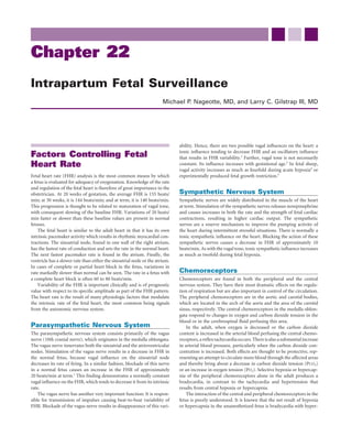
  • 11. CHAPTER 22 Intrapartum Fetal Surveillance 407 FIGURE 22-1 Normal fetal heart rate pattern. The tracing exhibits normal rate (approximately 130 beats/ min), normal variability (amplitude range about 15 beats/min), and absence of periodic changes. This pattern represents a nonacidemic fetus without evidence of hypoxic stress. Uterine contractions are 2 to 3 minutes apart and about 60 to 70 mm Hg in intensity. FIGURE 22-2 Prolonged fetal bradycardia resulting from excessive oxytocin-induced hyperstimulation of the uterus after intravenous infusion of meperidine (Demerol) and promethazine (Phenergan) into the same tubing. The heart rate is returning to normal at the end of the tracing, after appropriate treatment (signified by the notes “Pit off,” “O2 6 L/min,” and “side”). Note that fetal heart rate variability was maintained throughout this asphyxial stress, signifying adequate central oxygenation. fetal infection (e.g., chorioamnionitis). In most of these instances, the Classification and Significance of fetus is not hypoxic but has an elevated baseline FHR. Baseline Variability It is not uncommon for the FHR baseline to rise in the second stage As described earlier, FHR variability may be absent, minimal, moder- of labor. Certain drugs also cause tachycardia, such as β-mimetic ate, or marked, based on the amplitude range. The moderate (normal) agents used for attempted tocolysis or illicit drugs such as metham- amplitude range is between 6 and 25 beats/min. If the FHR variability phetamine and cocaine. is normal, regardless of what other FHR patterns may be present, the Tachycardia should not be confused with the rarer finding of a fetus is not experiencing cerebral tissue acidemia. This is because the fetal cardiac tachyarrhythmia, in which the FHR is faster than 240 fetus is able to centralize the available oxygen and is thus physiologi- beats/min. These arrhythmias may be intermittent or persistent and cally compensated. If excessive hypoxic stress persists, however, this are the result of abnormalities of the intrinsic determinants of cardiac compensation may break down, and the fetus may have progressive rhythm. Such findings of supraventricular tachyarrhythmias need to hypoxia in cerebral and myocardial tissues. In such cases, the FHR be monitored closely and possibly treated with medical therapy or variability decreases and eventually is lost. delivery, because they may be associated with deterioration of the fetal There are several possible nonhypoxic causes of decreased or absent status. FHR variability:
  • 12. 408 CHAPTER 22 Intrapartum Fetal Surveillance FIGURE 22-3 No variability of fetal heart rate. The mother had severe preeclampsia and was receiving magnesium sulfate and narcotics. The normal scalp blood pH (7.28) ensures that the absence of variability is nonasphyxic in origin and that the fetus is not chronically asphyxiated and decompensating. The uterine activity channel has an inaccurate trace in the first half. FIGURE 22-4 Unremitting fetal bradycardia. This tracing does not signify asphyxia, because this fetus had complete heart block, with a ventricular rate of about 55 beats/min. Note the absence of fetal heart rate variability. There were serious cardiac structural defects, and the fetus died shortly after birth. 1. Absence of the cortex (anencephaly) Reflex late deceleration is sometimes seen when an acute insult (e.g., 2. Narcotized or drugged higher centers (e.g., by morphine, meperi- reduced uterine blood flow resulting from maternal hypotension) is dine, diazepam) (Fig. 22-3) superimposed on a previously normally oxygenated fetus in the pres- 3. Vagal blockade (e.g., by atropine or scopolamine) ence of contractions. These late decelerations are caused by a decrease 4. Defective cardiac conduction system (e.g., complete heart block) in uterine blood flow (with the uterine contraction) beyond the capac- (Fig. 22-4) ity of the fetus to extract sufficient oxygen. The relatively deoxygenated blood is carried from the fetal placenta through the umbilical vein to Periodic Changes in Fetal Heart Rate the heart and is distributed to the aorta, neck vessels, and head. Here, LATE DECELERATIONS the low PO2 is sensed by chemoreceptors, and neuronal activity results Late decelerations (Fig. 22-5) are of two varieties: reflex and in a vagal discharge, which causes the transient deceleration. The decel- nonreflex.4,71-73 eration is presumed to be “late” because of the circulation time from
  • 13. CHAPTER 22 Intrapartum Fetal Surveillance 409 FIGURE 22-5 Late decelerations. These were recorded via Doppler ultrasound in the antepartum period in a severely growth-restricted (1700-g) term infant born to a 32-year-old preeclamptic primipara. Delivery was by cesarean section because neither a direct fetal electrocardiogram nor a fetal blood sample could be obtained due to a firm closed posterior cervix. The infant subsequently did well. FIGURE 22-6 Reflex late decelerations. The fetal heart rate pattern was previously normal, but late decelerations appeared after severe maternal hypotension (70/30 mm Hg), which was a result of sympathetic blockade caused by a caudal anesthetic agent. the fetal placental site to the chemoreceptors and because the progres- death.74 The animals were initially observed with normal blood gases, sively decreasing PO2 must reach a certain threshold before vagal activ- normal FHR variability, presence of accelerations, and no persistent ity occurs. There may also be baroreceptor activity causing the vagal periodic changes. After a variable period of time, they first demon- discharge.71 Between contractions, oxygen delivery is adequate and strated late decelerations and retained accelerations. This period was there is no additional vagal activity, so the baseline FHR is normal. associated with a small decline in ascending aortic PO2, from 28 to These reflex late decelerations are accompanied by normal FHR 24 mm Hg, and a normal acid-base state. These late decelerations were variability and thus signify normal central nervous system integrity probably of the vagal reflex type caused by chemoreceptor activity. At (i.e., the fetus is physiologically “compensated” in the vital organs) an average of more than 3 days after the onset of these reflex decelera- (Fig. 22-6). tions, accelerations were lost in the presence of worsening hypoxia The second type of late deceleration results from the same initial (PO2, 19 mm Hg) and acidemia (pH, 7.22). Fetal death followed an mechanism, except that the deoxygenated bolus of blood from the average of 36 hours of persistent late decelerations, and these latter placenta is presumed to be insufficient to support myocardial action. decelerations without accelerations were presumed to be of the non- Thus, for the period of the contraction, there is direct myocardial reflex myocardial depression type. hypoxic depression (or failure) and vagal activity.71,73 These nonreflex When late decelerations are present, one should make efforts to late decelerations are seen without variability (Fig. 22-7), signifying optimize placental blood flow and maternal oxygenation and ensure fetal “decompensation” (i.e., inadequate fetal cerebral and myocardial that maternal blood pressure is normal. oxygenation). They are seen most commonly in states of decreased placental reserve (e.g., preeclampsia, intrauterine growth restriction) VARIABLE DECELERATIONS or after prolonged hypoxic stress (e.g., a long period of severe reflex Variable decelerations (Fig. 22-8), have the following late decelerations). characteristics: Further support for the two etiologic mechanisms of late decelera- tions comes from observations on chronically catheterized fetal 1. They are variable in duration, depth, and shape. monkeys in spontaneous labor during the course of intrauterine 2. They are usually abrupt in onset and cessation.
  • 14. 410 CHAPTER 22 Intrapartum Fetal Surveillance FIGURE 22-7 Nonreflex late decelerations with virtual absence of fetal heart rate (FHR) variability. The decelerations represent transient asphyxic myocardial failure as well as intermittent vagal decreases in heart rate. The lack of FHR variability also signifies decreased cerebral oxygenation. Note the acidemia in fetal scalp blood (7.07). The infant, a 3340-g girl with Apgar scores of 3 (1 minute) and 4 (5 minutes) was delivered soon after this tracing. Cesarean section was considered to be contraindicated because of a severe preeclamptic coagulopathy. FIGURE 22-8 Variable decelerations. Intrapartum recording using fetal scalp electrode and tocodynamometer. The spikes in the uterine activity channel represent maternal pushing efforts in the second stage of labor. The normal baseline variability between contractions signifies normal central oxygenation despite the intermittent hypoxic stress represented by the moderate variable decelerations. 3. They are not reassuring when there is either a slow return to base- significant period), further diagnostic steps or delivery may be indi- line, a rise in baseline, or an absence of variability in the baseline cated. Certain patterns are of such a severe character that immediate between decelerations. delivery is warranted if they cannot rapidly be relieved (Figs. 22-9 and 22-10). EFFECT OF IN UTERO TREATMENT Fetal oxygenation can be improved, acidemia relieved, and variant Other Patterns FHR patterns abolished by certain modes of treatment. The events that SINUSOIDAL PATTERN result in fetal stress (recognized by FHR patterns) are presented in The sinusoidal pattern is a regular, smooth, sine wave–like baseline, Table 22-10 together with the recommended treatment maneuvers and with a frequency of approximately 3 to 6 cycles per minute and an presumed mechanisms for improving fetal oxygenation. These should amplitude range of up to 30 beats/min. The regularity of the waves be the primary maneuvers carried out; if the hypoxic event is acute distinguishes this pattern from long-term variability complexes, which and the fetus was previously normoxic, there is an excellent chance that are crudely shaped and irregular. Another distinguishing feature is the the undesired FHR pattern will be abolished. absence of beat-to-beat or short-term variability (Fig. 22-11). If the FHR pattern cannot be improved (i.e., if the stress patterns The sinusoidal pattern was first described in a group of severely indicative of peripheral tissue or central tissue hypoxia persist for a affected Rh-alloimmunized fetuses but was subsequently noted in
  • 15. CHAPTER 22 Intrapartum Fetal Surveillance 411 TABLE 22-10 INTRAUTERINE TREATMENT FOR VARIANT FETAL HEART RATE (FHR) PATTERNS Causes Possible Resulting FHR Patterns Corrective Maneuver Mechanism Hypotension (e.g., supine Bradycardia, late decelerations Intravenous fluids, position change, Return of uterine blood flow hypotension, conduction ephedrine toward normal anesthesia) Excessive uterine activity Bradycardia, late decelerations Decrease in oxytocin, lateral position Same as above Transient umbilical cord Variable decelerations Change in maternal position (e.g., Presumably removes fetal part compression left or right lateral, Trendelenburg) from cord Amnioinfusion Relieves compression of cord Head compression Early or variable decelerations Push only with alternate Allows fetal recovery contractions Decreased uterine blood flow Late decelerations Change in maternal position (e.g., Enhancement of uterine blood associated with uterine left lateral or Trendelenburg) flow toward optimum contraction Tocolytic agents (e.g., terbutaline) Decrease in contractions or tone Prolonged asphyxia Decreasing FHR variability* Change in maternal position (e.g., Enhancement of uterine blood left lateral or Trendelenburg), flow to optimum, increase establishment of maternal in maternal-fetal oxygen hyperoxia gradient *During labor, this is virtually always preceded by a heart rate pattern signifying asphyxial stress (e.g., late decelerations, usually severe), severe variable decelerations, or a prolonged bradycardia. This is not necessarily so in the antepartum period, before the onset of uterine contractions. FIGURE 22-9 Sinister heart rate pattern in a 28-week fetus (gestational age determined after delivery) with baseline tachycardia, absence of heart rate variability, and severe periodic changes. The scalp blood pH was 7.0, and the fetus died shortly after this tracing was made. Cesarean section was not performed because the fetus was believed to be previable, although in fact it weighed 1100 g. There is much artifact in the uterine activity channel. FIGURE 22-10 Bradycardia resulting from cord prolapse. The infant was delivered by cesarean section and did well.
  • 16. 412 CHAPTER 22 Intrapartum Fetal Surveillance A B FIGURE 22-11 Sinusoidal pattern. A and B, Sinusoidal pattern in a term fetus with severe hemolysis caused by Rh disease. Cord hematocrit was 20%, and the infant, delivered by cesarean section, was subsequently normal. Recording by direct fetal electrode. association with fetuses that were anemic for other reasons and in or lower. Many severely anemic Rh-affected fetuses do not have a severely depressed infants. It was also described in cases of normal sinusoidal pattern but rather have a rounded, blunted pattern, and infants born without depression or acid-base abnormalities, but in accelerations are usually absent. these cases there is dispute about whether the patterns were truly If a sinusoidal pattern is seen in an Rh-sensitized patient and severe sinusoidal or whether, because of the moderately irregular pattern, hemolysis is confirmed (by peak systolic velocity measurement of flow they were variants of long-term variability. Such patterns, often called in the middle cerebral artery of the fetus, by cordocentesis, or by the pseudosinusoidal, may also be seen after administration of narcotics deviation in optical density at 450 nm determination by spectropho- to the mother. tometry of amniotic fluid), rapid intervention is needed. This step may It is believed that an essential characteristic of the sinusoidal pattern take the form of delivery or intrauterine transfusion, depending on is extreme regularity and smoothness. Murata and colleagues75 impli- gestational age and the fetal status (see Chapter 26). cated arginine vasopressin in the sinusoidal pattern. The presence of a Management of a sinusoidal pattern in the absence of alloimmu- sinusoidal pattern or variant of this in an Rh-sensitized patient usually nization is somewhat more difficult to recommend. If the pattern is suggests anemia with a fetal hematocrit value of less than 30%. The persistent, monotonously regular, and unaccompanied by short-term presence of hydrops in such a fetus suggests a fetal hematocrit of 15% variability and cannot be abolished by the maneuvers just described,
  • 17. CHAPTER 22 Intrapartum Fetal Surveillance 413 FIGURE 22-12 Saltatory pattern. Saltatory pattern showing excessive fetal heart rate variability of up to 60 beats/min in brief intervals, probably representing mild hypoxic stress. further workup and evaluation of the adequacy of fetal oxygenation (e.g., contraction stress test, fetal stimulation test, biophysical profile, fetal blood sampling) are indicated. Nonalloimmune sinusoidal pat- Efficacy, Risks, and terns have been associated with severe fetal acidemia and with fetal anemia resulting from fetal-maternal bleeding. The latter diagnosis is Recommendations for Usage supported by the identification of fetal red blood cells in maternal Electronic Monitoring blood, detected by the Kleihauer-Betke test. If the pattern is irregularly sinusoidal or pseudosinusoidal, intermittently present, and not associ- versus Auscultation ated with intervening periodic decelerations, fetal compromise is Because there are no prospective randomized clinical trials comparing unlikely, and immediate delivery is not warranted. EFM with no fetal heart monitoring during labor, most efforts to suggest its efficacy have relied on research reports comparing EFM SALTATORY PATTERN with intermittent auscultation. Efficacy is generally held to be an The saltatory pattern consists of rapid variations in FHR with a expected decrease in complications, which, for FHR monitoring, could frequency of 3 to 6 cycles per minute and an amplitude range greater include fetal death in labor or severe neonatal and pediatric morbidity than 25 beats/min (Fig. 22-12). It is qualitatively described as a marked (e.g., neonatal seizures, cerebral palsy). Ideally, the improved outcomes variability, and the variations have a strikingly bizarre appearance. The would be accompanied by appropriate interventions and appropriate saltatory pattern is seen during labor rather than in the antepartum noninterventions. period. The etiology is uncertain, but it may be similar to that of the In a meta-analysis of the nine published clinical studies comparing increased FHR variability seen in animal experiments with brief and EFM with intermittent auscultation of the FHR, several conclusions acute hypoxia in a previously normoxic fetus. Therefore, efforts should were reached.77 (It should be noted that in several of these trials, be made to optimize placental blood flow and fetal oxygenation if such patients with conditions considered to be high risk were not random- a pattern appears during labor. ized for study inclusion.) The use of EFM was associated with signifi- cant increases in the rate of cesarean delivery for fetal intolerance to labor, the overall cesarean delivery rate, and the use of instrumenta- Congenital Anomalies tion for vaginal delivery (both vacuum and forceps). However, Except as described for the dysrhythmias, most fetuses with congenital there was no reduced overall perinatal mortality in these patients. anomalies have essentially normal FHR patterns and respond to As a result of such reports, either option of monitoring the fetus hypoxia in a manner similar to the normal fetus. There are several during labor is acceptable for patients not considered to be at high exceptions, including complete heart block and anencephaly. Aneu- risk.67 However, it should be noted that the optimal frequency for ploid fetuses and fetuses with aplastic lungs, meningomyelocele, and/ intermittent auscultation in such low-risk patients has not been estab- or hydrocephalus may give no FHR warning of such underlying defects, lished. At a minimum, the FHR should be assessed at least every 30 because they are not necessarily experiencing hypoxia or acidosis. minutes in the first stage of labor and every 15 minutes in the second However, even though there was no pathognomonic pattern in such stage.67 Another method is to auscultate and record FHR every fetuses, the rate of cesarean section for fetal intolerance to labor was 15 minutes in the active first stage of labor and every 5 minutes reported to be significantly increased, presumably because of nonreas- in the second stage, without limiting such a modality to low-risk suring patterns during or preceding labor.76 patients.78