SlideShare uma empresa Scribd logo
1 de 7
Baixar para ler offline
C L I N I C A L A N D LA B O R A T O R Y I N V E S T I G A T I O N S                DOI 10.1111/j.1365-2133.2005.06562.x

Human papillomavirus-associated plantar epidermoid cyst
related to epidermoid metaplasia of the eccrine duct
epithelium: a combined histological,
immunohistochemical, DNA–DNA in situ hybridization and
three-dimensional reconstruction analysis
K. Egawa, N. Egawa* and Y. Honda 
Departments of Dermatology and  Surgical Pathology, Kumamoto University School of Medicine, 1-1-1 Honjo, Kumamoto 860-0811, Japan
*Division of Cancer Cell Research, Institute of Medical Science, University of Tokyo, Minato-ku, Tokyo 108-8639, Japan

                                                Summary

Correspondence                                  Background We recently proposed that certain palmoplantar epidermoid cysts may
Kiyofumi Egawa.                                 be related to eccrine ducts and that human papillomavirus (HPV) 60 may play a
E-mail: kuroibo@kaiju.medic.kumamoto-u.ac.jp    role in their pathomechanism. However, the origin of palmoplantar epidermoid
                                                cysts is still controversial.
Accepted for publication
9 October 2004
                                                Objectives To examine the contribution of eccrine ducts and HPV 60 in the devel-
                                                opment of epidermoid cysts.
Key words:                                      Methods Five epidermoid cysts and four ridged warts that had developed on the
eccrine duct, epidermoid cyst, human            soles of a patient were studied histologically, immunohistochemically and by
papillomavirus 60, inclusion cyst, metaplasia   DNA–DNA in situ hybridization. Using serial sections obtained from its entire
                                                body, a three-dimensional reconstruction (3DR) analysis was performed on the
Conflicts of interest:
None declared.                                  smallest cyst to analyse the relationship between the epidermoid cyst, eccrine
                                                duct and the overlying epidermis.
                                                Results Histological and DNA–DNA in situ hybridization analyses demonstrated
                                                both homogeneous intracytoplasmic inclusion bodies pathognomonic for HPV
                                                60 infection and HPV 60 DNA sequences not only in all of the epidermoid cysts
                                                and ridged warts but also in the acrosyringeal portion of an eccrine duct, with
                                                the dermal portion of which the smallest cyst had been revealed to connect by
                                                3DR analysis. However, immunohistochemical analyses using antibodies against
                                                human carcinoembryonic antigen (CEA), involucrin and several cytokeratins
                                                (CKs) revealed that the immunoreactivity of the cyst was not identical to
                                                that of the eccrine dermal duct but was identical to that of suprabasal layers of
                                                the epidermis.
                                                Conclusions It was clearly demonstrated that an HPV 60-associated epidermoid cyst
                                                with immunoreactivities for CEA, involucrin and CKs which were identical to
                                                those of the epidermis connected with the eccrine dermal duct, supporting the
                                                idea that certain palmoplantar epidermoid cysts may develop following the epi-
                                                dermoid metaplasia of eccrine ducts with HPV 60 infection.


Epidermoid cysts of palmoplantar location (palmoplantar epi-                epidermoid cysts, with or without infection by human papil-
dermoid cysts) have been described as being caused by                       lomavirus (HPV) 60,5 and therefore proposed the alternative
implantation of epidermal fragments into the dermis as a                    pathogenic hypothesis that certain palmoplantar epidermoid
result of a penetrating or blunt injury.1 However, this assump-             cysts may develop from eccrine ducts and that HPV 60 may
tion has been questioned, and the necessity of a search for a               play a role in this development. However, on the basis of the
more common aetiology has been stressed.2                                   previous results,3–5 it is still likely that such epidermoid cysts
   We previously observed3,4 that eccrine sweat ducts were                  can develop as a result of the implantation of epidermal frag-
frequently found in and ⁄or connecting with palmoplantar                    ments that contain eccrine ducts with or without HPV 60

Ó 2005 British Association of Dermatologists • British Journal of Dermatology 2005 152, pp961–967                                         961
962 HPV-associated eccrine epidermoid cyst, K. Egawa et al.


infection from injuries as they do occasionally develop after
injuries3,6 and are mainly located beneath the weight-bearing
areas of the foot, i.e. the balls and heels.7
   Indeed, Abe et al.8 and Ohnishi and Watanabe9 questioned
the hypothesis of an ‘eccrine duct origin of the epidermoid
cyst’ on the basis of the immunoreactivities of several cyto-
keratins (CKs) which were not identical to those of sweat
glands and ducts but were identical to those of suprabasal
layers of the epidermis in plantar epidermoid cysts with HPV
infection.
   However, Egawa et al.10 proposed the idea that such a CK
profile in HPV-associated palmoplantar epidermoid cysts might
be a reflection of the epidermoid metaplasia of the eccrine
duct epithelium in the process of cyst formation in the eccrine
ducts, as it was revealed that the CK profile of cholesteatomas,
which are histologically defined as epidermoid cysts develop-
ing in the middle ear,11 cannot be used as a variable to decide
whether the origin of epidermoid cysts is epidermal or meta-
plastic.12 Thus, the origin of palmoplantar epidermoid cysts is
still controversial.
   Three-dimensional reconstruction (3DR) analysis from serial
sections is now a powerful tool for increasing our comprehen-
sion of the relationships among structural components, for the
indication of the connections between components, and for
revealing the distribution of components.13–15 In the present
study, using serial sections obtained from the entire body of
an epidermoid cyst, we attempted 3DR analysis to demon-                Fig 1. Three subcutaneous nodules (solid arrows) and two ridged
strate the fine structure of an HPV 60-associated epidermoid            warts (open arrows) on the left sole.
cyst of eccrine duct origin, in combination with histological,
immunohistochemical and DNA–DNA in situ hybridization
analyses.                                                              excised specimens were fixed in 10% formalin, and proc-
                                                                       essed with an automatic processor.

Materials and methods
                                                                       Histopathology
Patient
                                                                       Four-micrometre thick sections were obtained using a micro-
A 21-year-old Japanese woman presented to Kumamoto Uni-                tome from all of the biopsy specimens, stained with haema-
versity Hospital in April 1995, with several subcutaneous nod-         toxylin and eosin (H&E), and examined microscopically
ules and warts on the soles of her feet. She first noticed              (Figs 2 and 3). From the smallest cyst, gross serial sections
several miliary-sized asymptomatic nodules on her soles                were obtained from its entire body, and all sections were
3 months before presentation and 1 year after having had sev-          numbered in the order of sectioning, from one side to the
eral plantar warts treated with cryotherapy, although she could        other. Fifty serial sections were obtained from each of the
not say whether or not the nodules developed in the same               other cysts. Every alternate section was stained with H&E for
positions as the treated plantar warts. The nodules had gradu-         histological analysis (Fig. 3), whereas the remaining sections
ally enlarged and had come to cause tenderness during the              were used for further examinations such as immunohisto-
previous 3 months.                                                     chemical and DNA–DNA in situ hybridization analyses.
   At the time of her first visit, examination showed three
subcutaneous nodules of 5–8 mm diameter and two small
                                                                       Processing of histological and immunohistochemical data
flat warty lesions with clinical features of ridged warts16,17
                                                                       for computer analysis
on the left sole (Fig. 1) and one nodule of 7 mm diameter
on the right sole. Her health was otherwise good. After                Using photomicrographs of histopathological and immunohis-
informed consent was obtained, all the lesions were                    tochemically stained sections (Fig. 4a), we made montages by
removed successfully by scalpel under local anaesthesia.               tracing the outlines of the histological ductal structures expres-
However, about 6 months later, a further subcutaneous                  sing carcinoembryonic antigen (CEA) in serial sections in red,
nodule of similar size appeared on the left sole, with two             and those of the cyst wall and the overlying epidermis in blue
ridged warts. They were also removed by scalpel. The                   (Fig. 4b).

                                              Ó 2005 British Association of Dermatologists • British Journal of Dermatology 2005 152, pp961–967
HPV-associated eccrine epidermoid cyst, K. Egawa et al. 963


                                                                            (Fig. 4d). The three-dimensional images were rotated along
                                                                            various axes to analyse the relationship between the cyst,
                                                                            eccrine ducts and the overlying epidermis.


                                                                            Immunohistochemistry
                                                                            An immunohistochemical procedure for human CEA,3,4,15,18
                                                                            CKs8,9,19 and involucrin20 was performed on paraffin-embed-
                                                                            ded sections by the avidin–biotin–peroxidase complex (ABC)
                                                                            method,21 utilizing a Vectastain ABC kit (Vector Laboratories,
                                                                            Burlingame, CA, U.S.A.). A polyclonal antibody to human CEA
                                                                            (Dako, Glostrup, Denmark) and monoclonal antibodies to var-
                                                                            ious CKs (OV-TL 12 ⁄30, RCK108 and AE1 ⁄AE3, Dako; LL002,
                                                                            Biomeda, Foster City, CA, U.S.A.; and CAM5.2, Becton Dickin-
                                                                            son, San Jose, CA, U.S.A.) and involucrin (Novocastra, New-
Fig 2. Histological features of a ridged wart. Vacuolar structures are      castle-upon-Tyne, U.K.) were used as primary antibodies
seen in the thickened horny layer and homogeneous intracytoplasmic          (Table 1). In the case of CKs, the procedure was performed
inclusion bodies are seen in the granular and upper spinous cell layers     with or without proteinase K predigestion or autoclave heating
of the acanthotic epidermis. A tiny keratinous cyst is seen just beneath    for antigen retrieval as described previously.22 Negative con-
the acanthotic epidermis (arrow).                                           trols for immunostaining were performed by substituting the
                                                                            primary antibodies with phosphate-buffered saline or nonim-
                                                                            mune mouse or rabbit IgG.
Three-dimensional reconstruction analysis
An image scanner (IX-4025; Canon, Tokyo, Japan) and a per-
                                                                            DNA–DNA in situ hybridization
sonal computer (Apple Macintosh 8500) were used in the
3DR imaging. Montages of the serial sections obtained from                  HPV 60 DNA sequences were detected in formalin-fixed, paraf-
the entire body of the epidermoid cyst were displayed on a                  fin-embedded tissue sections using the DNA–DNA in situ hybrid-
television monitor (Multiscan 17s; Sony, Tokyo, Japan). The                 ization method as described previously.23,24 Hybridization was
outlines of the cysts and the eccrine ducts were digitized and              performed at 37 °C for 16 h using digoxigenin-labelled HPV
stored on the computer to make a wire-frame of these in vivo                60 complete genome DNA which we had previously cloned
structures (Fig. 4c). Based on the wire-frame, three-dimen-                 from a plantar epidermoid cyst.3 The hybridization mixture
sional images of the structures were reconstructed using an                 consisted of 10% dextran sulphate, 2 · saline sodium citrate
image analysis program (Specular Infini-D; Specular, Amherst,                (0Æ3% sodium chloride, 0Æ03 mol L)1 sodium citrate, pH 7Æ0),
MA, U.S.A.) and were displayed on the television monitor                    400 lg mL)1 sheared herring sperm DNA (Sigma, St Louis,


Fig 3. Representative histological pictures
selected from the serial sections obtained
from the entire body of an epidermoid
cyst. Section numbers appear at the lower
left corner of each picture. Varying
histological features coming from a
hypertrophied eccrine dermal duct (no.
200, inset) connecting the apical portion
of the cyst with an acrosyringium in the
overlying epidermis are seen in the serial
sections. The ecrine duct, appearing as a
ductal structure in the apical portion of
the cyst wall (nos 180–190), goes
upwards through the dermis to the
acrosyringium (nos 200–210) and
connects with it in the overlying
epidermis (no. 220). The acrosyringium is
extremely dilated in the horny layer (no.
230).


Ó 2005 British Association of Dermatologists • British Journal of Dermatology 2005 152, pp961–967
964 HPV-associated eccrine epidermoid cyst, K. Egawa et al.


                                                                                                      Fig 4. Computer-based, three-dimensional
                                                                                                      reconstruction (3DR) analysis of a human
                                                                                                      papillomavirus 60-associated plantar
                                                                                                      epidermoid cyst. (a) Histological section
                                                                                                      (the same as Figure 3, no.190). (b)
                                                                                                      Tracing of the outline of the histological
                                                                                                      ductal structures expressing
                                                                                                      carcinoembryonic antigen (red) and of the
                                                                                                      cyst wall and overlying epidermis (blue).
                                                                                                      (c) A wire-frame of the epidermoid cyst
                                                                                                      generated by computer using a montage
                                                                                                      of tracings obtained from all the serial
                                                                                                      sections. (d) 3DR analysis visualizing the
                                                                                                      fine structure of the epidermoid cyst
                                                                                                      connecting with the eccrine dermal duct.


Table 1 Immunohistochemical staining in a human papillomavirus (HPV) 60-associated plantar epidermoid cyst


                                                        Normal eccrine gland

                                                        Secretory         Eccrine
  Antibody             Specificity                       portion           dermal duct          Epidermis          Cyst wall          HD
  CEA                                                   +++               +++                  –                  –                  ++ (IML)
  AE1 ⁄ AE3            CKs 10, 14–16, 19 ⁄ 1–8          +++               +++                  +++                +++                +++
  LL002                CK14                             –                 ++                   ++ (LL)            ++ (OL)            + (OL)
  OV-TL 12 ⁄ 30        CK7                              +++               –                    –                  –                  –
  RCK108               CK19                             +                 –                    –                  –                  –
  CAM5.2               CKs 8, 18                        +++               –                    –                  –                  –
  Involucrin                                            –                 –                    +++ (UL)           +++ (IL)           ++ (IL)

  HD, hypertrophied eccrine dermal duct with which the HPV 60-associated epidermoid cyst connected; CEA, carcinoembryonic antigen;
  CK, cytokeratin; IML, innermost layers; LL, lower layers; OL, outer layers; UL, upper layers; IL, inner layers; –, negative; +, weak; ++,
  moderate; +++, strong.


MO, U.S.A.), 50% formamide and 2 lg mL)1 of probe DNA.                    (Figs 3 and 5c). The characteristic histological feature com-
The digoxigenin-labelled probe was detected by the streptavi-             monly seen in all the ridged warts and the epidermoid cysts
din-biotinylated polyalkaline phosphatase detection system                was the presence of eosinophilic homogeneous intracyto-
(DNA detection system; BRL, Gaithersburg, MD, U.S.A.). The                plasmic inclusion bodies pathognomonic for HPV 60
phaeochromes used were nitroblue tetrazolium and 5-bromo-                 infection3–7,16,17,24–27 in the granular and upper spinous cell
4-chloro-3-indolylphosphate (Fig. 5).                                     layers of the acanthotic epidermis of the ridged warts or in
                                                                          the inner cell layers of the cyst wall (Figs 2 and 5c). Vacuolar
                                                                          structures, also pathognomonic for HPV 60 infection,4,7 were
Results
                                                                          also seen both in the horny layer of the ridged warts and in
                                                                          keratinous material within the cyst cavity (Figs 2 and 5c).
Histology
                                                                             In the serial sections obtained from the entire body of the
In the ridged warts, the epidermis was acanthotic with hyper-             smallest cyst (Fig. 3), ductal structures coming from an eccrine
keratosis and partial hypergranulosis. In one lesion, a tiny              dermal duct were seen between the apical portion of the cyst
keratinous cyst was seen just beneath the acanthotic epidermis            and an acrosyringium in the overlying epidermis in the dermis
(Fig. 2). Epidermoid cysts were lined by stratified squamous               that connects the two histological components (Fig. 3, nos.
epithelium, showing keratinization of the epidermoid type                 190–220). The eccrine dermal duct was hypertrophied with

                                                 Ó 2005 British Association of Dermatologists • British Journal of Dermatology 2005 152, pp961–967
HPV-associated eccrine epidermoid cyst, K. Egawa et al. 965


                                                                            duct but not the secretory portion. In contrast, OV-TL 12 ⁄30
                                                                            (against CK7), RCK108 (against CK19) and CAM5.2 (against
                                                                            CKs 8 and 18) stained only the secretory portion. A monoclo-
                                                                            nal antibody against involucrin did not stain any portions of
                                                                            the normal eccrine sweat gland.


                                                                            Epidermis of the sole

                                                                            CEA was expressed only in the acrosyringeal epithelium in the
                                                                            epidermis. LL002 stained the lower cell layers; AE1 ⁄AE3
                                                                            stained whole cell layers; OV-TL 12 ⁄30, RCK108 and CAM5.2
                                                                            did not stain any cell layers; involucrin was expressed in the
                                                                            upper cell layers of the epidermis.


                                                                            Plantar epidermoid cyst

                                                                            CEA was not expressed in the cyst wall but was expressed in
                                                                            the acrosyringeal structures in the cyst cavity. LL002 stained
                                                                            the outer cell layers; AE1 ⁄AE3 stained whole cell layers; OV-
                                                                            TL 12 ⁄30, RCK108 and CAM5.2 did not stain any cell layers;
                                                                            involucrin was expressed in the inner cell layers of the cyst
                                                                            wall.


                                                                            Hypertrophied eccrine dermal duct with stratified
                                                                            squamous epithelium with which an epidermoid cyst
                                                                            connected
Fig 5. Histological localization of human papillomavirus (HPV)
60 DNA sequences. A high-power view of the same section                     The innermost cell layers of the stratified epithelium of the
(a,c; haematoxylin and eosin) and a serial section (b,d; DNA–DNA            hypertrophied eccrine dermal duct expressed CEA. Expression
in situ hybridization) of Figure 3, no. 230. Homogeneous                    of involucrin was also noted in the inner cell layers; LL002
intracytoplasmic inclusion bodies (a,c) and HPV 60 DNA sequences            stained the outer cell layers; AE1 ⁄AE3 stained whole cell
(b,d) are seen in the acrosyringeal epithelium (a,b) as well as in the
                                                                            layers; OV-TL 12 ⁄30, RCK108 and CAM5.2 did not stain any
cyst wall (c,d).
                                                                            cell layers of the duct.
                                                                               The results are summarized in Table 1.
stratified squamous epithelium (Fig. 3, no. 200, inset); the
acrosyringium was extremely dilated in the horny layer
                                                                            DNA–DNA in situ hybridization
(Fig. 3, no. 230; Fig. 5a); and the homogeneous intracytoplas-
mic inclusion bodies were seen in the acrosyringeal epithelium              HPV 60 DNA sequences were detected in all the epidermoid
(Fig. 5a) as well as in the epidermoid cyst (Fig. 5b).                      cysts and ridged warts. In the smallest epidermoid cyst ana-
   Ductal structures suggestive of eccrine ducts were observed              lysed with 3DR, the DNA sequences were identified both in
in three of the other four epidermoid cysts, using 50 serial                the acrosyringeal portion of the eccrine duct (Fig. 5b) and in
sections obtained from each cyst.                                           the cyst connecting with its dermal portion (Fig. 5d).


Three-dimensional reconstruction analysis                                   Discussion
The analysis clearly demonstrated that the smallest epidermoid              Implantation of epidermal fragments into the dermis has been
cyst connected with the eccrine dermal duct (Fig. 4d).                      thought to be the main cause of palmoplantar epidermoid
                                                                            cysts.1 Although we have proposed an alternative pathogenic
                                                                            hypothesis that certain palmoplantar epidermoid cysts may
Immunohistochemical observations
                                                                            develop from eccrine ducts and that HPV (especially HPV 60)
                                                                            may play a role in this development,3,4,10,17 there has been a
Normal eccrine sweat gland
                                                                            lack of direct evidence to support this hypothesis.
A polyclonal antibody against CEA and a monoclonal antibody                    HPV 60 was initially identified25,26 in and isolated5 from
AE1 ⁄AE3 (against CKs 10, 14–16, 19 ⁄1–8) stained all the                   plantar epidermoid cysts with homogeneous intracytoplasmic
secretory, dermal duct and acrosyringeal portions of the ec-                inclusion bodies, and was also subsequently found in flat
crine sweat gland. LL002 (against CK14) stained the dermal                  lesions retaining the appearance of dermal ridges on their

Ó 2005 British Association of Dermatologists • British Journal of Dermatology 2005 152, pp961–967
966 HPV-associated eccrine epidermoid cyst, K. Egawa et al.


surface (ridged warts).16,17 Thus, the clinical morphology of          appearance of CKs characteristic of stratified and cornifying
HPV 60-induced lesions also remains unclear. In the present            epithelia, indicating a true change in the differentiation of the
study, using 3DR analysis, it was clearly demonstrated that an         middle ear epithelium.12 Interestingly, HPV has also been
HPV 60-associated plantar epidermoid cyst connected with the           identified in cholesteatoma.31
dermal portion of an eccrine duct. HPV 60 DNA sequences                   Although the epidermoid cyst analysed with 3DR in this
were identified not only in the epidermoid cyst but also in             study connected with an eccrine dermal duct, the immunore-
the acrosyringeal portion of the eccrine duct by DNA–DNA               activities for CEA, involucrin and several CKs of the cyst wall
in situ hybridization. All the other plantar epidermoid cysts and      were not identical to those of normal eccrine dermal ducts
ridged warts that developed simultaneously on the same                 but were identical to those of the suprabasal layers of the
patient also harboured HPV 60 DNA sequences, suggesting a              epidermis, as observed by Abe et al.,8 Ohnishi and Watanabe9
close association between the lesions and HPV 60 infection. A          and Yokogawa et al.32 This suggests that the immunoreactivi-
tiny keratinous cyst, which might be a precursor of an HPV             ties of the palmoplanatar epidermoid cysts may also be an
60-associated plantar epidermoid cyst, was also seen just              indication of the metaplastic origin of the cysts from eccrine
beneath the acanthotic epidermis of a ridged wart.                     ducts. In the present case, the eccrine dermal duct with
   In a previous report, two plantar epidermoid cysts were             which the epidermoid cyst connected was hypertrophied
attached to the epidermis and open to the surface in a patient         with stratified squamous epithelium and expressed both CEA
with multiple plantar epidermoid cysts harbouring both HPV             and involucrin, suggesting a transitional form of the squa-
60 DNA sequences and eccrine ducts,3 suggesting that the               mous metaplasia of the eccrine duct epithelium.
cysts developed from the acrosyringeal or uppermost portions              However, as Epstein and Kligman have suggested that kera-
of eccrine ducts. As HPV 60 was frequently identified in                tinizing cysts may develop from any part of the epithelial sys-
palmoplantar epidermoid cysts concomitantly with eccrine               tem,33 further studies are still required to determine whether
ducts,3,4 we assumed that HPV 60 had a high affinity for                the cases in which neither HPV infection nor an association
eccrine duct epithelia and played a role in cyst formation.17          with eccrine ducts has been found in ordinary histological sec-
Supporting this idea, recent studies28,29 have suggested that          tions reflect a false-negative result or are an indication of epi-
papillomaviruses, including HPVs, may target epidermal stem            dermal inclusion cysts resulting from the implantation of
cells, which may exist in association with eccrine ducts in the        epidermal fragments.
human palmoplantar skin.29
   In the context of the previous findings and the results of
                                                                       Acknowledgments
the present case, we speculate that HPV 60 initially infects the
upper parts of the eccrine duct, such as the acrosyringeal epi-        We thank Ms Chiemi Shiotsu and Ms Yoshiko Sonoda for tech-
thelium, where ridged warts develop, and then migrates into            nical assistance and Gyohei Egawa, Haruki Egawa and Motoe
various parts of the dermal portions of the eccrine duct, where        Egawa for help in drawing montages of histological pictures.
the virus-associated epidermoid cysts develop.
   It has been revealed that HPV 60-associated palmoplantar
                                                                       References
epidermoid cysts occasionally develop in association with pre-
ceding blunt or penetrating injuries. The cysts are mainly              1 MacKie RM. Epidermal skin tumours. In: Textbook of Dermatology
located beneath the weight-bearing areas of the foot, i.e. the            (Champion RH, Burton JL, Ebling FJG, eds), 5th edn. Oxford:
                                                                          Blackwell Science, 1992; 1459–504.
balls and heels;7 an HPV 60-associated plantar epidermoid cyst
                                                                        2 Pinkus H. Epidermoid cysts or epidermal inclusion cysts? Arch Der-
developed just beneath a plantar wart when the wart was
                                                                          matol 1975; 111:130.
being treated with cryotherapy;6 multiple epidermoid cysts              3 Egawa K, Honda Y, Inaba Y et al. Multiple plantar epidermoid cysts
harbouring both HPV 60 DNA sequences and eccrine ducts                    harboring carcinoembryonic antigen and human papillomavirus
developed on the sole of a football player.3                              DNA sequences. J Am Acad Dermatol 1994; 30:494–6.
   In the present case, the cryotherapy performed on the pre-           4 Egawa K, Honda Y, Inaba Y et al. Detection of human papillomavi-
ceding plantar warts seemed to have a triggering role in the              ruses and eccrine ducts in palmoplantar epidermoid cysts. Br J Der-
                                                                          matol 1995; 132:533–42.
development of the multiple HPV 60-associated epidermoid
                                                                        5 Matsukura T, Iwasaki T, Kawashima M. Molecular cloning of a novel
cysts. Thus, traumatic injuries and mechanical pressure, which
                                                                          human papillomavirus (type 60) from a plantar epidermoid cyst
have long been thought to have a causative role in the implan-            with characteristic pathological changes. Virology 1992; 190:561–4.
tation of epidermal fragments following cyst formation in the           6 Inaba Y, Egawa K, Muto K, Arao T. Detection of papillomavirus
dermis,1 may have a triggering role in the progression of the             common antigens in plantar epidermoid cysts. Jpn J Dermatol 1990;
HPV 60-infected eccrine duct epithelium to the formation of               100:199–203.
the cyst.                                                               7 Egawa K, Inaba Y, Ono T, Arao T. ‘Cystic papilloma’ in humans?
                                                                          Demonstration of human papillomavirus in plantar epidermoid
   Squamous metaplasia is assumed to be a possible source of
                                                                          cysts. Arch Dermatol 1990; 126:1559–603.
cholesteatoma,12,30 which is histologically defined as an epi-
                                                                        8 Abe H, Ohnishi T, Watanabe S. Does plantar epidermoid cyst with
dermoid cyst developing in the middle ear.11 The squamous                 human papillomavirus infection originate from the eccrine dermal
cell metaplasia was immunohistochemically characterized by a              duct? Br J Dermatol 1999; 141:161–2.
loss of simple epithelial cell-related CKs accompanied by the

                                              Ó 2005 British Association of Dermatologists • British Journal of Dermatology 2005 152, pp961–967
HPV-associated eccrine epidermoid cyst, K. Egawa et al. 967

 9 Ohnishi T, Watanabe S. Immunohistochemical observation of cyto-                 between ABC and unlabeled antibody (PAP) procedures. J Histochem
   keratins in keratinous cysts including plantar epidermoid cyst.                 Cytochem 1981; 29:577–83.
   J Cutan Pathol 1999; 26:424–9.                                             22   Misumi S, Iyama K, Honda Y et al. Differential expression of base-
10 Egawa K, Kitasato H, Ono T. A palmar epidermoid cyst, showing                   ment membrane type-IV collagen a1, a2, a5 and a6 chains among
   histological features suggestive of eccrine duct origin, developing             the histological subtypes of adenoid cystic carcinoma. Virchows Arch
   after a bee-sting. Br J Dermatol 2000; 143:469–70.                              2004; 445:54–62.
11 Rosai J. Ear. In: Ackerman’s Surgical Pathology (Rosai L, ed.), 8th edn.   23   Egawa K, Sibasaki Y, de Villiers E-M. Double infection with human
   St Louis: Mosby, 1996; 2509–17.                                                 papillomavirus 1 and human papillomavirus 63 in a single cell of a
12 Kuijpers W, Vennix PP, Peters TA, Ramaekers FC. Squamous                        lesion displaying only an HPV 63-induced cytopathogenic effect.
   metaplasia of the middle ear epithelium. Acta Otolaryngol 1996;                 Lab Invest 1993; 69:583–8.
   116:293–8.                                                                 24   Kawase M, Honda M, Niimura M. Detection of human papilloma-
13 Braverman MS, Braverman IM. Three-dimensional reconstructions                   virus type 60 in plantar epidermoid cysts and verruca plantaris by
   of objects from serial sections using a microcomputer graphics sys-             the in situ hybridization method using digoxigenin labeled probe.
   tem. J Invest Dermatol 1986; 86:290–4.                                          J Dermatol 1994; 21:709–15.
14 Salisbury JR, Whimster WF. Progress in computer-generated three-           25   Egawa K, Ono T, Arao T. Epidermoid cyst of the sole and human
   dimensional reconstruction. J Pathol 1993; 170:223–7.                           papillomavirus. Jpn J Dermatol 1987; 97:493–5.
15 Honda Y, Egawa K, Baba Y, Ono T. Sweat duct milia—immuno-                  26   Kimura S, Sato N, Shigeeki H. A report of two cases of plantar epi-
   histological analysis of structure and three-dimensional reconstruc-            dermoid cyst which have papillomavirus antigen. Hifu-Rinsho 1987;
   tion. Arch Dermatol Res 1996; 288:133–9.                                        29:487–91.
16 Honda A, Iwasaki T, Sata T et al. Human papillomavirus type                27   Egawa K. New types of human papillomaviruses and intracytoplas-
   60-associated plantar wart: ridged wart. Arch Dermatol 1994;                    mic inclusion bodies, a classification of inclusion warts according
   130:1413–17.                                                                    to clinical features, histology and associated HPV types. Br J Dermatol
17 Egawa K, Kasai S, Hattori N et al. A case of a human papillomavirus             1994; 130:158–66.
   60-induced wart with clinical appearance of both pigmented and             28   Schmit A, Rochat A, Zeltner R et al. The primary target cells of the
   ridged warts. Dermatology 1998; 197:268–70.                                     high-risk cottontail rabbit papillomavirus colocalize with hair fol-
18 Pennys NS, Nadji M, McKinney EC. Carcinoembryonic antigen pre-                  licle stem cells. J Virol 1996; 70:1912–22.
   sent in human eccrine sweat. J Am Acad Dermatol 1981; 29:577–83.           29   Egawa K. Do human papillomaviruses target epidermal stem cells?
19 Watanabe S, Ichikawa E, Takanashi S, Takahashi H. Immunohisto-                  Dermatology 2003; 207:251–4.
   chemical localization of cytokeratins in normal eccrine glands, with       30   Sade J, Babiacki A, Pinkus G. The metaplastic and congenital origin
   monoclonal antibodies in routinely processed, formalin-fixed, par-               of cholesteatoma. Acta Otolaryngol 1983; 96:119–29.
   affin-embedded sections. J Am Acad Dermatol 1993; 28:203–12.                31   Bergmann K, Hoppe F, Yukai H et al. Human papillomavirus DNA
20 Hudson DL, Weiland KL, Dooley TP et al. Characterization of                     in cholesteatomas. Int J Cancer 1994; 59:463–6.
   eight monoclonal antibodies to involucrin. Hybridoma 1992; 11:             32   Yokogawa M, Egawa K, Dabenaka K et al. Multiple palmar epider-
   367–79.                                                                         moid cysts. Dermatology 2002; 205:398–400.
21 Hsu H-M, Raine L, Franer H. The use of avidin–biotin peroxidase            33   Epstein WL, Kligman AM. Epithelial cysts in buried human skin.
   complex (ABC) in immunoperoxidase technique: a comparison                       Arch Dermatol 1957; 76:437–45.


Ó 2005 British Association of Dermatologists • British Journal of Dermatology 2005 152, pp961–967

Mais conteúdo relacionado

Semelhante a Cisto metap ecrina hpv

Carcinoma neuroendocrino del setto un rarissimo tumore nasale
Carcinoma neuroendocrino del setto un rarissimo tumore nasaleCarcinoma neuroendocrino del setto un rarissimo tumore nasale
Carcinoma neuroendocrino del setto un rarissimo tumore nasaleMerqurio
 
Carcinoma neuroendocrino del setto un rarissimo tumore nasale
Carcinoma neuroendocrino del setto un rarissimo tumore nasaleCarcinoma neuroendocrino del setto un rarissimo tumore nasale
Carcinoma neuroendocrino del setto un rarissimo tumore nasaleMerqurio
 
Immune privileged tissues
Immune privileged tissuesImmune privileged tissues
Immune privileged tissuesCellGenuity
 
Propionibacterium acnes bacteriophage capsid protein structure determined by ...
Propionibacterium acnes bacteriophage capsid protein structure determined by ...Propionibacterium acnes bacteriophage capsid protein structure determined by ...
Propionibacterium acnes bacteriophage capsid protein structure determined by ...Joshua Chiou
 
Epidemiology and molecular typing of klebsiella pneumonia with the extended s...
Epidemiology and molecular typing of klebsiella pneumonia with the extended s...Epidemiology and molecular typing of klebsiella pneumonia with the extended s...
Epidemiology and molecular typing of klebsiella pneumonia with the extended s...BioMedSciDirect Publications
 
Comparative Study Of Pterygium Excision With Conjunctival Autograft,Wet Amnio...
Comparative Study Of Pterygium Excision With Conjunctival Autograft,Wet Amnio...Comparative Study Of Pterygium Excision With Conjunctival Autograft,Wet Amnio...
Comparative Study Of Pterygium Excision With Conjunctival Autograft,Wet Amnio...Dr. Jagannath Boramani
 

Semelhante a Cisto metap ecrina hpv (15)

Carcinoma neuroendocrino del setto un rarissimo tumore nasale
Carcinoma neuroendocrino del setto un rarissimo tumore nasaleCarcinoma neuroendocrino del setto un rarissimo tumore nasale
Carcinoma neuroendocrino del setto un rarissimo tumore nasale
 
Carcinoma neuroendocrino del setto un rarissimo tumore nasale
Carcinoma neuroendocrino del setto un rarissimo tumore nasaleCarcinoma neuroendocrino del setto un rarissimo tumore nasale
Carcinoma neuroendocrino del setto un rarissimo tumore nasale
 
Immune privileged tissues
Immune privileged tissuesImmune privileged tissues
Immune privileged tissues
 
ACVP poster
ACVP posterACVP poster
ACVP poster
 
Propionibacterium acnes bacteriophage capsid protein structure determined by ...
Propionibacterium acnes bacteriophage capsid protein structure determined by ...Propionibacterium acnes bacteriophage capsid protein structure determined by ...
Propionibacterium acnes bacteriophage capsid protein structure determined by ...
 
JoU
JoUJoU
JoU
 
Syringoma
SyringomaSyringoma
Syringoma
 
Syringoma
SyringomaSyringoma
Syringoma
 
Syringoma
SyringomaSyringoma
Syringoma
 
Austin Andrology
Austin AndrologyAustin Andrology
Austin Andrology
 
Epidemiology and molecular typing of klebsiella pneumonia with the extended s...
Epidemiology and molecular typing of klebsiella pneumonia with the extended s...Epidemiology and molecular typing of klebsiella pneumonia with the extended s...
Epidemiology and molecular typing of klebsiella pneumonia with the extended s...
 
Cyto-diagnosis of Papillary Hidradenoma of Vulva: A Case Report from A Tertia...
Cyto-diagnosis of Papillary Hidradenoma of Vulva: A Case Report from A Tertia...Cyto-diagnosis of Papillary Hidradenoma of Vulva: A Case Report from A Tertia...
Cyto-diagnosis of Papillary Hidradenoma of Vulva: A Case Report from A Tertia...
 
-28th ISPSR POSTER
-28th ISPSR POSTER-28th ISPSR POSTER
-28th ISPSR POSTER
 
Comparative Study Of Pterygium Excision With Conjunctival Autograft,Wet Amnio...
Comparative Study Of Pterygium Excision With Conjunctival Autograft,Wet Amnio...Comparative Study Of Pterygium Excision With Conjunctival Autograft,Wet Amnio...
Comparative Study Of Pterygium Excision With Conjunctival Autograft,Wet Amnio...
 
Reza Khorramirouz
Reza KhorramirouzReza Khorramirouz
Reza Khorramirouz
 

Último

Pondicherry Call Girls Book Now 9630942363 Top Class Pondicherry Escort Servi...
Pondicherry Call Girls Book Now 9630942363 Top Class Pondicherry Escort Servi...Pondicherry Call Girls Book Now 9630942363 Top Class Pondicherry Escort Servi...
Pondicherry Call Girls Book Now 9630942363 Top Class Pondicherry Escort Servi...GENUINE ESCORT AGENCY
 
Russian Call Girls Lucknow Just Call 👉👉7877925207 Top Class Call Girl Service...
Russian Call Girls Lucknow Just Call 👉👉7877925207 Top Class Call Girl Service...Russian Call Girls Lucknow Just Call 👉👉7877925207 Top Class Call Girl Service...
Russian Call Girls Lucknow Just Call 👉👉7877925207 Top Class Call Girl Service...adilkhan87451
 
Coimbatore Call Girls in Thudiyalur : 7427069034 High Profile Model Escorts |...
Coimbatore Call Girls in Thudiyalur : 7427069034 High Profile Model Escorts |...Coimbatore Call Girls in Thudiyalur : 7427069034 High Profile Model Escorts |...
Coimbatore Call Girls in Thudiyalur : 7427069034 High Profile Model Escorts |...chennailover
 
Jogeshwari ! Call Girls Service Mumbai - 450+ Call Girl Cash Payment 90042684...
Jogeshwari ! Call Girls Service Mumbai - 450+ Call Girl Cash Payment 90042684...Jogeshwari ! Call Girls Service Mumbai - 450+ Call Girl Cash Payment 90042684...
Jogeshwari ! Call Girls Service Mumbai - 450+ Call Girl Cash Payment 90042684...Anamika Rawat
 
Call Girls Rishikesh Just Call 9667172968 Top Class Call Girl Service Available
Call Girls Rishikesh Just Call 9667172968 Top Class Call Girl Service AvailableCall Girls Rishikesh Just Call 9667172968 Top Class Call Girl Service Available
Call Girls Rishikesh Just Call 9667172968 Top Class Call Girl Service Availableperfect solution
 
Call Girls Kolkata Kalikapur 💯Call Us 🔝 8005736733 🔝 💃 Top Class Call Girl Se...
Call Girls Kolkata Kalikapur 💯Call Us 🔝 8005736733 🔝 💃 Top Class Call Girl Se...Call Girls Kolkata Kalikapur 💯Call Us 🔝 8005736733 🔝 💃 Top Class Call Girl Se...
Call Girls Kolkata Kalikapur 💯Call Us 🔝 8005736733 🔝 💃 Top Class Call Girl Se...Namrata Singh
 
Model Call Girls In Chennai WhatsApp Booking 7427069034 call girl service 24 ...
Model Call Girls In Chennai WhatsApp Booking 7427069034 call girl service 24 ...Model Call Girls In Chennai WhatsApp Booking 7427069034 call girl service 24 ...
Model Call Girls In Chennai WhatsApp Booking 7427069034 call girl service 24 ...hotbabesbook
 
Models Call Girls In Hyderabad 9630942363 Hyderabad Call Girl & Hyderabad Esc...
Models Call Girls In Hyderabad 9630942363 Hyderabad Call Girl & Hyderabad Esc...Models Call Girls In Hyderabad 9630942363 Hyderabad Call Girl & Hyderabad Esc...
Models Call Girls In Hyderabad 9630942363 Hyderabad Call Girl & Hyderabad Esc...GENUINE ESCORT AGENCY
 
Call Girls Service Jaipur {8445551418} ❤️VVIP BHAWNA Call Girl in Jaipur Raja...
Call Girls Service Jaipur {8445551418} ❤️VVIP BHAWNA Call Girl in Jaipur Raja...Call Girls Service Jaipur {8445551418} ❤️VVIP BHAWNA Call Girl in Jaipur Raja...
Call Girls Service Jaipur {8445551418} ❤️VVIP BHAWNA Call Girl in Jaipur Raja...parulsinha
 
Trichy Call Girls Book Now 9630942363 Top Class Trichy Escort Service Available
Trichy Call Girls Book Now 9630942363 Top Class Trichy Escort Service AvailableTrichy Call Girls Book Now 9630942363 Top Class Trichy Escort Service Available
Trichy Call Girls Book Now 9630942363 Top Class Trichy Escort Service AvailableGENUINE ESCORT AGENCY
 
Manyata Tech Park ( Call Girls ) Bangalore ✔ 6297143586 ✔ Hot Model With Sexy...
Manyata Tech Park ( Call Girls ) Bangalore ✔ 6297143586 ✔ Hot Model With Sexy...Manyata Tech Park ( Call Girls ) Bangalore ✔ 6297143586 ✔ Hot Model With Sexy...
Manyata Tech Park ( Call Girls ) Bangalore ✔ 6297143586 ✔ Hot Model With Sexy...vidya singh
 
Call Girls Service Jaipur {9521753030} ❤️VVIP RIDDHI Call Girl in Jaipur Raja...
Call Girls Service Jaipur {9521753030} ❤️VVIP RIDDHI Call Girl in Jaipur Raja...Call Girls Service Jaipur {9521753030} ❤️VVIP RIDDHI Call Girl in Jaipur Raja...
Call Girls Service Jaipur {9521753030} ❤️VVIP RIDDHI Call Girl in Jaipur Raja...Sheetaleventcompany
 
Call Girls Hyderabad Just Call 8250077686 Top Class Call Girl Service Available
Call Girls Hyderabad Just Call 8250077686 Top Class Call Girl Service AvailableCall Girls Hyderabad Just Call 8250077686 Top Class Call Girl Service Available
Call Girls Hyderabad Just Call 8250077686 Top Class Call Girl Service AvailableDipal Arora
 
Call Girls Hosur Just Call 9630942363 Top Class Call Girl Service Available
Call Girls Hosur Just Call 9630942363 Top Class Call Girl Service AvailableCall Girls Hosur Just Call 9630942363 Top Class Call Girl Service Available
Call Girls Hosur Just Call 9630942363 Top Class Call Girl Service AvailableGENUINE ESCORT AGENCY
 
Night 7k to 12k Navi Mumbai Call Girl Photo 👉 BOOK NOW 9833363713 👈 ♀️ night ...
Night 7k to 12k Navi Mumbai Call Girl Photo 👉 BOOK NOW 9833363713 👈 ♀️ night ...Night 7k to 12k Navi Mumbai Call Girl Photo 👉 BOOK NOW 9833363713 👈 ♀️ night ...
Night 7k to 12k Navi Mumbai Call Girl Photo 👉 BOOK NOW 9833363713 👈 ♀️ night ...aartirawatdelhi
 
The Most Attractive Hyderabad Call Girls Kothapet 𖠋 9332606886 𖠋 Will You Mis...
The Most Attractive Hyderabad Call Girls Kothapet 𖠋 9332606886 𖠋 Will You Mis...The Most Attractive Hyderabad Call Girls Kothapet 𖠋 9332606886 𖠋 Will You Mis...
The Most Attractive Hyderabad Call Girls Kothapet 𖠋 9332606886 𖠋 Will You Mis...chandars293
 
Saket * Call Girls in Delhi - Phone 9711199012 Escorts Service at 6k to 50k a...
Saket * Call Girls in Delhi - Phone 9711199012 Escorts Service at 6k to 50k a...Saket * Call Girls in Delhi - Phone 9711199012 Escorts Service at 6k to 50k a...
Saket * Call Girls in Delhi - Phone 9711199012 Escorts Service at 6k to 50k a...BhumiSaxena1
 
Call Girls Jaipur Just Call 9521753030 Top Class Call Girl Service Available
Call Girls Jaipur Just Call 9521753030 Top Class Call Girl Service AvailableCall Girls Jaipur Just Call 9521753030 Top Class Call Girl Service Available
Call Girls Jaipur Just Call 9521753030 Top Class Call Girl Service AvailableJanvi Singh
 
Independent Call Girls In Jaipur { 8445551418 } ✔ ANIKA MEHTA ✔ Get High Prof...
Independent Call Girls In Jaipur { 8445551418 } ✔ ANIKA MEHTA ✔ Get High Prof...Independent Call Girls In Jaipur { 8445551418 } ✔ ANIKA MEHTA ✔ Get High Prof...
Independent Call Girls In Jaipur { 8445551418 } ✔ ANIKA MEHTA ✔ Get High Prof...parulsinha
 

Último (20)

Pondicherry Call Girls Book Now 9630942363 Top Class Pondicherry Escort Servi...
Pondicherry Call Girls Book Now 9630942363 Top Class Pondicherry Escort Servi...Pondicherry Call Girls Book Now 9630942363 Top Class Pondicherry Escort Servi...
Pondicherry Call Girls Book Now 9630942363 Top Class Pondicherry Escort Servi...
 
Russian Call Girls Lucknow Just Call 👉👉7877925207 Top Class Call Girl Service...
Russian Call Girls Lucknow Just Call 👉👉7877925207 Top Class Call Girl Service...Russian Call Girls Lucknow Just Call 👉👉7877925207 Top Class Call Girl Service...
Russian Call Girls Lucknow Just Call 👉👉7877925207 Top Class Call Girl Service...
 
Coimbatore Call Girls in Thudiyalur : 7427069034 High Profile Model Escorts |...
Coimbatore Call Girls in Thudiyalur : 7427069034 High Profile Model Escorts |...Coimbatore Call Girls in Thudiyalur : 7427069034 High Profile Model Escorts |...
Coimbatore Call Girls in Thudiyalur : 7427069034 High Profile Model Escorts |...
 
Jogeshwari ! Call Girls Service Mumbai - 450+ Call Girl Cash Payment 90042684...
Jogeshwari ! Call Girls Service Mumbai - 450+ Call Girl Cash Payment 90042684...Jogeshwari ! Call Girls Service Mumbai - 450+ Call Girl Cash Payment 90042684...
Jogeshwari ! Call Girls Service Mumbai - 450+ Call Girl Cash Payment 90042684...
 
Call Girls Rishikesh Just Call 9667172968 Top Class Call Girl Service Available
Call Girls Rishikesh Just Call 9667172968 Top Class Call Girl Service AvailableCall Girls Rishikesh Just Call 9667172968 Top Class Call Girl Service Available
Call Girls Rishikesh Just Call 9667172968 Top Class Call Girl Service Available
 
Call Girls Kolkata Kalikapur 💯Call Us 🔝 8005736733 🔝 💃 Top Class Call Girl Se...
Call Girls Kolkata Kalikapur 💯Call Us 🔝 8005736733 🔝 💃 Top Class Call Girl Se...Call Girls Kolkata Kalikapur 💯Call Us 🔝 8005736733 🔝 💃 Top Class Call Girl Se...
Call Girls Kolkata Kalikapur 💯Call Us 🔝 8005736733 🔝 💃 Top Class Call Girl Se...
 
Model Call Girls In Chennai WhatsApp Booking 7427069034 call girl service 24 ...
Model Call Girls In Chennai WhatsApp Booking 7427069034 call girl service 24 ...Model Call Girls In Chennai WhatsApp Booking 7427069034 call girl service 24 ...
Model Call Girls In Chennai WhatsApp Booking 7427069034 call girl service 24 ...
 
Models Call Girls In Hyderabad 9630942363 Hyderabad Call Girl & Hyderabad Esc...
Models Call Girls In Hyderabad 9630942363 Hyderabad Call Girl & Hyderabad Esc...Models Call Girls In Hyderabad 9630942363 Hyderabad Call Girl & Hyderabad Esc...
Models Call Girls In Hyderabad 9630942363 Hyderabad Call Girl & Hyderabad Esc...
 
Call Girls Service Jaipur {8445551418} ❤️VVIP BHAWNA Call Girl in Jaipur Raja...
Call Girls Service Jaipur {8445551418} ❤️VVIP BHAWNA Call Girl in Jaipur Raja...Call Girls Service Jaipur {8445551418} ❤️VVIP BHAWNA Call Girl in Jaipur Raja...
Call Girls Service Jaipur {8445551418} ❤️VVIP BHAWNA Call Girl in Jaipur Raja...
 
Call Girls in Gagan Vihar (delhi) call me [🔝 9953056974 🔝] escort service 24X7
Call Girls in Gagan Vihar (delhi) call me [🔝  9953056974 🔝] escort service 24X7Call Girls in Gagan Vihar (delhi) call me [🔝  9953056974 🔝] escort service 24X7
Call Girls in Gagan Vihar (delhi) call me [🔝 9953056974 🔝] escort service 24X7
 
Trichy Call Girls Book Now 9630942363 Top Class Trichy Escort Service Available
Trichy Call Girls Book Now 9630942363 Top Class Trichy Escort Service AvailableTrichy Call Girls Book Now 9630942363 Top Class Trichy Escort Service Available
Trichy Call Girls Book Now 9630942363 Top Class Trichy Escort Service Available
 
Manyata Tech Park ( Call Girls ) Bangalore ✔ 6297143586 ✔ Hot Model With Sexy...
Manyata Tech Park ( Call Girls ) Bangalore ✔ 6297143586 ✔ Hot Model With Sexy...Manyata Tech Park ( Call Girls ) Bangalore ✔ 6297143586 ✔ Hot Model With Sexy...
Manyata Tech Park ( Call Girls ) Bangalore ✔ 6297143586 ✔ Hot Model With Sexy...
 
Call Girls Service Jaipur {9521753030} ❤️VVIP RIDDHI Call Girl in Jaipur Raja...
Call Girls Service Jaipur {9521753030} ❤️VVIP RIDDHI Call Girl in Jaipur Raja...Call Girls Service Jaipur {9521753030} ❤️VVIP RIDDHI Call Girl in Jaipur Raja...
Call Girls Service Jaipur {9521753030} ❤️VVIP RIDDHI Call Girl in Jaipur Raja...
 
Call Girls Hyderabad Just Call 8250077686 Top Class Call Girl Service Available
Call Girls Hyderabad Just Call 8250077686 Top Class Call Girl Service AvailableCall Girls Hyderabad Just Call 8250077686 Top Class Call Girl Service Available
Call Girls Hyderabad Just Call 8250077686 Top Class Call Girl Service Available
 
Call Girls Hosur Just Call 9630942363 Top Class Call Girl Service Available
Call Girls Hosur Just Call 9630942363 Top Class Call Girl Service AvailableCall Girls Hosur Just Call 9630942363 Top Class Call Girl Service Available
Call Girls Hosur Just Call 9630942363 Top Class Call Girl Service Available
 
Night 7k to 12k Navi Mumbai Call Girl Photo 👉 BOOK NOW 9833363713 👈 ♀️ night ...
Night 7k to 12k Navi Mumbai Call Girl Photo 👉 BOOK NOW 9833363713 👈 ♀️ night ...Night 7k to 12k Navi Mumbai Call Girl Photo 👉 BOOK NOW 9833363713 👈 ♀️ night ...
Night 7k to 12k Navi Mumbai Call Girl Photo 👉 BOOK NOW 9833363713 👈 ♀️ night ...
 
The Most Attractive Hyderabad Call Girls Kothapet 𖠋 9332606886 𖠋 Will You Mis...
The Most Attractive Hyderabad Call Girls Kothapet 𖠋 9332606886 𖠋 Will You Mis...The Most Attractive Hyderabad Call Girls Kothapet 𖠋 9332606886 𖠋 Will You Mis...
The Most Attractive Hyderabad Call Girls Kothapet 𖠋 9332606886 𖠋 Will You Mis...
 
Saket * Call Girls in Delhi - Phone 9711199012 Escorts Service at 6k to 50k a...
Saket * Call Girls in Delhi - Phone 9711199012 Escorts Service at 6k to 50k a...Saket * Call Girls in Delhi - Phone 9711199012 Escorts Service at 6k to 50k a...
Saket * Call Girls in Delhi - Phone 9711199012 Escorts Service at 6k to 50k a...
 
Call Girls Jaipur Just Call 9521753030 Top Class Call Girl Service Available
Call Girls Jaipur Just Call 9521753030 Top Class Call Girl Service AvailableCall Girls Jaipur Just Call 9521753030 Top Class Call Girl Service Available
Call Girls Jaipur Just Call 9521753030 Top Class Call Girl Service Available
 
Independent Call Girls In Jaipur { 8445551418 } ✔ ANIKA MEHTA ✔ Get High Prof...
Independent Call Girls In Jaipur { 8445551418 } ✔ ANIKA MEHTA ✔ Get High Prof...Independent Call Girls In Jaipur { 8445551418 } ✔ ANIKA MEHTA ✔ Get High Prof...
Independent Call Girls In Jaipur { 8445551418 } ✔ ANIKA MEHTA ✔ Get High Prof...
 

Cisto metap ecrina hpv

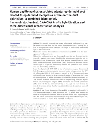
  • 1. C L I N I C A L A N D LA B O R A T O R Y I N V E S T I G A T I O N S DOI 10.1111/j.1365-2133.2005.06562.x Human papillomavirus-associated plantar epidermoid cyst related to epidermoid metaplasia of the eccrine duct epithelium: a combined histological, immunohistochemical, DNA–DNA in situ hybridization and three-dimensional reconstruction analysis K. Egawa, N. Egawa* and Y. Honda  Departments of Dermatology and  Surgical Pathology, Kumamoto University School of Medicine, 1-1-1 Honjo, Kumamoto 860-0811, Japan *Division of Cancer Cell Research, Institute of Medical Science, University of Tokyo, Minato-ku, Tokyo 108-8639, Japan Summary Correspondence Background We recently proposed that certain palmoplantar epidermoid cysts may Kiyofumi Egawa. be related to eccrine ducts and that human papillomavirus (HPV) 60 may play a E-mail: kuroibo@kaiju.medic.kumamoto-u.ac.jp role in their pathomechanism. However, the origin of palmoplantar epidermoid cysts is still controversial. Accepted for publication 9 October 2004 Objectives To examine the contribution of eccrine ducts and HPV 60 in the devel- opment of epidermoid cysts. Key words: Methods Five epidermoid cysts and four ridged warts that had developed on the eccrine duct, epidermoid cyst, human soles of a patient were studied histologically, immunohistochemically and by papillomavirus 60, inclusion cyst, metaplasia DNA–DNA in situ hybridization. Using serial sections obtained from its entire body, a three-dimensional reconstruction (3DR) analysis was performed on the Conflicts of interest: None declared. smallest cyst to analyse the relationship between the epidermoid cyst, eccrine duct and the overlying epidermis. Results Histological and DNA–DNA in situ hybridization analyses demonstrated both homogeneous intracytoplasmic inclusion bodies pathognomonic for HPV 60 infection and HPV 60 DNA sequences not only in all of the epidermoid cysts and ridged warts but also in the acrosyringeal portion of an eccrine duct, with the dermal portion of which the smallest cyst had been revealed to connect by 3DR analysis. However, immunohistochemical analyses using antibodies against human carcinoembryonic antigen (CEA), involucrin and several cytokeratins (CKs) revealed that the immunoreactivity of the cyst was not identical to that of the eccrine dermal duct but was identical to that of suprabasal layers of the epidermis. Conclusions It was clearly demonstrated that an HPV 60-associated epidermoid cyst with immunoreactivities for CEA, involucrin and CKs which were identical to those of the epidermis connected with the eccrine dermal duct, supporting the idea that certain palmoplantar epidermoid cysts may develop following the epi- dermoid metaplasia of eccrine ducts with HPV 60 infection. Epidermoid cysts of palmoplantar location (palmoplantar epi- epidermoid cysts, with or without infection by human papil- dermoid cysts) have been described as being caused by lomavirus (HPV) 60,5 and therefore proposed the alternative implantation of epidermal fragments into the dermis as a pathogenic hypothesis that certain palmoplantar epidermoid result of a penetrating or blunt injury.1 However, this assump- cysts may develop from eccrine ducts and that HPV 60 may tion has been questioned, and the necessity of a search for a play a role in this development. However, on the basis of the more common aetiology has been stressed.2 previous results,3–5 it is still likely that such epidermoid cysts We previously observed3,4 that eccrine sweat ducts were can develop as a result of the implantation of epidermal frag- frequently found in and ⁄or connecting with palmoplantar ments that contain eccrine ducts with or without HPV 60 Ó 2005 British Association of Dermatologists • British Journal of Dermatology 2005 152, pp961–967 961
  • 2. 962 HPV-associated eccrine epidermoid cyst, K. Egawa et al. infection from injuries as they do occasionally develop after injuries3,6 and are mainly located beneath the weight-bearing areas of the foot, i.e. the balls and heels.7 Indeed, Abe et al.8 and Ohnishi and Watanabe9 questioned the hypothesis of an ‘eccrine duct origin of the epidermoid cyst’ on the basis of the immunoreactivities of several cyto- keratins (CKs) which were not identical to those of sweat glands and ducts but were identical to those of suprabasal layers of the epidermis in plantar epidermoid cysts with HPV infection. However, Egawa et al.10 proposed the idea that such a CK profile in HPV-associated palmoplantar epidermoid cysts might be a reflection of the epidermoid metaplasia of the eccrine duct epithelium in the process of cyst formation in the eccrine ducts, as it was revealed that the CK profile of cholesteatomas, which are histologically defined as epidermoid cysts develop- ing in the middle ear,11 cannot be used as a variable to decide whether the origin of epidermoid cysts is epidermal or meta- plastic.12 Thus, the origin of palmoplantar epidermoid cysts is still controversial. Three-dimensional reconstruction (3DR) analysis from serial sections is now a powerful tool for increasing our comprehen- sion of the relationships among structural components, for the indication of the connections between components, and for revealing the distribution of components.13–15 In the present study, using serial sections obtained from the entire body of an epidermoid cyst, we attempted 3DR analysis to demon- Fig 1. Three subcutaneous nodules (solid arrows) and two ridged strate the fine structure of an HPV 60-associated epidermoid warts (open arrows) on the left sole. cyst of eccrine duct origin, in combination with histological, immunohistochemical and DNA–DNA in situ hybridization analyses. excised specimens were fixed in 10% formalin, and proc- essed with an automatic processor. Materials and methods Histopathology Patient Four-micrometre thick sections were obtained using a micro- A 21-year-old Japanese woman presented to Kumamoto Uni- tome from all of the biopsy specimens, stained with haema- versity Hospital in April 1995, with several subcutaneous nod- toxylin and eosin (H&E), and examined microscopically ules and warts on the soles of her feet. She first noticed (Figs 2 and 3). From the smallest cyst, gross serial sections several miliary-sized asymptomatic nodules on her soles were obtained from its entire body, and all sections were 3 months before presentation and 1 year after having had sev- numbered in the order of sectioning, from one side to the eral plantar warts treated with cryotherapy, although she could other. Fifty serial sections were obtained from each of the not say whether or not the nodules developed in the same other cysts. Every alternate section was stained with H&E for positions as the treated plantar warts. The nodules had gradu- histological analysis (Fig. 3), whereas the remaining sections ally enlarged and had come to cause tenderness during the were used for further examinations such as immunohisto- previous 3 months. chemical and DNA–DNA in situ hybridization analyses. At the time of her first visit, examination showed three subcutaneous nodules of 5–8 mm diameter and two small Processing of histological and immunohistochemical data flat warty lesions with clinical features of ridged warts16,17 for computer analysis on the left sole (Fig. 1) and one nodule of 7 mm diameter on the right sole. Her health was otherwise good. After Using photomicrographs of histopathological and immunohis- informed consent was obtained, all the lesions were tochemically stained sections (Fig. 4a), we made montages by removed successfully by scalpel under local anaesthesia. tracing the outlines of the histological ductal structures expres- However, about 6 months later, a further subcutaneous sing carcinoembryonic antigen (CEA) in serial sections in red, nodule of similar size appeared on the left sole, with two and those of the cyst wall and the overlying epidermis in blue ridged warts. They were also removed by scalpel. The (Fig. 4b). Ó 2005 British Association of Dermatologists • British Journal of Dermatology 2005 152, pp961–967
  • 3. HPV-associated eccrine epidermoid cyst, K. Egawa et al. 963 (Fig. 4d). The three-dimensional images were rotated along various axes to analyse the relationship between the cyst, eccrine ducts and the overlying epidermis. Immunohistochemistry An immunohistochemical procedure for human CEA,3,4,15,18 CKs8,9,19 and involucrin20 was performed on paraffin-embed- ded sections by the avidin–biotin–peroxidase complex (ABC) method,21 utilizing a Vectastain ABC kit (Vector Laboratories, Burlingame, CA, U.S.A.). A polyclonal antibody to human CEA (Dako, Glostrup, Denmark) and monoclonal antibodies to var- ious CKs (OV-TL 12 ⁄30, RCK108 and AE1 ⁄AE3, Dako; LL002, Biomeda, Foster City, CA, U.S.A.; and CAM5.2, Becton Dickin- son, San Jose, CA, U.S.A.) and involucrin (Novocastra, New- Fig 2. Histological features of a ridged wart. Vacuolar structures are castle-upon-Tyne, U.K.) were used as primary antibodies seen in the thickened horny layer and homogeneous intracytoplasmic (Table 1). In the case of CKs, the procedure was performed inclusion bodies are seen in the granular and upper spinous cell layers with or without proteinase K predigestion or autoclave heating of the acanthotic epidermis. A tiny keratinous cyst is seen just beneath for antigen retrieval as described previously.22 Negative con- the acanthotic epidermis (arrow). trols for immunostaining were performed by substituting the primary antibodies with phosphate-buffered saline or nonim- mune mouse or rabbit IgG. Three-dimensional reconstruction analysis An image scanner (IX-4025; Canon, Tokyo, Japan) and a per- DNA–DNA in situ hybridization sonal computer (Apple Macintosh 8500) were used in the 3DR imaging. Montages of the serial sections obtained from HPV 60 DNA sequences were detected in formalin-fixed, paraf- the entire body of the epidermoid cyst were displayed on a fin-embedded tissue sections using the DNA–DNA in situ hybrid- television monitor (Multiscan 17s; Sony, Tokyo, Japan). The ization method as described previously.23,24 Hybridization was outlines of the cysts and the eccrine ducts were digitized and performed at 37 °C for 16 h using digoxigenin-labelled HPV stored on the computer to make a wire-frame of these in vivo 60 complete genome DNA which we had previously cloned structures (Fig. 4c). Based on the wire-frame, three-dimen- from a plantar epidermoid cyst.3 The hybridization mixture sional images of the structures were reconstructed using an consisted of 10% dextran sulphate, 2 · saline sodium citrate image analysis program (Specular Infini-D; Specular, Amherst, (0Æ3% sodium chloride, 0Æ03 mol L)1 sodium citrate, pH 7Æ0), MA, U.S.A.) and were displayed on the television monitor 400 lg mL)1 sheared herring sperm DNA (Sigma, St Louis, Fig 3. Representative histological pictures selected from the serial sections obtained from the entire body of an epidermoid cyst. Section numbers appear at the lower left corner of each picture. Varying histological features coming from a hypertrophied eccrine dermal duct (no. 200, inset) connecting the apical portion of the cyst with an acrosyringium in the overlying epidermis are seen in the serial sections. The ecrine duct, appearing as a ductal structure in the apical portion of the cyst wall (nos 180–190), goes upwards through the dermis to the acrosyringium (nos 200–210) and connects with it in the overlying epidermis (no. 220). The acrosyringium is extremely dilated in the horny layer (no. 230). Ó 2005 British Association of Dermatologists • British Journal of Dermatology 2005 152, pp961–967
  • 4. 964 HPV-associated eccrine epidermoid cyst, K. Egawa et al. Fig 4. Computer-based, three-dimensional reconstruction (3DR) analysis of a human papillomavirus 60-associated plantar epidermoid cyst. (a) Histological section (the same as Figure 3, no.190). (b) Tracing of the outline of the histological ductal structures expressing carcinoembryonic antigen (red) and of the cyst wall and overlying epidermis (blue). (c) A wire-frame of the epidermoid cyst generated by computer using a montage of tracings obtained from all the serial sections. (d) 3DR analysis visualizing the fine structure of the epidermoid cyst connecting with the eccrine dermal duct. Table 1 Immunohistochemical staining in a human papillomavirus (HPV) 60-associated plantar epidermoid cyst Normal eccrine gland Secretory Eccrine Antibody Specificity portion dermal duct Epidermis Cyst wall HD CEA +++ +++ – – ++ (IML) AE1 ⁄ AE3 CKs 10, 14–16, 19 ⁄ 1–8 +++ +++ +++ +++ +++ LL002 CK14 – ++ ++ (LL) ++ (OL) + (OL) OV-TL 12 ⁄ 30 CK7 +++ – – – – RCK108 CK19 + – – – – CAM5.2 CKs 8, 18 +++ – – – – Involucrin – – +++ (UL) +++ (IL) ++ (IL) HD, hypertrophied eccrine dermal duct with which the HPV 60-associated epidermoid cyst connected; CEA, carcinoembryonic antigen; CK, cytokeratin; IML, innermost layers; LL, lower layers; OL, outer layers; UL, upper layers; IL, inner layers; –, negative; +, weak; ++, moderate; +++, strong. MO, U.S.A.), 50% formamide and 2 lg mL)1 of probe DNA. (Figs 3 and 5c). The characteristic histological feature com- The digoxigenin-labelled probe was detected by the streptavi- monly seen in all the ridged warts and the epidermoid cysts din-biotinylated polyalkaline phosphatase detection system was the presence of eosinophilic homogeneous intracyto- (DNA detection system; BRL, Gaithersburg, MD, U.S.A.). The plasmic inclusion bodies pathognomonic for HPV 60 phaeochromes used were nitroblue tetrazolium and 5-bromo- infection3–7,16,17,24–27 in the granular and upper spinous cell 4-chloro-3-indolylphosphate (Fig. 5). layers of the acanthotic epidermis of the ridged warts or in the inner cell layers of the cyst wall (Figs 2 and 5c). Vacuolar structures, also pathognomonic for HPV 60 infection,4,7 were Results also seen both in the horny layer of the ridged warts and in keratinous material within the cyst cavity (Figs 2 and 5c). Histology In the serial sections obtained from the entire body of the In the ridged warts, the epidermis was acanthotic with hyper- smallest cyst (Fig. 3), ductal structures coming from an eccrine keratosis and partial hypergranulosis. In one lesion, a tiny dermal duct were seen between the apical portion of the cyst keratinous cyst was seen just beneath the acanthotic epidermis and an acrosyringium in the overlying epidermis in the dermis (Fig. 2). Epidermoid cysts were lined by stratified squamous that connects the two histological components (Fig. 3, nos. epithelium, showing keratinization of the epidermoid type 190–220). The eccrine dermal duct was hypertrophied with Ó 2005 British Association of Dermatologists • British Journal of Dermatology 2005 152, pp961–967
  • 5. HPV-associated eccrine epidermoid cyst, K. Egawa et al. 965 duct but not the secretory portion. In contrast, OV-TL 12 ⁄30 (against CK7), RCK108 (against CK19) and CAM5.2 (against CKs 8 and 18) stained only the secretory portion. A monoclo- nal antibody against involucrin did not stain any portions of the normal eccrine sweat gland. Epidermis of the sole CEA was expressed only in the acrosyringeal epithelium in the epidermis. LL002 stained the lower cell layers; AE1 ⁄AE3 stained whole cell layers; OV-TL 12 ⁄30, RCK108 and CAM5.2 did not stain any cell layers; involucrin was expressed in the upper cell layers of the epidermis. Plantar epidermoid cyst CEA was not expressed in the cyst wall but was expressed in the acrosyringeal structures in the cyst cavity. LL002 stained the outer cell layers; AE1 ⁄AE3 stained whole cell layers; OV- TL 12 ⁄30, RCK108 and CAM5.2 did not stain any cell layers; involucrin was expressed in the inner cell layers of the cyst wall. Hypertrophied eccrine dermal duct with stratified squamous epithelium with which an epidermoid cyst connected Fig 5. Histological localization of human papillomavirus (HPV) 60 DNA sequences. A high-power view of the same section The innermost cell layers of the stratified epithelium of the (a,c; haematoxylin and eosin) and a serial section (b,d; DNA–DNA hypertrophied eccrine dermal duct expressed CEA. Expression in situ hybridization) of Figure 3, no. 230. Homogeneous of involucrin was also noted in the inner cell layers; LL002 intracytoplasmic inclusion bodies (a,c) and HPV 60 DNA sequences stained the outer cell layers; AE1 ⁄AE3 stained whole cell (b,d) are seen in the acrosyringeal epithelium (a,b) as well as in the layers; OV-TL 12 ⁄30, RCK108 and CAM5.2 did not stain any cyst wall (c,d). cell layers of the duct. The results are summarized in Table 1. stratified squamous epithelium (Fig. 3, no. 200, inset); the acrosyringium was extremely dilated in the horny layer DNA–DNA in situ hybridization (Fig. 3, no. 230; Fig. 5a); and the homogeneous intracytoplas- mic inclusion bodies were seen in the acrosyringeal epithelium HPV 60 DNA sequences were detected in all the epidermoid (Fig. 5a) as well as in the epidermoid cyst (Fig. 5b). cysts and ridged warts. In the smallest epidermoid cyst ana- Ductal structures suggestive of eccrine ducts were observed lysed with 3DR, the DNA sequences were identified both in in three of the other four epidermoid cysts, using 50 serial the acrosyringeal portion of the eccrine duct (Fig. 5b) and in sections obtained from each cyst. the cyst connecting with its dermal portion (Fig. 5d). Three-dimensional reconstruction analysis Discussion The analysis clearly demonstrated that the smallest epidermoid Implantation of epidermal fragments into the dermis has been cyst connected with the eccrine dermal duct (Fig. 4d). thought to be the main cause of palmoplantar epidermoid cysts.1 Although we have proposed an alternative pathogenic hypothesis that certain palmoplantar epidermoid cysts may Immunohistochemical observations develop from eccrine ducts and that HPV (especially HPV 60) may play a role in this development,3,4,10,17 there has been a Normal eccrine sweat gland lack of direct evidence to support this hypothesis. A polyclonal antibody against CEA and a monoclonal antibody HPV 60 was initially identified25,26 in and isolated5 from AE1 ⁄AE3 (against CKs 10, 14–16, 19 ⁄1–8) stained all the plantar epidermoid cysts with homogeneous intracytoplasmic secretory, dermal duct and acrosyringeal portions of the ec- inclusion bodies, and was also subsequently found in flat crine sweat gland. LL002 (against CK14) stained the dermal lesions retaining the appearance of dermal ridges on their Ó 2005 British Association of Dermatologists • British Journal of Dermatology 2005 152, pp961–967
  • 6. 966 HPV-associated eccrine epidermoid cyst, K. Egawa et al. surface (ridged warts).16,17 Thus, the clinical morphology of appearance of CKs characteristic of stratified and cornifying HPV 60-induced lesions also remains unclear. In the present epithelia, indicating a true change in the differentiation of the study, using 3DR analysis, it was clearly demonstrated that an middle ear epithelium.12 Interestingly, HPV has also been HPV 60-associated plantar epidermoid cyst connected with the identified in cholesteatoma.31 dermal portion of an eccrine duct. HPV 60 DNA sequences Although the epidermoid cyst analysed with 3DR in this were identified not only in the epidermoid cyst but also in study connected with an eccrine dermal duct, the immunore- the acrosyringeal portion of the eccrine duct by DNA–DNA activities for CEA, involucrin and several CKs of the cyst wall in situ hybridization. All the other plantar epidermoid cysts and were not identical to those of normal eccrine dermal ducts ridged warts that developed simultaneously on the same but were identical to those of the suprabasal layers of the patient also harboured HPV 60 DNA sequences, suggesting a epidermis, as observed by Abe et al.,8 Ohnishi and Watanabe9 close association between the lesions and HPV 60 infection. A and Yokogawa et al.32 This suggests that the immunoreactivi- tiny keratinous cyst, which might be a precursor of an HPV ties of the palmoplanatar epidermoid cysts may also be an 60-associated plantar epidermoid cyst, was also seen just indication of the metaplastic origin of the cysts from eccrine beneath the acanthotic epidermis of a ridged wart. ducts. In the present case, the eccrine dermal duct with In a previous report, two plantar epidermoid cysts were which the epidermoid cyst connected was hypertrophied attached to the epidermis and open to the surface in a patient with stratified squamous epithelium and expressed both CEA with multiple plantar epidermoid cysts harbouring both HPV and involucrin, suggesting a transitional form of the squa- 60 DNA sequences and eccrine ducts,3 suggesting that the mous metaplasia of the eccrine duct epithelium. cysts developed from the acrosyringeal or uppermost portions However, as Epstein and Kligman have suggested that kera- of eccrine ducts. As HPV 60 was frequently identified in tinizing cysts may develop from any part of the epithelial sys- palmoplantar epidermoid cysts concomitantly with eccrine tem,33 further studies are still required to determine whether ducts,3,4 we assumed that HPV 60 had a high affinity for the cases in which neither HPV infection nor an association eccrine duct epithelia and played a role in cyst formation.17 with eccrine ducts has been found in ordinary histological sec- Supporting this idea, recent studies28,29 have suggested that tions reflect a false-negative result or are an indication of epi- papillomaviruses, including HPVs, may target epidermal stem dermal inclusion cysts resulting from the implantation of cells, which may exist in association with eccrine ducts in the epidermal fragments. human palmoplantar skin.29 In the context of the previous findings and the results of Acknowledgments the present case, we speculate that HPV 60 initially infects the upper parts of the eccrine duct, such as the acrosyringeal epi- We thank Ms Chiemi Shiotsu and Ms Yoshiko Sonoda for tech- thelium, where ridged warts develop, and then migrates into nical assistance and Gyohei Egawa, Haruki Egawa and Motoe various parts of the dermal portions of the eccrine duct, where Egawa for help in drawing montages of histological pictures. the virus-associated epidermoid cysts develop. It has been revealed that HPV 60-associated palmoplantar References epidermoid cysts occasionally develop in association with pre- ceding blunt or penetrating injuries. The cysts are mainly 1 MacKie RM. Epidermal skin tumours. In: Textbook of Dermatology located beneath the weight-bearing areas of the foot, i.e. the (Champion RH, Burton JL, Ebling FJG, eds), 5th edn. Oxford: Blackwell Science, 1992; 1459–504. balls and heels;7 an HPV 60-associated plantar epidermoid cyst 2 Pinkus H. Epidermoid cysts or epidermal inclusion cysts? Arch Der- developed just beneath a plantar wart when the wart was matol 1975; 111:130. being treated with cryotherapy;6 multiple epidermoid cysts 3 Egawa K, Honda Y, Inaba Y et al. Multiple plantar epidermoid cysts harbouring both HPV 60 DNA sequences and eccrine ducts harboring carcinoembryonic antigen and human papillomavirus developed on the sole of a football player.3 DNA sequences. J Am Acad Dermatol 1994; 30:494–6. In the present case, the cryotherapy performed on the pre- 4 Egawa K, Honda Y, Inaba Y et al. Detection of human papillomavi- ceding plantar warts seemed to have a triggering role in the ruses and eccrine ducts in palmoplantar epidermoid cysts. Br J Der- matol 1995; 132:533–42. development of the multiple HPV 60-associated epidermoid 5 Matsukura T, Iwasaki T, Kawashima M. Molecular cloning of a novel cysts. Thus, traumatic injuries and mechanical pressure, which human papillomavirus (type 60) from a plantar epidermoid cyst have long been thought to have a causative role in the implan- with characteristic pathological changes. Virology 1992; 190:561–4. tation of epidermal fragments following cyst formation in the 6 Inaba Y, Egawa K, Muto K, Arao T. Detection of papillomavirus dermis,1 may have a triggering role in the progression of the common antigens in plantar epidermoid cysts. Jpn J Dermatol 1990; HPV 60-infected eccrine duct epithelium to the formation of 100:199–203. the cyst. 7 Egawa K, Inaba Y, Ono T, Arao T. ‘Cystic papilloma’ in humans? Demonstration of human papillomavirus in plantar epidermoid Squamous metaplasia is assumed to be a possible source of cysts. Arch Dermatol 1990; 126:1559–603. cholesteatoma,12,30 which is histologically defined as an epi- 8 Abe H, Ohnishi T, Watanabe S. Does plantar epidermoid cyst with dermoid cyst developing in the middle ear.11 The squamous human papillomavirus infection originate from the eccrine dermal cell metaplasia was immunohistochemically characterized by a duct? Br J Dermatol 1999; 141:161–2. loss of simple epithelial cell-related CKs accompanied by the Ó 2005 British Association of Dermatologists • British Journal of Dermatology 2005 152, pp961–967
  • 7. HPV-associated eccrine epidermoid cyst, K. Egawa et al. 967 9 Ohnishi T, Watanabe S. Immunohistochemical observation of cyto- between ABC and unlabeled antibody (PAP) procedures. J Histochem keratins in keratinous cysts including plantar epidermoid cyst. Cytochem 1981; 29:577–83. J Cutan Pathol 1999; 26:424–9. 22 Misumi S, Iyama K, Honda Y et al. Differential expression of base- 10 Egawa K, Kitasato H, Ono T. A palmar epidermoid cyst, showing ment membrane type-IV collagen a1, a2, a5 and a6 chains among histological features suggestive of eccrine duct origin, developing the histological subtypes of adenoid cystic carcinoma. Virchows Arch after a bee-sting. Br J Dermatol 2000; 143:469–70. 2004; 445:54–62. 11 Rosai J. Ear. In: Ackerman’s Surgical Pathology (Rosai L, ed.), 8th edn. 23 Egawa K, Sibasaki Y, de Villiers E-M. Double infection with human St Louis: Mosby, 1996; 2509–17. papillomavirus 1 and human papillomavirus 63 in a single cell of a 12 Kuijpers W, Vennix PP, Peters TA, Ramaekers FC. Squamous lesion displaying only an HPV 63-induced cytopathogenic effect. metaplasia of the middle ear epithelium. Acta Otolaryngol 1996; Lab Invest 1993; 69:583–8. 116:293–8. 24 Kawase M, Honda M, Niimura M. Detection of human papilloma- 13 Braverman MS, Braverman IM. Three-dimensional reconstructions virus type 60 in plantar epidermoid cysts and verruca plantaris by of objects from serial sections using a microcomputer graphics sys- the in situ hybridization method using digoxigenin labeled probe. tem. J Invest Dermatol 1986; 86:290–4. J Dermatol 1994; 21:709–15. 14 Salisbury JR, Whimster WF. Progress in computer-generated three- 25 Egawa K, Ono T, Arao T. Epidermoid cyst of the sole and human dimensional reconstruction. J Pathol 1993; 170:223–7. papillomavirus. Jpn J Dermatol 1987; 97:493–5. 15 Honda Y, Egawa K, Baba Y, Ono T. Sweat duct milia—immuno- 26 Kimura S, Sato N, Shigeeki H. A report of two cases of plantar epi- histological analysis of structure and three-dimensional reconstruc- dermoid cyst which have papillomavirus antigen. Hifu-Rinsho 1987; tion. Arch Dermatol Res 1996; 288:133–9. 29:487–91. 16 Honda A, Iwasaki T, Sata T et al. Human papillomavirus type 27 Egawa K. New types of human papillomaviruses and intracytoplas- 60-associated plantar wart: ridged wart. Arch Dermatol 1994; mic inclusion bodies, a classification of inclusion warts according 130:1413–17. to clinical features, histology and associated HPV types. Br J Dermatol 17 Egawa K, Kasai S, Hattori N et al. A case of a human papillomavirus 1994; 130:158–66. 60-induced wart with clinical appearance of both pigmented and 28 Schmit A, Rochat A, Zeltner R et al. The primary target cells of the ridged warts. Dermatology 1998; 197:268–70. high-risk cottontail rabbit papillomavirus colocalize with hair fol- 18 Pennys NS, Nadji M, McKinney EC. Carcinoembryonic antigen pre- licle stem cells. J Virol 1996; 70:1912–22. sent in human eccrine sweat. J Am Acad Dermatol 1981; 29:577–83. 29 Egawa K. Do human papillomaviruses target epidermal stem cells? 19 Watanabe S, Ichikawa E, Takanashi S, Takahashi H. Immunohisto- Dermatology 2003; 207:251–4. chemical localization of cytokeratins in normal eccrine glands, with 30 Sade J, Babiacki A, Pinkus G. The metaplastic and congenital origin monoclonal antibodies in routinely processed, formalin-fixed, par- of cholesteatoma. Acta Otolaryngol 1983; 96:119–29. affin-embedded sections. J Am Acad Dermatol 1993; 28:203–12. 31 Bergmann K, Hoppe F, Yukai H et al. Human papillomavirus DNA 20 Hudson DL, Weiland KL, Dooley TP et al. Characterization of in cholesteatomas. Int J Cancer 1994; 59:463–6. eight monoclonal antibodies to involucrin. Hybridoma 1992; 11: 32 Yokogawa M, Egawa K, Dabenaka K et al. Multiple palmar epider- 367–79. moid cysts. Dermatology 2002; 205:398–400. 21 Hsu H-M, Raine L, Franer H. The use of avidin–biotin peroxidase 33 Epstein WL, Kligman AM. Epithelial cysts in buried human skin. complex (ABC) in immunoperoxidase technique: a comparison Arch Dermatol 1957; 76:437–45. Ó 2005 British Association of Dermatologists • British Journal of Dermatology 2005 152, pp961–967