SlideShare uma empresa Scribd logo
1 de 11
Baixar para ler offline
Median Arcuate Ligament Syndrome
Literature Study and Osteopathic Considerations
Luc Peeters, MSc.Ost. and Grégoire Lason, MSc.Ost.
The International Academy of Osteopathy
http://www.osteopathy.eu


1. Introduction
Median Arcuate Ligament Syndrome (MALS) is also known as coeliac trunk
compression syndrome or Dunbar syndrome (Dunbar et al 1965). Occasionally it is
referred to as abdominal angina.
The median arcuate ligament can compress the coeliac trunk. This occurs frequently
but it is not understood why some cases are symptomatic while some remain
asymptomatic.
Various symptoms have been attributed to this syndrome, ranging from abdominal
symptoms to musculoskeletal symptoms to psychological symptoms (Carey et al
1969, Williams et al 1985).
Classical medicine will often advise operative intervention (the ligament is cut)
(Barcourt et al 1988, Ghosn et al 1982, Mihas et al 1977) but a clear indication for
such surgery is not present. Recently, laparoscopy has been used to relieve the
compression (Desmond & Roberts 2005). The results of these operations are highly
variable.
In the osteopathic practice patients with vague abdominal and subdiaphragmatic pain
- associated with eating or not - are frequently encountered. The osteopath will often
treat the stomach function, the diaphragm and the segments of the upper digestive
system. These patients often present with a very rigid and flattened thoracolumbar
spine. Decoaptation in this region will often improve the complaint.
Many of these complaints could be associated with median arcuate ligament
syndrome. Resultant ischemia of the organs in the upper digestive system and even
ischemia of the mesenteric root could also be the part of the problem. As well as the
arterial compression an irritation of the coeliac plexus could be contributing to the
complaint.


2. Anatomy
2.1. Median Arcuate Ligament
The median arcuate ligament is the tendinous junction between the right and left crus
of the diaphragm (Balaban 1997, Horton et al 2005). It is the anterosuperior
boundary of the aortic hiatus.
The histology of the ligament demonstrates dense connective tissue infiltrated by fat
cells, blood vessels and nerves. Striated muscle fibres are present together with
collagen fibres.


                                                                                    1
T12
              Median Arcuate lig.


                                Figure 1 – Median arcuate ligament

2.2. Coeliac Trunk
The coeliac trunk was found where the median arcuate ligament crosses the midline.
In12 of 83 cadavers (14.46%) the coeliac trunk was further from the ligament, in 35
cadavers (42.17%) the trunk touched the ligament and in 36 cases (43.37%) the
ligament crossed over the trunk. The average distance between both structures was
0.94 cm and when overlapping this distance became 0.42 cm (Dunbar et al 1965,
Petrella et al 2006). Certain authors suggest that the coeliac trunk is in fact
intrathoracic in some cases (Fadhli 1968, Warter et al 1970), which leads to a
congenital version of the compression syndrome.

                      Coeliac trunk                              Left gastric a.


    Right inferior phrenic a.                                    Left inferior phrenic a.

                                                                 Left subcostal a.
         Right subcostal a.
                                                                 Splenic a.
        Common hepatic a.
        communis
                                                                 Left median suprarenal a.
  Right median suprarenal a.
  right

                                                                 Superior mesenteric a.
                                                                 superior


                                      Figure 2 – Coeliac trunk


                                                                                             2
Relative to the spine the coeliac trunk is at the level of T12-L1.


                                                                     T12

                                                                     L1


                      Figure 3 – Topography of the coeliac trunk


                      Figure 4 – Compression of the coeliac trunk

The coeliac trunk is also the origin of the suspensory muscle of the duodenum. The
suspensory muscle of the duodenum is the bond between duodenum III and IV and
the coeliac trunk. This muscle consists of smooth muscle and connective tissue
(Saenco et al 1989).

2.3. Coeliac Plexus
The majority of the abdominal sympathetic nervous tissue is found anterior and
lateral upon the abdominal aorta. The upper part of this abdominal sympathetic
tissue is known as the coeliac plexus. The coeliac plexus is found at the level of T12-
L1 (Thomson et al 1977).
Studies of the individual variability of the location of the coeliac plexus show that in
32% of cases the coeliac plexus was an accumulation of numerous smaller ganglia
linked by a network of nerve fibres, relatively removed from the of the blood vessels
branching from the aorta. In 38 % of case the coeliac plexus was a grouping of


                                                                                      3
medium sized ganglia concentrated around the coeliac trunk, mostly on the left side
up to the branching of the superior mesenteric a. and without contact with the median
arcuate ligament. In 30% of cases the coeliac plexus was a grouping of 2 to 4 ganglia
inserting into the median arcuate ligament and forming a dense ring around the blood
vessels (Vlasova 2000).
The coeliac plexus is highly vascular (Promwikorn et al 1988). The arterial blood
comes from the aorta, inferior phrenic a. and the suprarenal arteries. The venous
drainage is via the inferior phrenic v. and the vena cava inferior.

3. Pathology
3.1. Arterial Compression
Due to compression of the coeliac trunk the arterial circulation of various digestive
organs is also likely to be compromised.
Anatomically, it is most likely that the arterial supply of the stomach, spleen and liver
are the most affected.
During exhalation the compression is increased due to the fact that the trunk ascends
(Reuter 1971, Reuter & Bernstein 1973). This is equally so in case of a diaphragm
high position dysfunction – due to fascial retractions in the thorax, congestion of
subdiaphragmatic organs or muscular weakness.
                                             Aorta
                                 Cardiac output = 5.000 ml/min

                          Superior mesenteric          Coeliac
                             700 ml/min                700 ml/min
                                   Inferior mesenteric           Gastric      Hepatic
                                   400 ml/min                                               500 ml/min

      Pancreas                                                         Splenic

                     Small                                 Stomach
                   Intestine

                               Colon                                       Spleen


                                                                                        Liver    Vena cava
                     Vena porta (1.300ml/min.)


         Figure 5 – Distribution of the arterial supply to the digestive organs


                                                                                                         4
Abdominal part of cardia

                                     branch to                                        Fundus
                                     Oesophagus
                                                                                      Short gastric arteries
                   Left gastric a.
                           Aorta
                   Coeliac trunk                                                      Corpus
             Gastroduodenal a.

                 Right gastric a.
                                                                                      Splenic a.
                        Pylorus

                                                                                      Left gastroepiploic a.


          Right gastroepiploic a.

Superior pancreaticoduodenal a.             Duodenum
                                                                    Greater omentum

                                                                 Atrium

                                     Figure 6 – Arterial supply to the stomach

      Via compression from the ligament ischemia can develop in the organs of the
      digestive system. Such ischemia can be mild but in serious cases can result in
      necrosis. In serious cases where the symptoms are confirmed with arteriography,
      urgent surgery is required as this situation can be life threatening, most certainly if
      combined with arteriosclerosis of the involved vessels (Sulkowski & Wolters 2000).
      In such serious cases the ischemia becomes constant and leads to the symptoms
      being independent of eating. In more mild cases the ischemic pain will most
      commonly occur 15 min after eating due to the fact that the arterial flow to the
      stomach cannot increase as required.

      3.2. Irritation of the Coeliac Plexus
      Balaban et al showed in 1997 that surgical reduction of the pressure upon the coeliac
      trunk affected the contractile rhythm of the stomach.
      Gastroparesis and gastric arrhythmia were diagnosed pre-operatively and were
      shown to be resolved post-operatively by way of a normal myoelectric activity of the
      stomach (of 3 cycles per minute). This finding indicates that the symptoms can be of
      neurogenic origin via irritation of the coeliac plexus.
      Trophic changes to the coeliac plexus could be the underlying cause of the coeliac
      trunk stenosis (Drapanas & Bron 1966, Harjola 1963 & 1968, Harjola & Lahtiharju
      1968, Jamieson 1986, Marable et al 1966, Of Gossun et al 1984).

      3.3. Dysfunction of the Duodenojejunal Angle
      This functional sphincter has no actual sphincter fibres but does suspend from the
      coeliac trunk and duo III and IV by way of a system consisting of smooth muscle
      (suspensory muscle of the duodenum), a striated muscle originating from the
      diaphragm and fibrous tissue. It acts to limit the transit so that the function of the
      duodenum is better controlled. If the functional sphincter system does not properly
      function then the evacuation of the duodenum is no longer exponential (first
      adequate filling must occur before the contents are allowed to advance further). The
      stomach-duodenal evacuation is interrupted.


                                                                                                               5
When transit is too fast the result is poor digestion.

4. Symptoms
The symptoms indicative of this syndrome are:
  • Abdominal pain not related to eating but due to the ischemia (in 71% of
      cases).
  • Systolic murmur (in 15% of cases).
  • Nausea (in 29% of cases).
  • Lower thoracic and thoracolumbar pain (in 22% of cases).
  • Acidic reflux (in 17% of cases).
  • Weight loss (in 15% of cases). The weight loss is due to the lack of normal
      increase in blood supply 15 min after beginning to eat. The pain is usually
      associated with eating.
  • Vomiting (in 15% of cases).
  • Diarrhoea (in 14% of cases). Occurs due to dysfunction of the coeliac plexus
      as is observed secondary to coeliac plexus block during pain therapy.
  • Breathlessness (in 14% of cases).
  • Murmur during auscultation due to the post-stenotic turbulence (Edwards et
      al 1970).

The syndrome occurs most commonly in adults but has been described in children
(Dubbins & Scholbach 2006) most likely due to abnormal congenital anatomy.


5. Functional Osteopathic Treatment
The osteopath must be aware of this syndrome. Treatment of the serious, structural
cases with associated arteriosclerosis is contraindicated but during the functional
stage osteopathic treatment is recommended.
Before osteopathic treatment commences arteriosclerosis must be discounted by
appropriate testing.

5.1. The osteopathic treatment has the following aims:
   •   Improving the mobility of the thoracolumbar junction: most important is
       improvement of the mobility into flexion via a thoracolumbar decoaptation
       technique.
   •   The normalisation of the diaphragm function: most important is increasing
       the inhalation capacity as it is during exhalation (or a dysfunction in high
       position of the diaphragm) when the coeliac trunk ascends and the chance of
       compression is most significant.
   •   The decongestion and stretching of the region ventral to T12-L1: the
       region of the coeliac plexus is the aim here.
   •   A fat-reducing diet is necessary for obese patients: the median arcuate
       ligament consists of fat cells and any increase will increase the chance of
       compression.

5.2. Specific Techniques
This article is written for professional osteopaths. It is assumed therefore that all the
necessary techniques required to achieve the aims above can be carried out in
safety. However, several specific techniques for this particular complaint are
presented below.


                                                                                       6
5.2.1. Active Mobilisation of the Coeliac Trunk
The patient is sitting on their knees and bent forward as far as possible so that the
abdomen approaches or even touched the knees. The patient abdominally inhales as
deeply as possible and is instructed to hold their breath while actively flexing their
thoracolumbar region (kyphosis). In this way the increase in abdominal pressure will
flex the thoracolumbar region and the coeliac trunk will descend. The contraction of
the diaphragm during the inhalation will place cranial traction upon the median
arcuate ligament. At the same time a drainage effect will occur in the abdominal
venous system.
This technique is repeated several times.


                  Figure 7 – Active mobilisation of the coeliac trunk

5.2.2. Defibrosing of the Coeliac Plexus (patient standing)
Stretch and mobilisation of the region will create a defibrosing action for the coeliac
plexus.


   Using the palmar side of 3 fingers,           While exhaling, the patient bends
   the osteopath contacts the zone of        forward and the osteopath strengthens
           the coeliac plexus.               the contact to cranial/posterior/right, in
                                              the direction of the vertebral bodies of
                                                               T12-L1.
                                              The osteopath stretches the region of
                                                the coeliac plexus several times to
                                              caudal while the patient remains bent
                                                  forwards (for optimal contact).


                                                                                          7
The osteopath holds the contact on
                     the coeliac plexus to posterior/caudal
                      while the patient exhales and stands
                                   up straight.
            Figure 8 – Defibrosing of the coeliac plexus (patient standing)

The effect of this technique is very strong and must be used with due consideration.
If neurovegetative signs such as sweating occur, the force of the contact should be
reduced. It should be clear that this technique is contraindicated in cases of
arteriosclerosis.
Problems of the coeliac plexus will often result in local pain with palpation. Avoid this
pain during the technique.
During or just after the technique orthostatic hypotension can occur. Inform the
patient that this frequently occurring reaction will spontaneously resolve.

5.2.3. Defibrosing of the Coeliac Plexus (patient supine)
The patient is supine with both legs bent.
The osteopath stands next to the patient and uses both hands to hook
lateral/posterior around the small intestine in the direction of the coeliac trunk. This is
in the direction of the bodies of spinal levels T12-L1. The osteopath hooks around
ventrally to the spine and mobilises towards him/herself. The technique is done
during exhalation otherwise the technique cannot be done deeply enough.
The technique is gentle, pain free and done several times on both sides.
It should be clear that this technique is contraindicated in cases of arteriosclerosis.


             Figure 9 – Defibrosing of the coeliac plexus (patient supine)

5.2.4. Lift and Mobilisation of the Duodenojejunal Angle
The patient is sitting and the osteopath stands behind the patient.


                                                                                         8
Using the fingers of both hands the osteopath hooks into the abdomen just left of the
midline, at the height of the navel, under the duodenojejunal angle. This structure is
not palpable as such but the anatomical location is used.
The region is lifted to cranial during an exhalation of the patient and then the region is
mobilised to mediolateral.
The technique is repeated several times.
This mobilisation will improve the circulatory supply and therefore the trophic
condition of the region around the coeliac trunk and plexus. Any adhesions and
retractions are also stretched.


            Figure 10 – Lift and mobilisation of the duodenojejunal angle


6. Bibliography
   •   Bacourt F., Goeau-Brissonnière O. & Koskas F. (1988) Compression associé
       du tronc coeliaque et the l'artère meséntérique supérieure par le diaphragme.
       Chirurgie, 114: 762-768.
   •   Balaban D.H., Chen J. Lin Z., Tribble C.G. & McCallum R.W. (1997) Median
       arcuate ligament syndrome: a possible cause of idiopathic gastroparesis. Am.
       J. Gastroenterol. 92(3): 519-523.
   •   Carey J. P., Stemmer E. A. & Connolly J. E. (1969) Median arcuate ligament
       syndrome. Arch. Surg. 99: 441-446.
   •   Desmond C.P. & Roberts S.K. (2004) Exercise - related abdominal pain as a
       manifestation of the median arcuate ligament syndrome. Scand. J.
       Gastroenterol, 39 (12): 1310 -1313.
   •   Drapanas T. & Bron K.M. (1966) Stenosis of the celiac artery. Ann. Surg., 164:
       1085-1088.
   •   Dubbins P.A. & Scholbach T. (2006) Celiac artery compression in children,
       adolescents, and young adults. J. Ultrasound Med. 25(8): 1108 - 1109.
   •   Dunbar J.D., Molnar W., Beman F.F. & Marable S.A. (1965) Compression of
       the celiac trunk and abdominal angina. Am. J. Roentgenol. Radium. Ther. Nucl.
       Med. 95: 731.
   •   Edwards A.J., Hamilton J.D., Nichol W.D., Taylor G.W. & Dawson A.M. (1970)
       Experience with Coeliac Axis Compression Syndrome. Br. Med. J. 1(5692):
       342–345.
   •   Fadhli H.A. (1968) Congenital diaphragmatic obstruction of the aorta and the
       celiac artery. J. Thorac. Cardiovasc. Surg., 55: 431-433.


                                                                                        9
•   Ghosn P.B., Rabbat A.G., Trudel J. D'Amico P. Lecours R. & Trudel J. (1982)
    Celiac compression syndrome. Can. J. Surg. 25(4): 377-379.
•   Harjola P.T. (1963) A rare obstruction of the coeliac artery. Ann. Chir.
    Gynaecol. Fenn. 52: 547-570.
•   Harjola P.T. (1968) Coeliac axis constriction and abdominal angina. Bull. Soc.
    Intern. Chir. 27: 464-467.
•   Harjola P.T. & Lahtiharju A. (1968) Celiac axis syndrome. Abdominal angina
    caused by external compression of the celiac artery. Am. J. Surg., 115: 864-
    869.
•   Horton K.M., Talamini M.A. & Fishman E.K. (2005) Median arcuate ligament
    syndrome: evaluation with CT angiography. Radiographics. 25: 1177.
•   Jamieson C.W. (1986) Coeliac axis compression syndrome. Br. Med. J.,
    293:159-160.
•   Marable S.A., Molnar W. & Beman F.M. (1966) Abdominal pain secondary to
    celiac axis compression. Am. J. Surg., 111: 493-495.
•   Mihas A.A., Laws H.L. & Jander H.P. (1977) Surgical treatment of the celiac
    axis compression syndrome. Am. J. Surg.133 (6): 688-691.
•   Petrella S., the Sousa Rodriguez C.F., Sgrott E.A., Medeiros Fernandes G.J.,
    Marques S.R. & Prates J.C. (2006) Relationship of the Celiac Trunk with
    Median Arcuate Ligament of the Diaphragm. Int. J. Morphol. 24(2): 263-274.
•   Promwikorn W., Thongpila S., Pradidarcheep W., Mingsakul T., Chunhabundit
    P. & Somana R. (1998) Angioarchitecture of the coeliac sympathetic ganglion
    complex in the common tree shrew (Tupaia glis) J. Anat. 193(Pt 3): 409–416.
•   Reuter S.R. (1971) Accentuation of celiac compression by the median arcuate
    ligament of the diaphragm during deep expiration. Diagn. Radiol. 98: 561-564.
•   Reuter S.R. & Bernstein E.F. (1973) The anatomic basis for respiratory
    variation in median ligament compression of the celiac artery. Surgery, 73:
    381-385.
•   Saenko V.F., Virchenko S.B., Kucherenko T.L. and Belianskii L.S. (1989) The
    role of Treitz' ligament in regulating the evacuation of solid food from the
    duodenum. Biull. Eksp. Biol. Med.108 (9): 263-266.
•   Sulkowski U. & Wolters U. (2000) Das Syndrom des Median arcuate ligament
    mit Zweigefäßbeteiligung. From the book Gefässchirurgie (Ausgabe NaN/2000)
    Springer Verlag.
•   Thompson G.E., Moore D.C., Bridenbaugh L.D. & Artin R.Y. (1977) Abdominal
    pain and alcohol celiac plexus nerve block. Anesth. Analg. 56: 1-5.
•   Of Gossun M., Morobe J., Of Laethem A., Burette A., Somerhausen J. &
    Schockert J. (1984) Syndrome the compression isolée du tronc coeliaque
    (SCITC). À propos the 8 CAS. Acta Chir. Belg., 84: 379-383.
•   Vlasova M.I. (2000) Individual variability in the structure and topography of the
    celiac plexus and its clinical significance. Morfologia. 117(1): 16-19.
•   Warter J., Storck D., Kieny R. & Tongio J. (1970) Sténoses congénitals du
    tronc coeliaque. Arch. Fr. Mal. Appar. Dig. 59: 765-780.
•   Williams S. Gillespie P. & Little J.M. (1985) Celiac axis compression syndrome:
    factors predicting a favorable outcome. Surgery 98(5): 879-887.


                                                                                 10
iNeuro App (App Store) A must have for every osteopath and health care
                     provider. Simple to use and no unnecessary information. It keeps your
                     knowledge sharp for daily patient care!

                     iCranialNerves App (App Store): The iCranialNerves App describes the 12
                     cranial nerves. The origin, course and different functions (somato-motor, somato-
                     sensory, viscero-motor, viscero-sensory, special senses) are described and
                     presented in clear drawings and the different neurological tests are shown on
                     streaming video.
                     This App gives students and professionals in medicine, osteopathy, chiropractic,
                     physiotherapy, manual therapy and other medical disciplines a practical tool for
study and daily use. The APP is unique in its completeness and ease of use.


                   Also look for iBooks in the iBook Store from Luc Peeters and Grégoire Lason.


                                                                                                   11

Mais conteúdo relacionado

Mais procurados

Biliary stricture ppt
Biliary stricture pptBiliary stricture ppt
Biliary stricture ppt
Sumer Yadav
 
Superior mesenteric artery syndrome
Superior mesenteric artery syndromeSuperior mesenteric artery syndrome
Superior mesenteric artery syndrome
Abhilash Cheriyan
 
Pancreatic pseudocyst
Pancreatic pseudocystPancreatic pseudocyst
Pancreatic pseudocyst
draakif
 

Mais procurados (20)

Biliary stricture ppt
Biliary stricture pptBiliary stricture ppt
Biliary stricture ppt
 
Superior mesenteric artery syndrome
Superior mesenteric artery syndromeSuperior mesenteric artery syndrome
Superior mesenteric artery syndrome
 
Pancreaticoduodenectomy (whipple procedure)
Pancreaticoduodenectomy (whipple procedure)Pancreaticoduodenectomy (whipple procedure)
Pancreaticoduodenectomy (whipple procedure)
 
Splenic trauma - Causes, Complications, Management
Splenic trauma - Causes, Complications, ManagementSplenic trauma - Causes, Complications, Management
Splenic trauma - Causes, Complications, Management
 
Splenectomy
Splenectomy Splenectomy
Splenectomy
 
Pancreatic pseudocyst
Pancreatic pseudocystPancreatic pseudocyst
Pancreatic pseudocyst
 
Rif mass
Rif massRif mass
Rif mass
 
Liver trauma
Liver traumaLiver trauma
Liver trauma
 
Lt hemicolectomy - Surgical Approach, Complications.
Lt hemicolectomy - Surgical Approach, Complications.Lt hemicolectomy - Surgical Approach, Complications.
Lt hemicolectomy - Surgical Approach, Complications.
 
Management of common bile duct stones
Management of common bile duct stonesManagement of common bile duct stones
Management of common bile duct stones
 
Internal hernia
Internal herniaInternal hernia
Internal hernia
 
Surgical Management of Chronic Pancreatitis
Surgical Management of Chronic PancreatitisSurgical Management of Chronic Pancreatitis
Surgical Management of Chronic Pancreatitis
 
Revised Atlanta classification of Acute Pancreatitis
Revised Atlanta classification of Acute PancreatitisRevised Atlanta classification of Acute Pancreatitis
Revised Atlanta classification of Acute Pancreatitis
 
Internal hernia
Internal herniaInternal hernia
Internal hernia
 
Choledochal cyst
Choledochal cystCholedochal cyst
Choledochal cyst
 
Post gastrectomy syndrome
Post gastrectomy syndrome   Post gastrectomy syndrome
Post gastrectomy syndrome
 
1.OGILVIE SYNDROME.pptx
1.OGILVIE SYNDROME.pptx1.OGILVIE SYNDROME.pptx
1.OGILVIE SYNDROME.pptx
 
Obstructive jaundice
Obstructive  jaundiceObstructive  jaundice
Obstructive jaundice
 
Surgical Jaundice
Surgical JaundiceSurgical Jaundice
Surgical Jaundice
 
Obstructive jaundice
Obstructive jaundiceObstructive jaundice
Obstructive jaundice
 

Semelhante a Median Arcuate Ligament Syndrome

The gastrointestinal system
The gastrointestinal systemThe gastrointestinal system
The gastrointestinal system
Dr Motawei
 
Esophagus stomach-dr.gosai
Esophagus stomach-dr.gosaiEsophagus stomach-dr.gosai
Esophagus stomach-dr.gosai
Dr.B.B. Gosai
 
Acs0904 Urologic Considerations For The General Surgeon
Acs0904 Urologic Considerations For The General SurgeonAcs0904 Urologic Considerations For The General Surgeon
Acs0904 Urologic Considerations For The General Surgeon
medbookonline
 
Chapter22 digestivepart2marieb
Chapter22 digestivepart2mariebChapter22 digestivepart2marieb
Chapter22 digestivepart2marieb
Lawrence James
 
Anterolateral abdominal wall (rectus sheath) & hernia
Anterolateral abdominal wall (rectus sheath) & herniaAnterolateral abdominal wall (rectus sheath) & hernia
Anterolateral abdominal wall (rectus sheath) & hernia
Irma Suntoo
 
Liver, biliary system, pancreas and spleen
Liver, biliary system, pancreas and spleenLiver, biliary system, pancreas and spleen
Liver, biliary system, pancreas and spleen
ess_online
 

Semelhante a Median Arcuate Ligament Syndrome (20)

The gastrointestinal system
The gastrointestinal systemThe gastrointestinal system
The gastrointestinal system
 
Anatomy of the stomach
Anatomy of the stomachAnatomy of the stomach
Anatomy of the stomach
 
Anatomy Of The Stomach
Anatomy Of The StomachAnatomy Of The Stomach
Anatomy Of The Stomach
 
Esophagus stomach-dr.gosai
Esophagus stomach-dr.gosaiEsophagus stomach-dr.gosai
Esophagus stomach-dr.gosai
 
Acs0904 Urologic Considerations For The General Surgeon
Acs0904 Urologic Considerations For The General SurgeonAcs0904 Urologic Considerations For The General Surgeon
Acs0904 Urologic Considerations For The General Surgeon
 
Chapter 4-stomach
Chapter 4-stomachChapter 4-stomach
Chapter 4-stomach
 
Splanchnic circulation
Splanchnic circulationSplanchnic circulation
Splanchnic circulation
 
PERITONEUM.pptx
PERITONEUM.pptxPERITONEUM.pptx
PERITONEUM.pptx
 
Stomach
StomachStomach
Stomach
 
peritoneum
 peritoneum peritoneum
peritoneum
 
Chapter22 digestivepart2marieb
Chapter22 digestivepart2mariebChapter22 digestivepart2marieb
Chapter22 digestivepart2marieb
 
H
HH
H
 
Chapter23 digestivepart2marieb
Chapter23 digestivepart2mariebChapter23 digestivepart2marieb
Chapter23 digestivepart2marieb
 
Anterolateral abdominal wall (rectus sheath) & hernia
Anterolateral abdominal wall (rectus sheath) & herniaAnterolateral abdominal wall (rectus sheath) & hernia
Anterolateral abdominal wall (rectus sheath) & hernia
 
Liver, biliary system, pancreas and spleen
Liver, biliary system, pancreas and spleenLiver, biliary system, pancreas and spleen
Liver, biliary system, pancreas and spleen
 
Development of stomach
Development of stomachDevelopment of stomach
Development of stomach
 
Summary: Anatomy of Abdomen Anterior Abdominal Wall, Esophagus and Stomach
Summary: Anatomy of Abdomen Anterior Abdominal Wall, Esophagus and StomachSummary: Anatomy of Abdomen Anterior Abdominal Wall, Esophagus and Stomach
Summary: Anatomy of Abdomen Anterior Abdominal Wall, Esophagus and Stomach
 
Peritoneal cavity and relations
Peritoneal cavity and relationsPeritoneal cavity and relations
Peritoneal cavity and relations
 
Anatomy of Pancreas
Anatomy of PancreasAnatomy of Pancreas
Anatomy of Pancreas
 
pancreas-180218085812.pdf
pancreas-180218085812.pdfpancreas-180218085812.pdf
pancreas-180218085812.pdf
 

Mais de IAO The International Academy of Osteopathy

Mais de IAO The International Academy of Osteopathy (20)

Opleiding Osteopathie België Nederland
Opleiding Osteopathie België NederlandOpleiding Osteopathie België Nederland
Opleiding Osteopathie België Nederland
 
Ausbildung Osteopathie Deutschland - Österreich - Schweiz
Ausbildung Osteopathie Deutschland - Österreich - SchweizAusbildung Osteopathie Deutschland - Österreich - Schweiz
Ausbildung Osteopathie Deutschland - Österreich - Schweiz
 
Master opleiding tot osteopaat - Modulair - Gent-Antwerpen-Zeist
Master opleiding tot osteopaat - Modulair - Gent-Antwerpen-ZeistMaster opleiding tot osteopaat - Modulair - Gent-Antwerpen-Zeist
Master opleiding tot osteopaat - Modulair - Gent-Antwerpen-Zeist
 
Ausbildung osteopathie
Ausbildung osteopathieAusbildung osteopathie
Ausbildung osteopathie
 
Flyer de juni_17
Flyer de juni_17Flyer de juni_17
Flyer de juni_17
 
Introduction course june_2017
Introduction course june_2017Introduction course june_2017
Introduction course june_2017
 
Pediatric Osteopathy Copenhagen 2017 2019
Pediatric Osteopathy Copenhagen  2017 2019Pediatric Osteopathy Copenhagen  2017 2019
Pediatric Osteopathy Copenhagen 2017 2019
 
IAO Flyer Deutsch
IAO Flyer DeutschIAO Flyer Deutsch
IAO Flyer Deutsch
 
Poster app journal_de
Poster app journal_dePoster app journal_de
Poster app journal_de
 
Why do clever people believe stupid things.docx
Why do clever people believe stupid things.docxWhy do clever people believe stupid things.docx
Why do clever people believe stupid things.docx
 
The Vertebral Artery Test
The Vertebral Artery TestThe Vertebral Artery Test
The Vertebral Artery Test
 
Modern Principles of Osteopathy
Modern Principles of OsteopathyModern Principles of Osteopathy
Modern Principles of Osteopathy
 
Diaphragm and posture
Diaphragm and postureDiaphragm and posture
Diaphragm and posture
 
Research event iao prof. comhaire
Research event iao prof. comhaireResearch event iao prof. comhaire
Research event iao prof. comhaire
 
Persbericht research avond the international academy of osteopathy
Persbericht research avond the international academy of osteopathyPersbericht research avond the international academy of osteopathy
Persbericht research avond the international academy of osteopathy
 
Vertebral Artery Pathology
Vertebral Artery PathologyVertebral Artery Pathology
Vertebral Artery Pathology
 
Common Referred Pain Patterns – Low Back
Common Referred Pain Patterns – Low BackCommon Referred Pain Patterns – Low Back
Common Referred Pain Patterns – Low Back
 
Preventive Paediatric Osteopathy
Preventive Paediatric OsteopathyPreventive Paediatric Osteopathy
Preventive Paediatric Osteopathy
 
The Place of Osteopathy
The Place of OsteopathyThe Place of Osteopathy
The Place of Osteopathy
 
Pelvic, Hip and Core Stability
Pelvic, Hip and Core StabilityPelvic, Hip and Core Stability
Pelvic, Hip and Core Stability
 

Último

Russian Call Girls Lucknow Just Call 👉👉7877925207 Top Class Call Girl Service...
Russian Call Girls Lucknow Just Call 👉👉7877925207 Top Class Call Girl Service...Russian Call Girls Lucknow Just Call 👉👉7877925207 Top Class Call Girl Service...
Russian Call Girls Lucknow Just Call 👉👉7877925207 Top Class Call Girl Service...
adilkhan87451
 
Call Girls in Gagan Vihar (delhi) call me [🔝 9953056974 🔝] escort service 24X7
Call Girls in Gagan Vihar (delhi) call me [🔝  9953056974 🔝] escort service 24X7Call Girls in Gagan Vihar (delhi) call me [🔝  9953056974 🔝] escort service 24X7
Call Girls in Gagan Vihar (delhi) call me [🔝 9953056974 🔝] escort service 24X7
9953056974 Low Rate Call Girls In Saket, Delhi NCR
 

Último (20)

Russian Call Girls Lucknow Just Call 👉👉7877925207 Top Class Call Girl Service...
Russian Call Girls Lucknow Just Call 👉👉7877925207 Top Class Call Girl Service...Russian Call Girls Lucknow Just Call 👉👉7877925207 Top Class Call Girl Service...
Russian Call Girls Lucknow Just Call 👉👉7877925207 Top Class Call Girl Service...
 
💕SONAM KUMAR💕Premium Call Girls Jaipur ↘️9257276172 ↙️One Night Stand With Lo...
💕SONAM KUMAR💕Premium Call Girls Jaipur ↘️9257276172 ↙️One Night Stand With Lo...💕SONAM KUMAR💕Premium Call Girls Jaipur ↘️9257276172 ↙️One Night Stand With Lo...
💕SONAM KUMAR💕Premium Call Girls Jaipur ↘️9257276172 ↙️One Night Stand With Lo...
 
9630942363 Genuine Call Girls In Ahmedabad Gujarat Call Girls Service
9630942363 Genuine Call Girls In Ahmedabad Gujarat Call Girls Service9630942363 Genuine Call Girls In Ahmedabad Gujarat Call Girls Service
9630942363 Genuine Call Girls In Ahmedabad Gujarat Call Girls Service
 
Call Girls in Gagan Vihar (delhi) call me [🔝 9953056974 🔝] escort service 24X7
Call Girls in Gagan Vihar (delhi) call me [🔝  9953056974 🔝] escort service 24X7Call Girls in Gagan Vihar (delhi) call me [🔝  9953056974 🔝] escort service 24X7
Call Girls in Gagan Vihar (delhi) call me [🔝 9953056974 🔝] escort service 24X7
 
Saket * Call Girls in Delhi - Phone 9711199012 Escorts Service at 6k to 50k a...
Saket * Call Girls in Delhi - Phone 9711199012 Escorts Service at 6k to 50k a...Saket * Call Girls in Delhi - Phone 9711199012 Escorts Service at 6k to 50k a...
Saket * Call Girls in Delhi - Phone 9711199012 Escorts Service at 6k to 50k a...
 
Call Girls Service Jaipur {9521753030} ❤️VVIP RIDDHI Call Girl in Jaipur Raja...
Call Girls Service Jaipur {9521753030} ❤️VVIP RIDDHI Call Girl in Jaipur Raja...Call Girls Service Jaipur {9521753030} ❤️VVIP RIDDHI Call Girl in Jaipur Raja...
Call Girls Service Jaipur {9521753030} ❤️VVIP RIDDHI Call Girl in Jaipur Raja...
 
Call Girls Hyderabad Just Call 8250077686 Top Class Call Girl Service Available
Call Girls Hyderabad Just Call 8250077686 Top Class Call Girl Service AvailableCall Girls Hyderabad Just Call 8250077686 Top Class Call Girl Service Available
Call Girls Hyderabad Just Call 8250077686 Top Class Call Girl Service Available
 
Most Beautiful Call Girl in Bangalore Contact on Whatsapp
Most Beautiful Call Girl in Bangalore Contact on WhatsappMost Beautiful Call Girl in Bangalore Contact on Whatsapp
Most Beautiful Call Girl in Bangalore Contact on Whatsapp
 
Low Rate Call Girls Bangalore {7304373326} ❤️VVIP NISHA Call Girls in Bangalo...
Low Rate Call Girls Bangalore {7304373326} ❤️VVIP NISHA Call Girls in Bangalo...Low Rate Call Girls Bangalore {7304373326} ❤️VVIP NISHA Call Girls in Bangalo...
Low Rate Call Girls Bangalore {7304373326} ❤️VVIP NISHA Call Girls in Bangalo...
 
Call Girls Kolkata Kalikapur 💯Call Us 🔝 8005736733 🔝 💃 Top Class Call Girl Se...
Call Girls Kolkata Kalikapur 💯Call Us 🔝 8005736733 🔝 💃 Top Class Call Girl Se...Call Girls Kolkata Kalikapur 💯Call Us 🔝 8005736733 🔝 💃 Top Class Call Girl Se...
Call Girls Kolkata Kalikapur 💯Call Us 🔝 8005736733 🔝 💃 Top Class Call Girl Se...
 
Premium Bangalore Call Girls Jigani Dail 6378878445 Escort Service For Hot Ma...
Premium Bangalore Call Girls Jigani Dail 6378878445 Escort Service For Hot Ma...Premium Bangalore Call Girls Jigani Dail 6378878445 Escort Service For Hot Ma...
Premium Bangalore Call Girls Jigani Dail 6378878445 Escort Service For Hot Ma...
 
Manyata Tech Park ( Call Girls ) Bangalore ✔ 6297143586 ✔ Hot Model With Sexy...
Manyata Tech Park ( Call Girls ) Bangalore ✔ 6297143586 ✔ Hot Model With Sexy...Manyata Tech Park ( Call Girls ) Bangalore ✔ 6297143586 ✔ Hot Model With Sexy...
Manyata Tech Park ( Call Girls ) Bangalore ✔ 6297143586 ✔ Hot Model With Sexy...
 
Independent Call Girls Service Mohali Sector 116 | 6367187148 | Call Girl Ser...
Independent Call Girls Service Mohali Sector 116 | 6367187148 | Call Girl Ser...Independent Call Girls Service Mohali Sector 116 | 6367187148 | Call Girl Ser...
Independent Call Girls Service Mohali Sector 116 | 6367187148 | Call Girl Ser...
 
(Low Rate RASHMI ) Rate Of Call Girls Jaipur ❣ 8445551418 ❣ Elite Models & Ce...
(Low Rate RASHMI ) Rate Of Call Girls Jaipur ❣ 8445551418 ❣ Elite Models & Ce...(Low Rate RASHMI ) Rate Of Call Girls Jaipur ❣ 8445551418 ❣ Elite Models & Ce...
(Low Rate RASHMI ) Rate Of Call Girls Jaipur ❣ 8445551418 ❣ Elite Models & Ce...
 
Call Girls in Delhi Triveni Complex Escort Service(🔝))/WhatsApp 97111⇛47426
Call Girls in Delhi Triveni Complex Escort Service(🔝))/WhatsApp 97111⇛47426Call Girls in Delhi Triveni Complex Escort Service(🔝))/WhatsApp 97111⇛47426
Call Girls in Delhi Triveni Complex Escort Service(🔝))/WhatsApp 97111⇛47426
 
Call Girls Ahmedabad Just Call 9630942363 Top Class Call Girl Service Available
Call Girls Ahmedabad Just Call 9630942363 Top Class Call Girl Service AvailableCall Girls Ahmedabad Just Call 9630942363 Top Class Call Girl Service Available
Call Girls Ahmedabad Just Call 9630942363 Top Class Call Girl Service Available
 
Best Rate (Guwahati ) Call Girls Guwahati ⟟ 8617370543 ⟟ High Class Call Girl...
Best Rate (Guwahati ) Call Girls Guwahati ⟟ 8617370543 ⟟ High Class Call Girl...Best Rate (Guwahati ) Call Girls Guwahati ⟟ 8617370543 ⟟ High Class Call Girl...
Best Rate (Guwahati ) Call Girls Guwahati ⟟ 8617370543 ⟟ High Class Call Girl...
 
Call Girls Raipur Just Call 9630942363 Top Class Call Girl Service Available
Call Girls Raipur Just Call 9630942363 Top Class Call Girl Service AvailableCall Girls Raipur Just Call 9630942363 Top Class Call Girl Service Available
Call Girls Raipur Just Call 9630942363 Top Class Call Girl Service Available
 
Premium Call Girls In Jaipur {8445551418} ❤️VVIP SEEMA Call Girl in Jaipur Ra...
Premium Call Girls In Jaipur {8445551418} ❤️VVIP SEEMA Call Girl in Jaipur Ra...Premium Call Girls In Jaipur {8445551418} ❤️VVIP SEEMA Call Girl in Jaipur Ra...
Premium Call Girls In Jaipur {8445551418} ❤️VVIP SEEMA Call Girl in Jaipur Ra...
 
Russian Call Girls Service Jaipur {8445551418} ❤️PALLAVI VIP Jaipur Call Gir...
Russian Call Girls Service  Jaipur {8445551418} ❤️PALLAVI VIP Jaipur Call Gir...Russian Call Girls Service  Jaipur {8445551418} ❤️PALLAVI VIP Jaipur Call Gir...
Russian Call Girls Service Jaipur {8445551418} ❤️PALLAVI VIP Jaipur Call Gir...
 

Median Arcuate Ligament Syndrome

  • 1. Median Arcuate Ligament Syndrome Literature Study and Osteopathic Considerations Luc Peeters, MSc.Ost. and Grégoire Lason, MSc.Ost. The International Academy of Osteopathy http://www.osteopathy.eu 1. Introduction Median Arcuate Ligament Syndrome (MALS) is also known as coeliac trunk compression syndrome or Dunbar syndrome (Dunbar et al 1965). Occasionally it is referred to as abdominal angina. The median arcuate ligament can compress the coeliac trunk. This occurs frequently but it is not understood why some cases are symptomatic while some remain asymptomatic. Various symptoms have been attributed to this syndrome, ranging from abdominal symptoms to musculoskeletal symptoms to psychological symptoms (Carey et al 1969, Williams et al 1985). Classical medicine will often advise operative intervention (the ligament is cut) (Barcourt et al 1988, Ghosn et al 1982, Mihas et al 1977) but a clear indication for such surgery is not present. Recently, laparoscopy has been used to relieve the compression (Desmond & Roberts 2005). The results of these operations are highly variable. In the osteopathic practice patients with vague abdominal and subdiaphragmatic pain - associated with eating or not - are frequently encountered. The osteopath will often treat the stomach function, the diaphragm and the segments of the upper digestive system. These patients often present with a very rigid and flattened thoracolumbar spine. Decoaptation in this region will often improve the complaint. Many of these complaints could be associated with median arcuate ligament syndrome. Resultant ischemia of the organs in the upper digestive system and even ischemia of the mesenteric root could also be the part of the problem. As well as the arterial compression an irritation of the coeliac plexus could be contributing to the complaint. 2. Anatomy 2.1. Median Arcuate Ligament The median arcuate ligament is the tendinous junction between the right and left crus of the diaphragm (Balaban 1997, Horton et al 2005). It is the anterosuperior boundary of the aortic hiatus. The histology of the ligament demonstrates dense connective tissue infiltrated by fat cells, blood vessels and nerves. Striated muscle fibres are present together with collagen fibres. 1
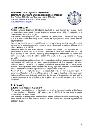
  • 2. T12 Median Arcuate lig. Figure 1 – Median arcuate ligament 2.2. Coeliac Trunk The coeliac trunk was found where the median arcuate ligament crosses the midline. In12 of 83 cadavers (14.46%) the coeliac trunk was further from the ligament, in 35 cadavers (42.17%) the trunk touched the ligament and in 36 cases (43.37%) the ligament crossed over the trunk. The average distance between both structures was 0.94 cm and when overlapping this distance became 0.42 cm (Dunbar et al 1965, Petrella et al 2006). Certain authors suggest that the coeliac trunk is in fact intrathoracic in some cases (Fadhli 1968, Warter et al 1970), which leads to a congenital version of the compression syndrome. Coeliac trunk Left gastric a. Right inferior phrenic a. Left inferior phrenic a. Left subcostal a. Right subcostal a. Splenic a. Common hepatic a. communis Left median suprarenal a. Right median suprarenal a. right Superior mesenteric a. superior Figure 2 – Coeliac trunk 2
  • 3. Relative to the spine the coeliac trunk is at the level of T12-L1. T12 L1 Figure 3 – Topography of the coeliac trunk Figure 4 – Compression of the coeliac trunk The coeliac trunk is also the origin of the suspensory muscle of the duodenum. The suspensory muscle of the duodenum is the bond between duodenum III and IV and the coeliac trunk. This muscle consists of smooth muscle and connective tissue (Saenco et al 1989). 2.3. Coeliac Plexus The majority of the abdominal sympathetic nervous tissue is found anterior and lateral upon the abdominal aorta. The upper part of this abdominal sympathetic tissue is known as the coeliac plexus. The coeliac plexus is found at the level of T12- L1 (Thomson et al 1977). Studies of the individual variability of the location of the coeliac plexus show that in 32% of cases the coeliac plexus was an accumulation of numerous smaller ganglia linked by a network of nerve fibres, relatively removed from the of the blood vessels branching from the aorta. In 38 % of case the coeliac plexus was a grouping of 3
  • 4. medium sized ganglia concentrated around the coeliac trunk, mostly on the left side up to the branching of the superior mesenteric a. and without contact with the median arcuate ligament. In 30% of cases the coeliac plexus was a grouping of 2 to 4 ganglia inserting into the median arcuate ligament and forming a dense ring around the blood vessels (Vlasova 2000). The coeliac plexus is highly vascular (Promwikorn et al 1988). The arterial blood comes from the aorta, inferior phrenic a. and the suprarenal arteries. The venous drainage is via the inferior phrenic v. and the vena cava inferior. 3. Pathology 3.1. Arterial Compression Due to compression of the coeliac trunk the arterial circulation of various digestive organs is also likely to be compromised. Anatomically, it is most likely that the arterial supply of the stomach, spleen and liver are the most affected. During exhalation the compression is increased due to the fact that the trunk ascends (Reuter 1971, Reuter & Bernstein 1973). This is equally so in case of a diaphragm high position dysfunction – due to fascial retractions in the thorax, congestion of subdiaphragmatic organs or muscular weakness. Aorta Cardiac output = 5.000 ml/min Superior mesenteric Coeliac 700 ml/min 700 ml/min Inferior mesenteric Gastric Hepatic 400 ml/min 500 ml/min Pancreas Splenic Small Stomach Intestine Colon Spleen Liver Vena cava Vena porta (1.300ml/min.) Figure 5 – Distribution of the arterial supply to the digestive organs 4
  • 5. Abdominal part of cardia branch to Fundus Oesophagus Short gastric arteries Left gastric a. Aorta Coeliac trunk Corpus Gastroduodenal a. Right gastric a. Splenic a. Pylorus Left gastroepiploic a. Right gastroepiploic a. Superior pancreaticoduodenal a. Duodenum Greater omentum Atrium Figure 6 – Arterial supply to the stomach Via compression from the ligament ischemia can develop in the organs of the digestive system. Such ischemia can be mild but in serious cases can result in necrosis. In serious cases where the symptoms are confirmed with arteriography, urgent surgery is required as this situation can be life threatening, most certainly if combined with arteriosclerosis of the involved vessels (Sulkowski & Wolters 2000). In such serious cases the ischemia becomes constant and leads to the symptoms being independent of eating. In more mild cases the ischemic pain will most commonly occur 15 min after eating due to the fact that the arterial flow to the stomach cannot increase as required. 3.2. Irritation of the Coeliac Plexus Balaban et al showed in 1997 that surgical reduction of the pressure upon the coeliac trunk affected the contractile rhythm of the stomach. Gastroparesis and gastric arrhythmia were diagnosed pre-operatively and were shown to be resolved post-operatively by way of a normal myoelectric activity of the stomach (of 3 cycles per minute). This finding indicates that the symptoms can be of neurogenic origin via irritation of the coeliac plexus. Trophic changes to the coeliac plexus could be the underlying cause of the coeliac trunk stenosis (Drapanas & Bron 1966, Harjola 1963 & 1968, Harjola & Lahtiharju 1968, Jamieson 1986, Marable et al 1966, Of Gossun et al 1984). 3.3. Dysfunction of the Duodenojejunal Angle This functional sphincter has no actual sphincter fibres but does suspend from the coeliac trunk and duo III and IV by way of a system consisting of smooth muscle (suspensory muscle of the duodenum), a striated muscle originating from the diaphragm and fibrous tissue. It acts to limit the transit so that the function of the duodenum is better controlled. If the functional sphincter system does not properly function then the evacuation of the duodenum is no longer exponential (first adequate filling must occur before the contents are allowed to advance further). The stomach-duodenal evacuation is interrupted. 5
  • 6. When transit is too fast the result is poor digestion. 4. Symptoms The symptoms indicative of this syndrome are: • Abdominal pain not related to eating but due to the ischemia (in 71% of cases). • Systolic murmur (in 15% of cases). • Nausea (in 29% of cases). • Lower thoracic and thoracolumbar pain (in 22% of cases). • Acidic reflux (in 17% of cases). • Weight loss (in 15% of cases). The weight loss is due to the lack of normal increase in blood supply 15 min after beginning to eat. The pain is usually associated with eating. • Vomiting (in 15% of cases). • Diarrhoea (in 14% of cases). Occurs due to dysfunction of the coeliac plexus as is observed secondary to coeliac plexus block during pain therapy. • Breathlessness (in 14% of cases). • Murmur during auscultation due to the post-stenotic turbulence (Edwards et al 1970). The syndrome occurs most commonly in adults but has been described in children (Dubbins & Scholbach 2006) most likely due to abnormal congenital anatomy. 5. Functional Osteopathic Treatment The osteopath must be aware of this syndrome. Treatment of the serious, structural cases with associated arteriosclerosis is contraindicated but during the functional stage osteopathic treatment is recommended. Before osteopathic treatment commences arteriosclerosis must be discounted by appropriate testing. 5.1. The osteopathic treatment has the following aims: • Improving the mobility of the thoracolumbar junction: most important is improvement of the mobility into flexion via a thoracolumbar decoaptation technique. • The normalisation of the diaphragm function: most important is increasing the inhalation capacity as it is during exhalation (or a dysfunction in high position of the diaphragm) when the coeliac trunk ascends and the chance of compression is most significant. • The decongestion and stretching of the region ventral to T12-L1: the region of the coeliac plexus is the aim here. • A fat-reducing diet is necessary for obese patients: the median arcuate ligament consists of fat cells and any increase will increase the chance of compression. 5.2. Specific Techniques This article is written for professional osteopaths. It is assumed therefore that all the necessary techniques required to achieve the aims above can be carried out in safety. However, several specific techniques for this particular complaint are presented below. 6
  • 7. 5.2.1. Active Mobilisation of the Coeliac Trunk The patient is sitting on their knees and bent forward as far as possible so that the abdomen approaches or even touched the knees. The patient abdominally inhales as deeply as possible and is instructed to hold their breath while actively flexing their thoracolumbar region (kyphosis). In this way the increase in abdominal pressure will flex the thoracolumbar region and the coeliac trunk will descend. The contraction of the diaphragm during the inhalation will place cranial traction upon the median arcuate ligament. At the same time a drainage effect will occur in the abdominal venous system. This technique is repeated several times. Figure 7 – Active mobilisation of the coeliac trunk 5.2.2. Defibrosing of the Coeliac Plexus (patient standing) Stretch and mobilisation of the region will create a defibrosing action for the coeliac plexus. Using the palmar side of 3 fingers, While exhaling, the patient bends the osteopath contacts the zone of forward and the osteopath strengthens the coeliac plexus. the contact to cranial/posterior/right, in the direction of the vertebral bodies of T12-L1. The osteopath stretches the region of the coeliac plexus several times to caudal while the patient remains bent forwards (for optimal contact). 7
  • 8. The osteopath holds the contact on the coeliac plexus to posterior/caudal while the patient exhales and stands up straight. Figure 8 – Defibrosing of the coeliac plexus (patient standing) The effect of this technique is very strong and must be used with due consideration. If neurovegetative signs such as sweating occur, the force of the contact should be reduced. It should be clear that this technique is contraindicated in cases of arteriosclerosis. Problems of the coeliac plexus will often result in local pain with palpation. Avoid this pain during the technique. During or just after the technique orthostatic hypotension can occur. Inform the patient that this frequently occurring reaction will spontaneously resolve. 5.2.3. Defibrosing of the Coeliac Plexus (patient supine) The patient is supine with both legs bent. The osteopath stands next to the patient and uses both hands to hook lateral/posterior around the small intestine in the direction of the coeliac trunk. This is in the direction of the bodies of spinal levels T12-L1. The osteopath hooks around ventrally to the spine and mobilises towards him/herself. The technique is done during exhalation otherwise the technique cannot be done deeply enough. The technique is gentle, pain free and done several times on both sides. It should be clear that this technique is contraindicated in cases of arteriosclerosis. Figure 9 – Defibrosing of the coeliac plexus (patient supine) 5.2.4. Lift and Mobilisation of the Duodenojejunal Angle The patient is sitting and the osteopath stands behind the patient. 8
  • 9. Using the fingers of both hands the osteopath hooks into the abdomen just left of the midline, at the height of the navel, under the duodenojejunal angle. This structure is not palpable as such but the anatomical location is used. The region is lifted to cranial during an exhalation of the patient and then the region is mobilised to mediolateral. The technique is repeated several times. This mobilisation will improve the circulatory supply and therefore the trophic condition of the region around the coeliac trunk and plexus. Any adhesions and retractions are also stretched. Figure 10 – Lift and mobilisation of the duodenojejunal angle 6. Bibliography • Bacourt F., Goeau-Brissonnière O. & Koskas F. (1988) Compression associé du tronc coeliaque et the l'artère meséntérique supérieure par le diaphragme. Chirurgie, 114: 762-768. • Balaban D.H., Chen J. Lin Z., Tribble C.G. & McCallum R.W. (1997) Median arcuate ligament syndrome: a possible cause of idiopathic gastroparesis. Am. J. Gastroenterol. 92(3): 519-523. • Carey J. P., Stemmer E. A. & Connolly J. E. (1969) Median arcuate ligament syndrome. Arch. Surg. 99: 441-446. • Desmond C.P. & Roberts S.K. (2004) Exercise - related abdominal pain as a manifestation of the median arcuate ligament syndrome. Scand. J. Gastroenterol, 39 (12): 1310 -1313. • Drapanas T. & Bron K.M. (1966) Stenosis of the celiac artery. Ann. Surg., 164: 1085-1088. • Dubbins P.A. & Scholbach T. (2006) Celiac artery compression in children, adolescents, and young adults. J. Ultrasound Med. 25(8): 1108 - 1109. • Dunbar J.D., Molnar W., Beman F.F. & Marable S.A. (1965) Compression of the celiac trunk and abdominal angina. Am. J. Roentgenol. Radium. Ther. Nucl. Med. 95: 731. • Edwards A.J., Hamilton J.D., Nichol W.D., Taylor G.W. & Dawson A.M. (1970) Experience with Coeliac Axis Compression Syndrome. Br. Med. J. 1(5692): 342–345. • Fadhli H.A. (1968) Congenital diaphragmatic obstruction of the aorta and the celiac artery. J. Thorac. Cardiovasc. Surg., 55: 431-433. 9
  • 10. Ghosn P.B., Rabbat A.G., Trudel J. D'Amico P. Lecours R. & Trudel J. (1982) Celiac compression syndrome. Can. J. Surg. 25(4): 377-379. • Harjola P.T. (1963) A rare obstruction of the coeliac artery. Ann. Chir. Gynaecol. Fenn. 52: 547-570. • Harjola P.T. (1968) Coeliac axis constriction and abdominal angina. Bull. Soc. Intern. Chir. 27: 464-467. • Harjola P.T. & Lahtiharju A. (1968) Celiac axis syndrome. Abdominal angina caused by external compression of the celiac artery. Am. J. Surg., 115: 864- 869. • Horton K.M., Talamini M.A. & Fishman E.K. (2005) Median arcuate ligament syndrome: evaluation with CT angiography. Radiographics. 25: 1177. • Jamieson C.W. (1986) Coeliac axis compression syndrome. Br. Med. J., 293:159-160. • Marable S.A., Molnar W. & Beman F.M. (1966) Abdominal pain secondary to celiac axis compression. Am. J. Surg., 111: 493-495. • Mihas A.A., Laws H.L. & Jander H.P. (1977) Surgical treatment of the celiac axis compression syndrome. Am. J. Surg.133 (6): 688-691. • Petrella S., the Sousa Rodriguez C.F., Sgrott E.A., Medeiros Fernandes G.J., Marques S.R. & Prates J.C. (2006) Relationship of the Celiac Trunk with Median Arcuate Ligament of the Diaphragm. Int. J. Morphol. 24(2): 263-274. • Promwikorn W., Thongpila S., Pradidarcheep W., Mingsakul T., Chunhabundit P. & Somana R. (1998) Angioarchitecture of the coeliac sympathetic ganglion complex in the common tree shrew (Tupaia glis) J. Anat. 193(Pt 3): 409–416. • Reuter S.R. (1971) Accentuation of celiac compression by the median arcuate ligament of the diaphragm during deep expiration. Diagn. Radiol. 98: 561-564. • Reuter S.R. & Bernstein E.F. (1973) The anatomic basis for respiratory variation in median ligament compression of the celiac artery. Surgery, 73: 381-385. • Saenko V.F., Virchenko S.B., Kucherenko T.L. and Belianskii L.S. (1989) The role of Treitz' ligament in regulating the evacuation of solid food from the duodenum. Biull. Eksp. Biol. Med.108 (9): 263-266. • Sulkowski U. & Wolters U. (2000) Das Syndrom des Median arcuate ligament mit Zweigefäßbeteiligung. From the book Gefässchirurgie (Ausgabe NaN/2000) Springer Verlag. • Thompson G.E., Moore D.C., Bridenbaugh L.D. & Artin R.Y. (1977) Abdominal pain and alcohol celiac plexus nerve block. Anesth. Analg. 56: 1-5. • Of Gossun M., Morobe J., Of Laethem A., Burette A., Somerhausen J. & Schockert J. (1984) Syndrome the compression isolée du tronc coeliaque (SCITC). À propos the 8 CAS. Acta Chir. Belg., 84: 379-383. • Vlasova M.I. (2000) Individual variability in the structure and topography of the celiac plexus and its clinical significance. Morfologia. 117(1): 16-19. • Warter J., Storck D., Kieny R. & Tongio J. (1970) Sténoses congénitals du tronc coeliaque. Arch. Fr. Mal. Appar. Dig. 59: 765-780. • Williams S. Gillespie P. & Little J.M. (1985) Celiac axis compression syndrome: factors predicting a favorable outcome. Surgery 98(5): 879-887. 10
  • 11. iNeuro App (App Store) A must have for every osteopath and health care provider. Simple to use and no unnecessary information. It keeps your knowledge sharp for daily patient care! iCranialNerves App (App Store): The iCranialNerves App describes the 12 cranial nerves. The origin, course and different functions (somato-motor, somato- sensory, viscero-motor, viscero-sensory, special senses) are described and presented in clear drawings and the different neurological tests are shown on streaming video. This App gives students and professionals in medicine, osteopathy, chiropractic, physiotherapy, manual therapy and other medical disciplines a practical tool for study and daily use. The APP is unique in its completeness and ease of use. Also look for iBooks in the iBook Store from Luc Peeters and Grégoire Lason. 11